- “Monster Stars” existed in the early universe Astronomy Magazine
- Astronomers find first direct evidence of “Monster Stars” from the cosmic dawn Center for Astrophysics | Harvard & Smithsonian
- James Webb Space Telescope finds 1st evidence…
Author: admin
-
“Monster Stars” existed in the early universe – Astronomy Magazine
-
African States Explore Incentives, Challenges and Solutions to Advancing Strategic Trade Management of Dual-Use Items to Prevent WMD Proliferation
From 2 to 4 December 2025, the United Nations Office for Disarmament Affairs (UNODA), in partnership with the Republic of South Africa, the European Union Partner-to-Partner Export Control Programme for Dual-Use Goods (EU P2P), and the German…
Continue Reading
-
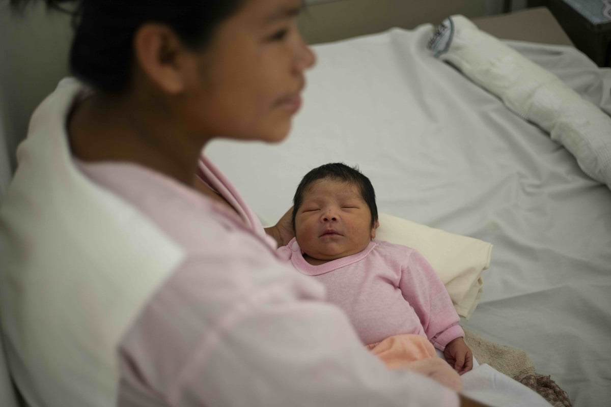
WHO validates Brazil for eliminating mother-to-child transmission of HIV
The World Health Organization (WHO) has validated Brazil for the elimination of mother-to-child transmission (EMTCT) of HIV, making it the most populous country in the Americas to achieve this historic milestone. This accomplishment reflects…
Continue Reading
-
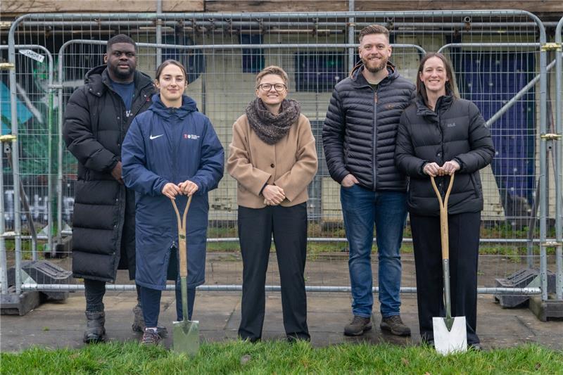
Refurbishment of Changing Pavilion at Cringle Playing Fields begins
A significant refurbishment of the changing pavilion at Cringle Playing Fields is underway.
Manchester City Council, in partnership with the Premier League, The FA and Government’s Football Foundation, is spearheading the modernisation,…
Continue Reading
-

King Charles pours ‘perfect pint’ at new brewery
The King was opening the Guinness Open Gate Brewery, a £73m investment which will be a new London visitor attraction as well as producing a range of beers.
There was a lesson in how to pour a pint, with the King shown how to tilt the glass to 45…
Continue Reading
-

Astronomers just watched a black hole twist spacetime
The universe has delivered a rare breakthrough for researchers chasing one of the hardest effects to catch in the night sky.
In findings reported in Science Advances, scientists describe the first observations of a spiraling swirl in spacetime…
Continue Reading
-

Moving further, faster: Our research highlights of 2025
2025 was another impactful year for our research, as we poured over £5.5m into pioneering brain tumour projects around the world.
We also announced 16 new grants, including four Quality of Life Awards, three…
Continue Reading
-

Soccer unties the world – SuperJoost Playlist
Despite having criticized The Game Awards in the past, I really enjoyed the 2025 edition.
My long-standing concern has been that the games industry shouldn’t emulate other forms of entertainment merely for the sake of perceived legitimacy….
Continue Reading
-
UNGA at 80: From 1946 to Our Future
The half-day programme will feature speeches and panel discussions focused on practical solutions and charting a constructive path forward for the UN. Topics will include the UN’s role in promoting international peace, security, and…
Continue Reading
-
UNODA Vienna Marks 25 Years of Women, Peace and Security
2025 marks a year of important anniversaries in the global effort to achieve gender equality. The UNODA Vienna Office celebrated the 25th anniversary of Security Council Resolution 1325 and the Women, Peace and Security (WPS) agenda with an…
Continue Reading