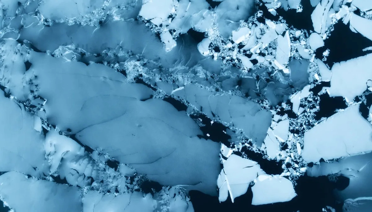
Author: admin

  • F1-inspired aerodynamics with an aggressive appearance

    F1-inspired aerodynamics with an aggressive appearance

    The 2025 season has been full of action and intrigue as teams and drivers learn more and more about the new car. From how to set it up to the best way to get pace out of it, everyone’s understanding has taken a step forward since Melbourne.

    The new car is a striking departure from the last generation, and new 16-inch profile tyres add up to create an impressive-looking machine. But what is the thinking behind its design?

    FIA Formula 3 Technical Director Pierre-Alain Michot breaks down the philosophy behind the ’25 car and the process that went in to designing it.

    “The way we designed the new car has been to follow in the footsteps of F2, so mainly making sure that the car has better capability to follow the one ahead in an efficient manner and so they don’t lose too much downforce,” he explains.

    “That was the main target we had. But we also wanted to keep the ‘family look’ in line with F1, but with a more conventional rear wing design.”
    Michot says that the design started out from a completely new beginning point compared to the old generation of car.

    From the “blank page” the new car started from, there were key design elements in mind based around ensuring the drivers were able to experience a step-up from what they would have driven before, along with preparing them up for what’s to come in Formula 2 and beyond.

    Michot says the philosophy of the new F3 car was to simplify but emulate the F2 design

    “We had a blank page beginning with this car. But we benefitted from the previous generation and the F2 car design in the process. But in the end, it’s a completely brand-new car, and none of the components are the same as before.

    “We wanted to have a smaller car that looked like an F2 one, but with a simpler floor, less complexity and downforce than in F2.

    “That was also to make setting the car up simpler and easier as well. It’s less complex than the F2 car and in terms of design, it’s easier for the teams but we’ve still been able to make it look like an F2 car, it’s just easier to maintain which is good for the teams and the drivers.”

    READ MORE

    Those points had to be hit whilst still keeping the complexity relatively simple, enabling teams to learn the car quickly.

    It also had to be similar enough to the F2 and F1 cars of the current generation that the lineage of the F3 car was obvious, though with its own unique identity.

    Michot adds that from the front to the rear of the car, it has all been designed with that FIA pyramid pathway in mind.

    “In the end, the general philosophy we ended up having at the beginning of the project and in the initial sketches were still present in the end.

    The rear wing is designed for efficiency but to still retain the F1-family appearance
    The rear wing is designed for efficiency but to still retain the ‘F1-family’ appearance

    “For sure, there was some iterating to make sure that it was as efficient as planned and performed as expected, because from the first drawing to the last produced part, we had to make sure everything worked well together.

    “From the front wing to the floor to the rear wing, it all had to be efficient as was part of the overall package and process. So, we still had to have a front wing looking similar to F1, and the sidepod mimicking the latest F1-spec without being as complex, along with having a rear wing which worked in terms of downforce and DRS efficiency.”

    The current generation F2 car introduced last season came with two rear wing specifications, standard and low downforce configurations that teams could choose between based on circuit requirements.

    READ MORE: Bruno Michel Double-Header Debrief: The twists keep coming in closely fought title battle

    While there was a consideration for the new F3 car to utilise a similar design philosophy, ultimately it was decided that things should be kept simple, with the focus on complexity based around other areas of the car.

    “We evaluated it at the time, but in the end, we wanted to keep it as a single concept for the rear wing.

    “We had to make sure that we could go from a low downforce set-up used at Monza to the highest downforce set-up. So, in the end, the wing that we designed was just to make sure we could accomplish this with a single configuration.”

    The revamped F3 car is able to corner faster as a result of the changes made for 2025
    The revamped F3 car is able to corner faster as a result of the changes made for 2025

    The design has revamped the aerodynamic behaviour of the car, with a simpler version of the F2 floor, which itself is based closer to those used in Formula 1, and changed how teams and drivers reach the limits.

    Cornering speeds in particular have been improved with the new car, as drivers have noted they’re able to push more through the turns compared to the previous generation.

    “When the drivers arrive in F3 from other categories further down the pyramid, they reach this level and it’s another level of downforce.

    “They need to accommodate for that added corner speed, and when they go up, it’s another step in a car that’s even more efficient. It’s about being able to give them a bit of a step-up and more to account for in their driving.

    “The positivity we’ve had is really good”

    “So, in the end, the car is slightly more efficient overall as a package. On the chassis side, we have a better handling car, and that’s also down to the low-profile tyres, because of the lower side wall. It means that the cars are better through mainly high-speed corners.”

    Overall, there has been a positive reaction to the appearance and performance of the new car, with the larger wheels and striking wing designs modernising the F3 car and moving it closer to its older siblings in F2 and F1.

    Michot says that it has been gratifying to see the enthusiasm for the new car, and that the ’25 machine is performing as expected.

    “The positivity we’ve had is really good. Because the balance of the car, the dimensions plus the 16-inch wheels, the shape of the sidepods mean it looks quite aggressive, but it is still good looking and most importantly, efficient.

    “It’s still in its first year, so we will see if we have to do some evolution or modification. But from what we’ve seen on track so far, it looks to be as efficient and working as expected.”

    Continue Reading

  • Phillips ruled out of Zimbabwe tour with injury | Robinson to remain as T20 cover

    Phillips ruled out of Zimbabwe tour with injury | Robinson to remain as T20 cover

    BLACKCAPS batting allrounder Glenn Phillips has been withdrawn from the tour of Zimbabwe after injuring his right groin.

    Phillips suffered the injury during the Major League Cricket (MLC) final and was assessed on arrival in Zimbabwe where it was determined he would require a number of weeks to rehabilitate.

    Wellington Firebirds batter Tim Robinson, who joined the squad as cover for players involved in the MLC final, will stay with the squad for the remainder of the T20 Tri-Series. 

    BLACKCAPS head coach Rob Walter said he felt for Phillips.

    “It’s obviously disappointing to lose someone of Glenn’s calibre,” said Walter.

    “Much like Finn, we really feel for Glenn and that he misses out on this series.

    “We know he was eager to get out on the field for the BLACKCAPS and unfortunately, he won’t be able to do that for this series.

    “We know he’ll work hard to get himself back onto the park, and I look forward to when that happens.”

    Phillips’ replacement in the Test squad will be named in due course.

    He will return to New Zealand with Mitch Hay and Jimmy Neesham who were also called in as cover alongside Robinson.

    Continue Reading

  • Security forces arrest suspected suicide bombers – RADIO PAKISTAN

    1. Security forces arrest suspected suicide bombers  RADIO PAKISTAN
    2. Security forces foil infiltration bid by terrorists on Pak-Afghan border: state media  Dawn
    3. Five ‘suicide bombers’ arrested during infiltration attempt at Pak-Afghan border  Geo.tv
    4. Security forces foil infiltration bid, arrest five Afghan militants in North Waziristan  nation.com.pk
    5. Security forces capture five suicide bombers at Pak-Afghan border  Aaj English TV

    Continue Reading

  • SKYCO Leasing orders six additional Airbus H175 helicopters

    Zhuhai, China, 18 July 2025 – Airbus Helicopters and SKYCO International Financial Leasing Co., Ltd. (hereinafter referred to as SKYCO) have signed a contract for six offshore transport H175 helicopters. This order is in addition to the initial six H175s SKYCO ordered in early 2024 for Guangdong Emergency Management and Public Service missions. 

    This new order reinforces the commitment of the strategic cooperation between Airbus Helicopters and Guangdong Province in promoting the regional development of general aviation and low-altitude economy, strongly supported by SKYCO and China Southern Airlines General Aviation Company Limited (hereinafter referred to as CSAGA). 

    These six offshore transport helicopters purchased by SKYCO are to be leased and operated by CSAGA, one of China’s leading helicopter operators, to serve China’s offshore energy market needs. This marks another milestone for the H175 in the Chinese energy market.

    “We are proud of our ongoing partnership with Guangdong Province and we thank SKYCO for their purchase of these additional six H175 helicopters, which reflects our joint commitment to fostering the development of the helicopter industry in Guangdong,” said Colin James, Managing Director of Airbus Helicopters in China. “The H175 helicopter is a state-of-the-art product that performs a wide variety of helicopter missions around the world, and will strongly enhance CSAGA’s operations with its unparalleled performance, efficiency and reliability.”

    In service since 2014, Airbus’ H175 belongs to the super-medium class of helicopters, combining long-range and payload with smooth flight qualities, making it the optimal solution for a wide range of onshore and offshore mission profiles, including disaster relief, search and rescue and other public services, as well as crew change and private and business aviation. 71 H175s have been delivered to date and have accumulated more than 250,000 flight hours. 

    @airbusheli  #H175 #MakingMissionsPossible

    Continue Reading

  • MolecuLight “Lights the Way to Equitable Wound Care” at National Medical Association Conference

    MolecuLight “Lights the Way to Equitable Wound Care” at National Medical Association Conference

    PITTSBURGH, July 18, 2025 /PRNewswire/ – MolecuLight Corp., a global pioneer in point-of-care fluorescence imaging for wound care, today announced its participation in the National Medical Association (NMA) Annual Convention and Scientific Assembly, being held from July 19-23, 2025, in Chicago, IL. Attendees are invited to visit MolecuLight at Booth #800 to learn how the MolecuLightDX® multimodal imaging platform is contributing to more equitable wound care.

    The NMA Annual Convention and Scientific Assembly is acclaimed as the nation’s premier forum on medical science and African American health, gathering healthcare providers to discuss critical topics, including the role of novel technologies in advancing healthcare equity. MolecuLight is eager to demonstrate how its innovative and inclusive imaging technology can “Light the Way to Equitable Wound Care” by providing objective, real-time information on bacterial presence, wound measurement, and skin temperature changes for all patients. These clinical insights support precise treatment and enhanced wound infection management, crucial for improving healthcare equity and combating the disproportionately poor healing outcomes faced by African American patients1. Clinical signs and symptoms of infection, such as erythema, are often missed by standard of care in patients with dark skin tones. Studies show 12x more wounds containing high bacterial loads are detected in dark skin2 using MolecuLight, and MolecuLight-informed care has been associated with an 11% reduction in amputations3.

    “The National Medical Association is deeply committed to improving access to care and reducing health disparities, especially for minority communities and those impacted by chronic conditions like diabetes,” says Dr. Johnathan Johnson, a distinguished member of the NMA and Founder and Surgical Director for Comprehensive Woundcare Services in Washington, DC. “Wounds in patients with complicated conditions can lead to mortality rates even higher than some cancers, making effective wound care a critical focus. MolecuLight’s advanced imaging technology offers a powerful tool to address these challenges by providing real-time, objective information at the point of care. This is essential for preventing severe complications and ultimately saving lives, aligning perfectly with the NMA’s mission to advance health equity and improve patient outcomes.”

    “We are incredibly proud to showcase the growing body of evidence demonstrating how MolecuLight’s technology supports equitable wound care practices,” says Anil Amlani, CEO of MolecuLight. “Our devices provide vital visual data for patients with all skin tones at the point of care, helping to standardize wound assessment and optimize patient outcomes across diverse populations. We are committed to empowering clinicians with the tools they need to deliver the highest standard of care.”

    MolecuLight will highlight key clinical evidence at the conference, including peer-reviewed publications and compelling statistics that illustrate the impact of its integrated platform in improving wound care among diverse patient populations.

    ________________________________

    1 Tan T, Armstrong DG, Concha-Moore CC, et al. Association between race/ethnicity and the risk of amputation of lower extremities among medicare beneficiaries with diabetic foot ulcers and diabetic foot infections: BMJ Open Diabetes Research & Care 2020;8:e001328.

    2 Johnson J, Johnson AR Jr, Andersen CA, et al. Skin Pigmentation Impacts the Clinical Diagnosis of Wound Infection: Imaging of Bacterial Burden to Overcome Diagnostic Limitations. J Racial Ethn Health Disparities. 2024 Apr;11(2):1045-1055. 

    3 Price N. Routine Fluorescence Imaging to Detect Wound Bacteria Reduces Antibiotic Use and Antimicrobial Dressing Expenditure While Improving Healing Rates: Retrospective Analysis of 229 Foot Ulcers. Diagnostics (Basel). 2020;10(11).

    About MolecuLight Corp.
    MolecuLight Corp. is a privately owned medical imaging company with a global presence that manufactures the MolecuLight i:X® and DX® wound imaging devices. These are the only commercially available point-of-care imaging devices with Class II FDA-clearance for the real-time detection of elevated bacterial burden in wounds. They also provide accurate digital wound measurement for comprehensive wound management, supported by strong clinical evidence including over 100 peer-reviewed publications.

    About the National Medical Association (NMA)
    The National Medical Association (NMA) is the largest and oldest national organization representing African American physicians and health professionals in the United States. The NMA is committed to improving the quality of health among racial and ethnic minority populations through its membership, professional development, community education, advocacy, research, and partnerships.

    SOURCE MolecuLight

    Continue Reading

  • Three science-backed steps to help reverse biological ageing

    Three science-backed steps to help reverse biological ageing

    How old are you? No, really. We’re not asking whether you sneakily round the number down (this is a judgement-free zone). In fact chronological age, or how many birthdays we’ve had, isn’t even the most important factor in our ageing. Experts now think what really matters is our biological age – how well our body is functioning as the years pass.

    There are now at least 12 scientifically recognised markers of biological ageing, but low-grade chronic inflammation – which can be caused by our lifestyle choices and can go on for years and damage our cells – is the main culprit when it comes to making our bodies age faster than usual.

    ‘Our inflammatory response is designed to be activated for short periods,’ explains neuroscientist Dr Julia Jones. ‘But our modern lifestyles are frequently triggering it. I’m commonly seeing clients in their 50s with a biological age in their 70s.’

    Without having the tests, are there any telltale signs that signify you’re ageing more rapidly on the inside? ‘Ultimately, it’s about how well your body is functioning,’ explains Dr Richard Siow, director of ageing research at King’s College London. ‘So, simple examples are: how well are you eating and sleeping? Are you stressed? Do you have aches and pains, or feel more breathless than usual after exercising? These all give you an idea of what’s going on under the surface.’

    Obviously, the experts aren’t suggesting you can be 45 with the biological age of a 25-year-old. ‘That isn’t realistic, but having a biological age that’s two or three years below your chronological age means you’re ageing at a slower rate, which could increase the number of years you remain in good health,’ says Dr Siow. ‘So any decrease in your biological age will be beneficial to your health and lifespan.’ Here’s what the latest science suggests you try…

    Tweak the times you eat

    When it comes to longevity calorie restriction seems to slow the pace of ageing and improve our cardiometabolic profile (which determines our risk factor for cardiovascular disease and metabolic issues). A study published in the journal Nature Aging found that healthy adults who cut calories by a quarter slowed their biological age by 2-3%, which translates into a 10-15% reduction in the likelihood of dying early. But cutting calories long-term isn’t easy.

    ‘Most people find it difficult to sustain weight loss beyond a year and constantly practising some form of calorie restriction could be problematic,’ explains registered nutritionist and author of The Vegetarian Athlete’s Cookbook Anita Bean. ‘But it’s important to keep body fat levels at a healthy level – excess body fat increases inflammation and this underlies many age-related conditions, such as heart disease, certain cancers and type 2 diabetes.’

    One option to make this more achievable is simply to move breakfast to later in the morning, so you fast for 14 hours and eat during a 10-hour window – something that’s known as time-restricted eating. This has been shown to have anti-ageing benefits in animal studies, while in a three-month American trial, people who ate during a 10-hour window shed abdominal fat and improved their blood pressure and cholesterol levels.

    The theory with time- restricted eating is that by having less time to eat, you automatically incur a calorie reduction. It’s also believed that it minimises the time your body spends on digestion, maximises the time for repair and maintenance, and keeps your blood sugar more stable (one of the markers of biological ageing).

    ‘When it comes to slowing ageing, it’s not clear whether calorie intake is really important or having a period of fasting matters more,’ says Anita. ‘Whichever you choose, ensure you eat high-quality, nutrition-rich foods that will keep you fit and healthy, such as a Mediterranean-style diet based on fruit and vegetables, wholegrains, nuts, seeds, beans, lentils and fish. Also, plant diversity is really important – aim to eat at least 30 different plants a week.’

    Move more and faster

    Researchers at the University of Leicester who studied genetic data from 405,981 people found that a lifetime of walking faster in everyday life, over 3-4mph, could help you be the equivalent of 16 years younger by midlife. The reason? Those with a faster walking pace tended to have longer telomeres – protective caps on the end of each chromosome – which are another biological age marker. The researchers also noted that walking is good for your muscles, bones, heart and lungs, as well as thinking skills and mental health.

    Even better, you don’t have to up the pace all the time. In one of the largest studies of its kind, experts at Cambridge University found that an 11-minute brisk walk every day could help prevent one in 10 premature deaths worldwide.

    Sleep like goldilocks

    Both too little and too much sleep have been linked to an increased risk of biological ageing, according to research published last year in the journal Sleep. More than 300,000 people filled out sleep behaviour questionnaires, with the results showing short sleepers have a 7% higher risk of biological ageing and long sleepers an 18% higher risk than normal sleepers who snoozed between six and eight hours a day. The reason? Poor sleep is associated with shorter telomeres, and it’s sleep consistency that seems to be even more important. Sticking to a fixed sleep schedule lowers the risk of chronic diseases and DNA damage. The recommendation is to sleep at least seven hours, but not more than nine, a night, as that’s when your sleeping times could become inconsistent.

    ‘It’s the amount of core sleep we get that’s important,’ explains sleep expert Dr Nerina Ramlakhan (drnerina.com). ‘It’s vital to have six to seven hours, and nice to have an additional hour or so, but any longer and you get into oversleeping territory.’

    ‘Just being aware of pollution and its ageing effects is a powerful tool,’ she adds. ‘For instance, try to sleep have been linked to an increased biological ageing. ‘Whether it’s air, noise or nutritional pollution, anything going into your body that it wasn’t designed to receive is known to cause damage across most of the biological ageing markers,’ explains Dr Julia Jones.

    To achieve the right balance, stick to a regular sleep/wake pattern. However, evidence suggests it’s good to rise early, around 7am – or earlier if that works for you. ‘This will get you the natural light you need to optimize your internal body clock,’ says Dr Ramlakhan. ‘Also, avoid weekend lie-ins. Oversleeping at the weekend is the equivalent of junk food and can disrupt your sleep cycle for the rest of the week, so you’re better off getting up at your normal weekday time.’

    Continue Reading

  • Refining the Pocketable Foldable – Samsung Newsroom South Africa

    Refining the Pocketable Foldable – Samsung Newsroom South Africa

    On July 09, Samsung Electronics unveiled the Galaxy Z Flip7 and Galaxy Z Flip7 FE at Galaxy Unpacked 2025 in Brooklyn — marking another step forward in foldable innovation.


    The Galaxy Z Flip7 raises the bar for flip-style smartphones, combining a compact, pocketable design with intuitive AI experiences and the largest cover screen in the Galaxy Z Flip series to date. Samsung Newsroom got an exclusive early look at the Galaxy Z Flip7 during Galaxy Unpacked 2025.


    Design: Larger Window in a Slimmer Frame

    The Galaxy Z Flip7 introduces the Galaxy Z Flip series’ largest cover screen and its slimmest design yet. The standout 4.1-inch FlexWindow features ultra-thin 1.25-millimeter bezels, allowing the display to span nearly the entire cover — excluding the camera lens, for an expanded viewing experience. Everyday tasks such as checking schedules, replying to messages or using AI-powered features can be done without unfolding the device. 


    ▲ Ultra-thin bezels allow the FlexWindow to stretch from edge to edge.


    When opened, a 6.9-inch Dynamic AMOLED 2X main display is revealed. Both the FlexWindow and main display support up to 2,600 nits of brightness, a 120-hertz refresh rate and Vision Booster technology for improved outdoor visibility — delivering clear, vivid content anytime, anywhere.


    ▲ A larger main display delivers sharper visuals.


    Measuring 6.5 millimetres when unfolded, 13.7 millimetres when folded and weighing 188 grams, the Galaxy Z Flip7 is the slimmest yet — refining the compactness and portability that define the Galaxy Z Flip series.


    ▲ The Galaxy Z Flip7 is 6.5 millimetres thick when unfolded, shown next to a pen for scale.


     A durable Armor Aluminum frame and a thinner, reinforced Armor FlexHinge improve resilience while maintaining the device’s sleek silhouette.


    ▲ The Armor FlexHinge provides smooth, reliable folding over extended use.


    Inside, a 4, 300 mAh battery — the largest ever in the Galaxy Z Flip series — supports up to 31 hours of video playback for extended use with less worry about charging.


    Multi-modal AI and One UI 8: Ready-to-Go Personal Assistant

    In today’s fast-paced world, relying on AI has become second nature. The Galaxy Z Flip7 takes those interactions further — offering a more intuitive, seamless experience through One UI 8, optimised for the expanded FlexWindow and advanced multimodal AI technology.


    For example, users planning a trip can check their schedule directly from the FlexWindow through Now Brief.


    ▲ The FlexWindow displays information such as schedules via Now Brief.


    Long pressing the power button launches Google’s Gemini Live, offering travel-ready help including restaurant recommendations, weather updates and outfit suggestions using the camera.


    ▲ Gemini Live supports camera screen sharing for real-time assistance.


    The Galaxy Z Flip7 moves beyond design innovation, offering an AI experience that fits naturally into everyday routines. With its pocket-friendly size and built-for-anywhere features, the Galaxy Z Flip7 elevates the standard for foldables.


    Continue Reading

  • Germany presses ahead with deportations to Afghanistan

    Germany presses ahead with deportations to Afghanistan

    Afghan refugees are being processed inside Hangar 5 at Ramstein Air Base in Germany, September 8, 2021. — Reuters
    • Interior ministry says plane carrying men took off Friday.
    • Human rights org criticises deportations to Afghanistan.
    • Berlin had only indirect contact with Taliban through third parties.

    BERLIN: Germany said on Friday it had deported 81 Afghan men convicted of crimes to their Taliban-controlled homeland, as Chancellor Friedrich Merz’s government looks to signal a hard line on immigration.

    The interior ministry said a plane carrying the men took off Friday morning bound for Afghanistan, adding that all the deportees were under expulsion orders and were convicted by the criminal justice system.

    The government of Europe’s top economy was forging ahead with a “policy change”, said Interior Minister Alexander Dobrindt, who was hosting several European counterparts for a migration meeting.

    “Deportations to Afghanistan must continue to be carried out safely in the future. There is no right of residence for serious criminals in our country.”

    Berlin has had only indirect contact with the Taliban authorities through third parties with Friday’s operation executed with the help of Qatar, said the German interior ministry.

    Germany had stopped deportations to Afghanistan and closed its embassy in Kabul following the Taliban’s return to power in 2021.

    But expulsions resumed last year for the first time since the Taliban came to power, when the previous government of Social Democrat chancellor Olaf Scholz expelled a group of 28 Afghans convicted of crimes.

    Rights concerns

    Human rights group Amnesty International strongly criticised the renewed deportations to Afghanistan saying the situation in the country was “catastrophic”.

    “Extrajudicial executions, enforced disappearances, and torture are commonplace,” the group said in a statement.

    At the beginning of the month, the International Criminal Court issued arrest warrants for two senior Taliban leaders in Afghanistan, accusing them of crimes against humanity over the persecution of women and girls.

    Merz told a press conference on Friday that Germany wants to be an “attractive country for immigration”, to attract the best talent and fill gaping labour shortages,

    The previous government’s policies had however left local administrations with too large a burden to carry, Merz said.

    His government had “initiated corrections” to put migration policy on the right course, he said, including tightening border controls and limiting family reunifications rights for some refugees.

    Merz said that policing Germany´s borders with its neighbours was only a “temporary” solution and a durable solution was needed at the European level.

    Migration summit

    Dobrindt was meeting his French, Polish, Austrian, Danish and Czech counterparts, as well as European Commissioner for Home Affairs Magnus Brunner, in southern Germany on Friday.

    The objective of the meeting is to “strengthen European migration policy,” Dobrindt told the Augsburger Allgemeine daily.

    A debate over resuming expulsions has flared as migration has risen up the political agenda in tandem with the rise of the far-right Alternative for Germany (AfD) party.

    The AfD scored a historic election result of over 20% in February — its highest score at a national level — leaving the party nipping on the heels of Merz’s conservative CDU/CSU bloc.

    The controversy over immigration was fuelled by a series of deady attacks last year where the suspects were asylum seekers — including several from Afghanistan.

    Germany’s new government, a coalition between the CDU/CSU and the centre-left Social Democrats (SPD), has promised to expel more foreign criminals alongside a crackdown on irregular migration.

    As well as carrying out deportations to Afghanistan, Dobrindt has said he was in contact with authorities to enable deportations to Syria, which have been suspended since 2012.

    Longtime Syrian ruler Bashar al-Assad was toppled in December. The country is now under the control of leaders, some of whom were once linked with the Al-Qaeda jihadist network.


    Continue Reading

  • A case report of acute transverse myelitis in a child following oral l

    A case report of acute transverse myelitis in a child following oral l

    Introduction

    Transverse myelitis is a major medical problem worldwide. It affects approximately from 1 to 8 million people annually,1 or one in three per 100,000 population.2 Acute transverse myelitis (ATM) is five times more common in children than in adults.3

    After the disease, approximately 33% of patients recover completely, 33% of patients have long-term mild disorders, and 33% of patients remain with persistent neurological disorders leading to disability.4 The prognosis in children is somewhat better: almost 50% of children with acute myelitis recover within 2 years, and 10–20% of children have long-term severe disorders.3,5

    The etiology of ATM has not been finally established. In some patients, the development of myelitis may be associated with the influence of various bacterial and viral infections (Lyme disease, syphilis, tuberculosis, Mycoplasma pneumonia, COVID-19, herpes virus and enterovirus infections, poliomyelitis, etc)., injuries, autoimmune diseases, multiple sclerosis, vasculitis and vaccine preparations.1,6 When the cause of myelitis cannot be established, it is called idiopathic or primary myelitis, the frequency of which ranges from 30% to 60% of cases.7

    According to the literature, among children with ATM, 66% had an infectious disease and 28–30% received vaccination within a month before the appearance of the first manifestations of the disease.3,5,8

    Postvaccinal transverse myelitis is a rare complication of vaccination.8 There are no separate statistics on ATM associated with vaccination. According to the literature from 1970 to 2009, only 43 cases of postvaccinal ATM were described after the administration of vaccines against viral hepatitis B, measles-mumps-rubella, diphtheria-tetanus-pertussis, rabies, tetanus, poliomyelitis, influenza, typhoid fever, and cholera.9,10 In most cases, ATM developed within a few days to 3 months after the vaccine administration.11

    The literature sources there is information about 11 cases of transverse myelitis per 33 million doses of the Janssen/Johnson&Johnson (J/J&J) COVID-19 vaccine administered, which is 3 cases per 10 million doses. Therefore, ATM has been recognized as a possible, very rare adverse event after the J/J&J vaccine, but a causal relationship has not been definitively established.12 The Netherlands Pharmacovigilance Center Lareb has registered 48 reports of ATM after vaccination against COVID-19. ATM has been identified as a rare adverse event of the COVID-19 vaccines from AstraZeneca and Janssen, but a relationship between ATM and other COVID-19 vaccines (Pfizer/BioNTech and Moderna) has not been established.13

    The mechanism of vaccine-mediated damage to the nervous system has not been fully established. There is a hypothesis of “molecular mimicry”, which states that immunization can lead to autoimmune disease due to structural similarity between a viral antigen (or other vaccine component) and an auto antigen.14,15 This similarity may trigger an autoimmune reaction. The hypothesis of a neuroinflammatory disorder is also proposed, when a cross-reaction of antibodies and T-cells in epitopes of the central and peripheral nervous system is created after vaccination.15

    Cases of polio vaccine-associated paralysis are closely monitored worldwide. Oral polio vaccine (OPV) contains attenuated vaccine poliovirus, which in rare cases can re-acquire virulence and cause vaccine-associated paralytic poliomyelitis (VAPP) in the vaccine recipient or in contacts.11,16 The incidence of VAPP is 1 case per 2.6 million doses of vaccine, after the first dose of OPV the incidence of VAPP reaches 1 case per 520 thousand doses and decreases with repeated administration of the vaccine (on average 1 case per 12.3 million doses).17 In European countries, the WHO has determined the annual incidence of VAPP at 0.4–3.0 cases per million vaccinated children, with significant differences between different countries.8 In Romania, the incidence of VAPP was significantly higher, reaching one case per 183,000 doses of OPV,10 which is associated with the frequent intramuscular injections in this region. According to the literature, intramuscular injections within 30 days after OPV administration are one of the risk factors for VAPP.11,18,19 Individuals with primary immunodeficiency have an almost 3000 time higher risk of developing VAPP than the general population.20 Recognized risk factors for VAPP after OPV include impaired B-cell immunity in children and primary immunodeficiency.19

    In populations with low immunization rates, persistently circulating vaccine-derived poliovirus (cVDPV) can cause paralytic poliomyelitis, similar to wild-type strains. Among cVDPV, type 2 (cVDPV2) is the most common: by 2015, more than 90% of cVDPV cases were associated with cVDPV2. According to WHO, since 2000, more than 10 billion doses of OPV have been administered to almost 3 billion children worldwide. During this time, 24 outbreaks of cVDPV have been reported in 21 countries, resulting in 760 cases of paralysis. WHO attributes this to low polio immunization coverage and emphasizes that if the population is fully immunized, it will be protected against both vaccine-derived viruses and wild polioviruses.16

    In single cases, people with rare immunodeficiency disorders have experienced prolonged replication of the virus in the intestines with prolonged release into the environment after OPV administration. This variant is called vaccine-associated immunodeficiency poliovirus (iVDPV). Since 1962, 111 cases of iVDPV have been reported worldwide. Most of these patients either ceased shedding the virus within six months or died.

    VAPP occurs due to mutation reversion in the viral genome of vaccine strains of poliovirus with the acquisition of virulence.21 For example, a U to C reversion at nucleotide 472 is observed in the virus isolated from patients with VAPP caused by vaccine virus type 3. After the administration of OPV, the virus multiplies mainly in the intestinal mucosa, then enters the mesenteric lymph nodes and then into the blood. In 99% of cases, the immune system response (mainly due to interferon alpha/beta (IFNα/β)) limits poliovirus replication.12 However, in 1–2% of people, this response may be insufficient, as a result of which the virus reaches the central nervous system and can cause paralysis.21

    The median time between OPV administration and the onset of VAPP symptoms was usually 2 weeks.19 VAPP usually presents with acute flaccid paralysis (AFP). Specific symptoms of VAPP described in the literature include irritability, muscle weakness, hypotension, atrophy, and absence of tendon reflexes in the absence of sensory deficits.19,22 In addition to VAPP, one case of postvaccinal flaccid monoparesis after the first dose of OPV has been described, which presented with muscle weakness, hypotension, and atrophy of the lower limb.22

    In VAPP, complete recovery occurs in approximately 15% of cases. According to Suares J.E. (2024) among the registered cases of VAPP, fatal outcomes were in 6.15% of cases, and permanent disability was in 36.9% of cases.19

    Inactivated oral polio vaccine (IPV) does not carry a risk of VAPP, and many countries have therefore abandoned the use of OPV. No case of VAPP after IPV administration has been described in the available literature, even when OPV vaccination was continued.11,23

    Rare neurological complications associated with OPV include encephalitis, seizures, transverse myelitis, and Guillain-Barré syndrome (GBS).24

    The cause-and-effect relationship between the administration of OPV and VAPP/ATM is proven by determining the neurovirulence of selected viruses in a mouse model in vivo. Neurovirulence is expressed in PD (50). PD (50) is the dose of virus that causes paralysis in 50% of mice. The PD (50) for wild-type poliovirus was 3.83, for the attenuated vaccine strain was 7.63, for the virus from a patient with transverse myelitis was 4.81, and for the poliovirus that caused VAPP was 4.96.25

    Molecular biology studies of polioviruses isolated from feces and the central nervous system of patients with VAPP confirmed the vaccine origin of the isolates and revealed genomic modifications that enhance their neurovirulence. However, similar genomic modifications have been found in strains isolated from healthy OPV recipients and healthy contacts. This indicates that susceptibility and immune response of the host is also involved in the development of VAPP.24

    Case Presentation

    A 12-year-old child has become acutely ill. Symptoms appeared suddenly while fishing. The boy squatted down, suddenly felt numbness and weakness first in the left, and then in the right leg and lower third of the torso, lower back pain and acute urinary retention appeared. 3–4 days before that, the child felt pain in the legs and a feeling of “tingles running”.

    According to the immunization record, the child was vaccinated with IPV at the age of 6 months and 8 months and OPV at the age of 1 year and 3 months. Revaccination against poliomyelitis at the age of 6 years, according to the vaccination calendar in the country, was not carried out. The child has not been vaccinated against COVID-19.

    Eighteen days before the onset of the disease (at the age of 12 years), the child was revaccinated against polio with OPV.

    During hospitalization in the infectious disease department, the child’s general condition was severe. Tone in the lower extremities was reduced. Tendon reflexes: from the hands – normotonic; from the feet – knee, Achilles were not evoked; cremasteric and abdominal reflexes were absent. There were no active movements in the lower limbs. All types of deep and superficial sensitivity were absent. Sensitivity disorders by conductive type from level T −10. Muscle tone in the hands was preserved, in the legs was absent. Does not control the work of the pelvic organs, disorders of the central type. Consciousness was clear, according to the Glasgow Coma Scale (GCS) – 15 points. Eyes were symmetrical, eye movement was full, photoreaction was lively, symmetrical. Cranial nerves without pathological changes. Meningeal symptoms were absent.

    A magnetic resonance imaging (MRI) scan of the spine (spinal cord) was performed.

    The result of the MRI: Local uneven expansion of the central spinal canal at the levels of Th3-Th9. Limbus vertebra (bone tubercle) of the L2 vertebra. MR signs of myelopathy at the level of the spinal cord cone. MR picture corresponds to transverse myelitis (Figure 1A and B).

    Figure 1 (A) A magnetic resonance imaging (MRI) scan of the spine (spinal cord) of the patient (frontal projection). (B) A magnetic resonance imaging (MRI) scan of the spine (spinal cord) of the patient (sagittal projection).

    Serological examination of blood was performed using polymerase chain reaction (PCR) for herpesvirus DNA: HSV ½. Epstein-Barr, CMV – not detected.

    Virological study of feces on cell culture was conducted and intratypic differentiation for polioviruses in the reference laboratory of the Center for HIV/AIDS Diagnostics, Viral and Particularly Dangerous Pathogens with confirmation in the WHO regional reference laboratory. Vaccine poliovirus type 3-Sabin-3 (PV-SL3) was detected in the first sample. Serological examination of blood by the paired serum method: antibody titers to poliovirus PV1 – 1:32 and PV type 3–1:16; in the dynamics of the study after 3 weeks, antibody titers to poliovirus PV1 – 1:32 and PV type 3–1:16.

    Due to the lack of positive dynamics, the child was directed to the State Institution “Institute of Neurosurgery” (Kyiv) to clarify the diagnosis and determine further treatment tactics.

    On the 18-th day after the onset of acute lower paraplegia there were complaints about the inability to make active movements in the legs, loss of all types of sensitivity in the legs, lower abdomen and lower lumbar area, perineum area; uncontrolled work of the pelvic organs and complete absence of sensations during urination and defecation. On the heels – initial signs of bedsores. Clearly expressed symptoms of oral automatism: proboscis, naso-labial. Periosteal and tendon reflexes from the hands were lively, symmetrical, from the legs were absent. Abdominal reflexes: upper was evoked, torpid, middle and lower were absent. Cremasteric reflexes were absent. Absence of all types of sensitivity (deep and superficial) in the legs and lower parts of the trunk (lower abdomen, lumbar zone, perineum) – from the level T 10 by conductive type. Muscle strength in the hands was preserved, absent in the legs. Muscle tone in the hands was preserved, normal, absent in the legs. No pathological foot signs were detected. Finger-nose test in the supine position was performed clearly. Did not sit independently, can sit for some time with support (the end of the bed is raised). Did not control the work of the pelvic organs. Acute lower paraplegia was diagnosed. Transverse myelitis Th11-L5. Isolation of vaccine poliovirus type 3.

    A spinal tap was performed, revealing: cytosis 3 cells per μL, protein 0.99 g/l; RNA/DNA of cerebrospinal fluid enteroviruses, EBV, CMV, herpes viruses of the group – 5,2, 6, 8 types were not detected. Due to the fact that the child did not urinate on his own, a urinary catheter was installed. Urinary tract infection was diagnosed. An MRI of the spine (spinal cord) was performed in dynamics (Figure 2).

    Figure 2 An MRI of the spine (spinal brain) in dynamics.

    The spinal cord is located in the center of the spinal canal. In comparison with the previous MR study (Figure 2), there is a moderate positive MR dynamics in relation to previously detected changes at the level of the spinal cord cone (Thll-L2) in the form of a moderate decrease in the volume of the lesion due to a moderate weakening of edematous changes, a decrease in the intensity of the accumulation of paramagnetic in this area. The zone of the changed MR signal, as before, extended to the entire diameter of the spinal cord, is accompanied by a restriction of diffusion and an increase in the MR signal on TIWI, which may be due to diffuse hemorrhagic infiltration. The degree of hydromyelia at the level of Th6-Th9 has somewhat decreased, now the width of the central canal is approximately 0.27 cm (previously it was 0.38 cm). In the anterior cords of the spinal cord, more to the right, at the level of Th7-Th9, small foci of hyperintense MR signal on 2WI are determined, which were not clearly visualized during the previous study. At the level of Th11, there is moderate local thinning of the spinal cord.

    Considering the clinical data, anamnesis, and MRI dynamics of the process, the indicated intramedullary changes at the level of the spinal cord cone and at the level of Th6-Th9 may be due to a combination of two processes: transverse myelitis and impaired spinal blood circulation.

    On the 27-th day after the onset of acute lower paraplegia the consciousness was clear. Emotionally labile: episodic euphoria alternates with episodes of depression. Could sit independently for some time (up to 5 min), but could not sit up on his own. Minimal active efforts in the lower extremities appeared: with passive bending of the legs in the knee joints in the supine position – could tilt the legs to the right and left. The face was symmetrical. Full eye movement. Photoreaction was lively, symmetrical. Bulbar disorders were absent. Periosteal and tendon reflexes from the hands were lively, symmetrical, reflexogenic zones were not disturbed; from the feet were absent. Meningeal symptoms were absent. No pathological foot signs were found. Abdominal and cremaster reflexes were absent.

    Partial restoration of vibration and temperature sensitivity at the level of Th11-Th12 was noted; complete absence of all types of sensitivity according to the conductive type from the level of L1 and mosaic violation of all types of sensitivity at the level of T10-T12. Involuntary muscle contractions – fasciculation and fibrillation appeared in the paralytic limbs. Pronounced symptoms of oral automatism were preserved: proboscis, naso-labial and palmar-chin reflexes. The work of the pelvic organs is not controlled by the control; violation of the work of the pelvic organs according to the central type. When examining the amplitude of passive movements in the lower limbs, initial signs of restriction were found, which might be a prerequisite for the formation of contractures.

    Another MRI of the spine (spinal cord) was performed in dynamics with intravenous contrast (Figure 3).

    Figure 3 An MRI of the spine (spinal brain) in dynamics with intravenous contrast.

    The result of the MRI (Figure 3): In comparison with the previous MR study, atrophic changes in the spinal cord were observed at the level of Th11-L1 in the form of a decrease in volume, which extends over its entire diameter, the MR signal from its structure was heterogeneously increased on T2WI, STIR. After pre-contrast, areas of paramagnetic accumulation were observed. At the level of the spinal cord cone, as before, a diffuse increase in the MR signal on T1WI was observed, which might be due to hemorrhagic infiltration. Objectively analyzing the dynamics of the disease, the MRI data could not be assessed as positive. Against the background of a decrease in edema, extensive areas of atrophy and gliosis were visualized in numerous segments of the spinal cord, and as a result, the increase in hydromyelia. Clear correlations could be noted between the dramatic MRI indicators of the spine, pathological neurological status and pessimistic development of the clinical picture.

    For further treatment and rehabilitation, the child was referred to the neurological department at his place of residence.

    Discussion

    A case of VAPP is defined as a case of acute flaccid paralysis with residual paralysis (consistent with paralytic poliomyelitis) lasting at least 60 days and occurring in an OPV recipient between 4 and 40 days after vaccine administration.19,26 On the other hand, it can develop in a person who had known contact with a vaccine recipient between 7 and 60–75 days after a dose of OPV.

    Isolation of vaccine-associated poliovirus from any stool specimen and absence of wild poliovirus isolation is often used as VAPP diagnosis criteria, but virus isolation alone cannot be the basis for the diagnosis.27

    In VAPP, the virus mosaically infects the motor neurons of the anterior horns of the spinal cord, resulting in asymmetric paralysis of predominantly proximal muscles. VAPP is clinically indistinguishable from poliomyelitis caused by the wild poliovirus strain. Impaired sensitivity and function of the pelvic organs is not typical for VAPP.10,16

    Clinical manifestations of ATM associated with OPV vaccination, include motor, sensory and autonomic dysfunctions, often with pelvic organ dysfunction.1,10,28

    In ATM, regardless of etiology, spinal MRI reveals a widespread, centrally located intramedullary lesion extending into 3 vertebral segments, predominantly thoracic, and involving all or most of the cord diameter.29 Involvement of smaller number of segments is more typical of transverse myelitis associated with multiple sclerosis, while involvement of more than three vertebral segments increases the likelihood of idiopathic ATM or neuromyelitis optica.10

    The case of ATM in a 12-year-old child described by us corresponds to the existing definition of a VAPP case: a temporal relationship between the onset of flaccid paralysis and the administration of OPV and the duration of paralysis. The child developed flaccid paralysis 18 days after the administration of OPV and persisted for more than 60 days from the onset of the disease. Vaccine virus type 3 was isolated from the feces. However, the child received 2 doses of IPV and 1 dose of OPV before the disease. It is known from the literature that the use of IPV allows to level the development of VAPP with the sequential use of IPV-OPV.23,30 The child did not have an increase in the titer of antibodies in paired sera to polioviruses types 1 and 3. These facts minimize the causal relationship between vaccination and ATM.

    The patient had sensory and motor disorders in the distal and proximal leg muscles with pelvic organ dysfunction, which was not typical for VAPP. But this fact was often found in ATM of various etiologies.

    Spinal cord MRI revealed an intramedullary focus with hyperintense MR signal on 2WI, 2FS at the level of the spinal cord cone (Th11-L2), which spread across the entire diameter and unevenly accumulated paramagnetic. These changes were characteristic of ATM. In addition, the first images revealed local uneven expansion of the central spinal canal at the levels of Th3-Th9. In the dynamics of spinal cord MRI, atrophic changes were detected at the level of Th11-L1, which spread across the entire diameter, the MR signal was heterogeneously increased on T2WI, STIR with accumulation of paramagnetic after contrast. At the level of the spinal cord cone, a diffuse increase in the MR signal on T1WI was observed, which might be due to hemorrhagic infiltration.

    The clinical picture and MRI data were described: sensory, motor and autonomic disorders below the Th11 level, pelvic organ dysfunction, spinal cord lesions throughout the entire diameter, hyperintense intramedullary lesions on T2WI, STIR at the level of the spinal cord cone (Th11-L2) with paramagnetic accumulation after contrast, meeting the criteria for ATM.

    The case was considered by an expert commission of specialists. An expert commission emphasized that the presence of prior administration of 2 doses of IPV, one dose of OPV, the absence of an increase in the titer of antibodies to type 3 poliovirus isolated from feces, did not make it possible to unequivocally consider ATM in a child as a vaccine-associated complication of OPV.

    By presenting this clinical case, we aimed to emphasize the importance of differential diagnosis of myelitis in a child who received OPV. This is important from the point of view of epidemiological surveillance of poliomyelitis and possible adverse reactions after vaccine administration.

    The presented study has certain limitations. First of all, this is a clinical case that described the condition of one child and does not represent a sample of a certain cohort of patients. We also have some limitations in conducting laboratory studies, such as genotyping and studying the virus for neuropathogenicity due to the war in our country. But this does not eliminate the need to conduct differential diagnostics of myelitis. In further cases, conducting a detailed laboratory study, virological sequencing, and a comprehensive immunological assessment is desirable.

    Abbreviations

    ATM, acute transverse myelitis; OPV, oral polio vaccine; IPV, inactivated oral polio vaccine; VAPP, vaccine-associated paralytic poliomyelitis; cVDPV, circulating vaccine-derived poliovirus; iVDPV, vaccine-associated immunodeficiency poliovirus; GCS, Glasgow Coma Scale; AFP, acute flaccid paralysis; MRI, magnetic resonance imaging.

    Data Sharing Statement

    Data sharing is not applicable to this article as no datasets were generated or analyzed.

    Ethical Approval

    Ethical approval is not required to publish the case details in accordance with local or national guidelines.

    Consent for Publication

    Written informed consent was obtained from the parents of the minor patient, including for the publication of the article and its content.

    Author Contributions

    All authors made a significant contribution to the work reported, whether that is in the conception, study design, execution, acquisition of data, analysis and interpretation, or in all these areas; took part in drafting, revising or critically reviewing the article; gave final approval of the version to be published; have agreed on the journal to which the article has been submitted; and agree to be accountable for all aspects of the work.

    Funding

    This study did not receive any external funding.

    Disclosure

    The authors report no conflicts of interest for this work.

    References

    1. Tahir N, Koorapati G, Prasad S, et al. SARS-CoV-2 vaccination-induced transverse myelitis. Cureus. 2021;13(7):e16624. doi:10.7759/cureus.16624

    2. West TW, Hess C, Cree BA. Acute transverse myelitis: demyelinating, inflammatory, and infectious myelopathies. Semin Neurol. 2012;32(2):97–113. doi:10.1055/s-0032-1322586

    3. Pidcock FS, Krishnan C, Crawford TO, Salorio CF, Trovato M, Kerr DA. Acute transverse myelitis in childhood: center-based analysis of 47 cases. Neurology. 2007;68(18):1474–1480. doi:10.1212/01.wnl.0000260609.11357.6f

    4. Krishnan C, Kaplin AI, Pardo CA, Kerr DA, Keswani SC. Demyelinating disorders: update on transverse myelitis. CurrNeurolNeurosci Rep. 2006;6(3):236–243.

    5. Tavasoli A, Tabrizi A.Acute transverse myelitis in children, literature review. Iran J Child Neurol. 2018;12(2):7–16.

    6. Neri VC, Xavier MF, Barros PO, Melo Bento C, Marignier R, Papais Alvarenga R. Case report: acute transverse myelitis after Zika virus infection. Am J Trop Med Hyg. 2018;99(6):1419–1421. doi:10.4269/ajtmh.17-0938

    7. Christopher G, Simone, Prabhu D. Emmady transverse myelitis. 2022. Available from: https://www.ncbi.nlm.nih.gov/books/NBK559302/. Accessed July 8, 2025.

    8. John TJ.A developing country perspective on vaccine-associated paralytic poliomyelitis. Bull World Health Organ. 2004;82(1):53–57.

    9. Agmon-Levin N, Kivity S, Szyper-Kravitz M, Shoenfeld Y. Transverse myelitis and vaccines: a multi-analysis. Lupus. 2009;18(13):1198–1204. doi:10.1177/0961203309345730

    10. Marx A, Glass JD, Roland W. Sutter differential diagnosis of acute flaccid paralysis and its role in poliomyelitis surveillance. Epidemiologic Reviews. 2000;22(2):298–316. doi:10.1093/OXFORDJOURNALS.EPIREV.A018041

    11. Platt LR, Estívariz CF, Sutter RW. Vaccine-associated paralytic poliomyelitis: a review of the epidemiology and estimation of the global burden get access arrow. J Infect Dis. 2014;210(suppl_1):S380–S389. doi:10.1093/infdis/jiu184

    12. COVID19 vaccine safety update for COVID19 Vaccine Janssen. 2021. Available from: https://www.ema.europa.eu/en/documents/covid19vaccinesafetyupdate/covid19vaccinesafetyupdatecovid19vaccinejanssen6october2021_en.pdf. Accessed July 8, 2025.

    13. Available from: https://www.lareb.nl/Knowledge/FilePreview?id=42797&p=33550. Accessed July 8, 2025.

    14. Goh C, Phal PM, Desmond PM. Neuroimaging in acute transverse myelitis. Neuroimaging Clin N Am. 2011;21(4):951–973. PMID: 22032509. doi:10.1016/j.nic.2011.07.010

    15. Chandra A, Rajbhandari R, Acharya S, Gurung P, Pant B. Vaccine induced acute transverse myelitis: case report. J Neurol Stroke. 2017;6(2):00197. doi:10.15406/jnsk.2017.06.00197

    16. World Health Organization. Circulating vaccine-derived poliovirus type 2 (cVDPV2) – Papua New Guinea. Disease Outbreak News; 2025. Available from: https://www.who.int/emergencies/disease-outbreak-news/item/2025-DON571#:~:text=The%20occurrence%20of%20cVDPV%20outbreaks,to%20provide%20assistance%2C%20as%20required. Accessed July 8, 2025.

    17. Chen Y, Yue T, Zhang Z. The pathology of poliomyelitis and the vaccines and nonvaccine therapy. E3S Web of Conferences; 2021; MSETEE. doi: 10.1051/e3sconf/202130802018.

    18. Strebel PM, Ion-Nedelcu N, Baughman AL, Sutter RW, Cochi SL. Intramuscular injections within 30 days of immunization with oral poliovirus vaccine—a risk factor for vaccine-associated paralytic poliomyelitis. N Engl J Med. 1995;332(8):500–506. doi:10.1056/NEJM199502233320804

    19. Suares JE, Khan S, Aadrika A, Poojari PG, Rashid M, Thunga G. Vaccine-associated paralytic poliomyelitis in oral polio vaccine recipients: disproportionality analysis using VAERS and systematic review. Expert Opin Drug Saf. 2024;23(7):855–867. doi:10.1080/14740338.2024.2359616

    20. Shahmahmoodi S, Mamishi S, Aghamohammadi A, et al. Vaccine-associated paralytic poliomyelitis in immunodeficient children, Iran, 1995-2008. Emerg Infect Dis. 2010;16(7):1133–1136. PMID: 20587188; PMCID: PMC3321898. doi:10.3201/eid1607.091606

    21. Vincent R. Racaniell One hundred years of poliovirus pathogenesis. Virology. 2006;344(1):9–16. doi:10.1016/j.virol.2005.09.015

    22. Kim SJ, Kim SH, Jee YM, et al. Vaccine-associated paralytic poliomyelitis: a case report of flaccid monoparesis after oral polio vaccine. J Korean Med Sci. 2007;22(2):362–364. doi:10.3346/jkms.2007.22.2.362

    23. Wattigney WA, Mootrey GT, Braun MM, Chen RT. Surveillance for poliovirus vaccine adverse events, 1991 to 1998: impact of a sequential vaccination schedule of inactivated poliovirus vaccine followed by oral poliovirus vaccine. Pediatrics. 2001;107(5):E83. doi:10.1542/peds.107.5.e83

    24. Friedrich F.Neurologic complications associated with oral poliovirus vaccine and genomic variability of the vaccine strains after multiplication in humans. Acta Virol. 1998;42(3):187–194.

    25. Thorley B, Kelly H, Nishimura Y, et al. Oral poliovirus vaccine type 3 from a patient with transverse myelitis is neurovirulent in a transgenic mouse model. J Clin Virol. 2009;44(4):268–271. doi:10.1016/j.jcv.2009.01.014.

    26. Centers for Disease Control and Prevention. Vaccine-associated paralytic polio [Internet]. Centers for Disease Control and Prevention; 2022. Available from: https://www.cdc.gov/vaccines/vpd/polio/hcp/vaccine-associated-paralytic-polio-faq.html. Accessed February 15, 2024.

    27. Nkowane BM, Wassilak SG, Orenstein WA, et al. Vaccine-associated paralytic poliomyelitis: United States: 1973 through 1984. JAMA. 1987;257(10):1335–1340. doi:10.1001/jama.1987.03390100073029

    28. Naeem F, Hasan S, Ram M, et al. The association between SARS-CoV-2 vaccines and transverse myelitis: a review. Ann Med Surg. 2022;79(1):03870. doi:10.1016/j.amsu.2022.103870

    29. Kozic D, Turkulov V, Bjelan M, Petrovic K, Popovic-Petrovic S, Vanhoenacker FM. Extensive myelitis after oral polio vaccination: MRI features. JBR-BTR. 2014;97(6):358–360. doi:10.5334/jbr-btr.134.

    30. World Health Organization. Polio vaccines: WHO position paper, January 2014. Wkly Epidemiol Rec. 2014;89(9):73–92.

    Continue Reading

  • Evolving Pulmonary Care Strategies for Lung Cancer Patients

    Evolving Pulmonary Care Strategies for Lung Cancer Patients

    Lung cancer poses a potential smoke screen for today’s pulmonologists. Overall, incidence rates continue to decline in the US as other cancers are on the rise, yet a growing number of patients are being diagnosed with lung cancer without any history of smoking. While this could partly be attributed to airborne pollutants, making a lung cancer diagnosis as early as possible is essential in reducing the rate of fatalities.

    According to the American Lung Association (ALA), many lung cancer diagnoses are not happening early enough for many of today’s patients. Recent ALA findings claim that only 27.4% of patients are being diagnosed at a point when their chances of 5-year survival are optimal. “We are making progress against lung cancer, but there’s still tremendous opportunity for improvement,” said Peter Olivieri III, MD, director of interventional pulmonary at the University of Maryland (UM) Baltimore Washington Medical Center, Glen Burnie, Maryland. “Early diagnosis is key, but most studies that have been done suggest that screening is vastly underutilized.”

    Although insurance does not usually cover routine lung screenings among never smokers, Olivieri and other providers believe there are strategies that can help identify lung cancer early and help treat comorbid infections in patients with lung cancer.

    The Pulmonologist’s Place Supporting Patients

    With stage I lung cancer being asymptomatic, improving outcomes hinges on the detection of “incidental” pulmonary nodules, said Olivieri. “The majority of pulmonary nodules that are actually detected today are not found through lung cancer screenings — they’re found on CT scans that are done for other reasons,” he said. “For instance, patients who come into the ED [emergency department] after a trauma or with chest pain.”

    Although the majority of these nodules are found to be noncancerous, there are enough early-stage cancer nodules found this way to make an impact. “We need to develop an infrastructure to identify those patients who are found incidentally and get them care quickly because many of them actually would not have qualified to undergo screening, and it represents an opportunity to detect cancer early,” said Olivieri.

    At UM Baltimore Washington Medical Center, an incidental lung nodule program launched in 2024 by interventional pulmonary and thoracic surgery specialists associated with the Lung Center and Tate Cancer Center reviews ED scans to better identify nodules. Olivieri encourages others to consider a similar approach. “There should be some process for how to identify these nodules, whether it’s in the ED, the hospital, or your local outpatient radiology center where primary care doctors may be ordering scans for various reasons,” he said. “It’s a big logistical and resource endeavor, but we think it’s worth the investment if you can make that a priority and then funnel patients into a clinic where they can be evaluated.”

    At the University of Iowa, a group of pulmonologists in the Holden Comprehensive Cancer Center’s Lung Cancer Clinic perform same-day lung function testing, point-of-care ultrasound, and bronchoscopy to assist in patient management, according to Thomas J. Gross, MD, professor of internal medicine-pulmonary, critical care, and occupational medicine. “We stay involved with and perform procedural interventions in cases that may need physical tumor debulking or airway stenting, or patients who suffer from bleeding complications related to tumor invasion,” he said.

    Iowa’s pulmonologists are also involved in advanced bronchoscopic biopsy techniques, including robotic-guided navigation to the lung periphery that allows for a safe biopsy of small peripheral nodules to assist in curative surgical resection planning. “We also perform measurements of pulmonary function and cardiopulmonary fitness to assess risk for lung resection,” said Gross.

    Adapting Disease Treatments With Cancer

    Pulmonary disease treatments often need to be modified when lung cancer is present, especially for chronic conditions such as chronic obstructive pulmonary disease (COPD), interstitial lung disease (ILD), asthma, or pulmonary hypertension, said Amina Pervaiz, MD, pulmonologist, thoracic oncologist, and member of the thoracic oncology multidisciplinary team at Karmanos Cancer Institute, Detroit. According to Pervaiz, COPD complicates surgery and standard cancer treatments, limiting surgical options, such as lobectomy; increasing postoperative risks, such as failure of extubation; and increasing respiratory infections, especially with the forced expiratory volume in 1 second < 50%.

    “As an alternative, stereotactic body radiation therapycan offer similar outcomes to surgery in early-stage patients with poor lung function,” said Pervaiz. “ILD patients face higher risks of lung toxicity from chemo- and radiotherapy. Regimens like carboplatin and paclitaxel are preferred. Antifibrotic drugs, such as pirfenidone and nintedanib, added to chemotherapy can reduce postoperative exacerbations and improve outcomes.”

    With asthma, systemic steroids can interfere with immune checkpoint inhibitors, necessitating careful balancing of control and immunotherapy risks, while pulmonary hypertension or reduced diffusing capacity of the lungs for carbon monoxide (DLCO) can affect candidacy for curative-intent surgery or radiotherapy, Pervaiz said.

    According to Kathleen McAvoy, MD, assistant professor at Yale School of Medicine, New Haven, Connecticut, “it is incredibly important for any newly diagnosed patient who’s scheduled to undergo lung cancer treatment to have other pulmonary conditions defined and under good control.”

    New or worsening respiratory symptoms often lead to treatment interruptions in lung cancer, said McAvoy. “Controlling underlying lung diseases can help streamline a patient’s treatments,” she said.

    All medications should be reviewed with a pharmacist due to the possibility of interactions with cancer treatments, McAvoy advises. “For those with underlying pulmonary disease who are at higher risk for cancer treatment-related complications, frequent monitoring of symptoms, lung function, and pulse oximetry, including with ambulation, is also strongly encouraged,” she said.

    Jeffrey D. Marshall, MD, a pulmonologist and critical care medicine physician at UM Baltimore Washington Medical Center, agrees that pulmonary diseases change certain approaches to lung cancer care. “Management of infectious processes may be different given concerns around resistance or more opportunistic infections in the face of chemotherapy or other immunomodulating therapies used to treat lung cancer,” he said. “And though we already consider patients with structural lung disease or COPD to be at risk for pseudomonas aeruginosa, patients on chemotherapy are at risk of other gram-negative organisms, invasive aspergillosis, and less common organisms such as nocardia, necessitating a lower threshold for thorough diagnostic workup.”

    Other considerations include the use of steroids for management of COPD, asthma exacerbations, or community-acquired pneumonia. “Though steroids are the backbone for the treatment of reactive airway disease flares or exacerbations, when patients are on immunomodulatory therapies their use is controversial,” said Marshall. “Many of our newest treatments for all cancers work by revving up the immune system to use our own mechanisms for defense against the cancer cells. The use of steroids necessarily inhibits this immune response. We know from laboratory experiments and clinical trials that the use of steroids can dramatically impair the ability of checkpoint inhibitors to fight cancer.”

    Immunotherapy or small molecule-targeted therapies can cause pneumonitis and secondary infections related to impaired immunity. “And we manage medications to palliate symptoms,” said Gross.

    At the same time, determining whether a patient has infectious pneumonia vs pneumonitis secondary to therapy can prove diagnostically challenging and make it difficult to determine the need for steroids vs antibiotics, said Marshall.

    Current Research and Best Practices

    Earlier diagnosis of lung cancer could be on the horizon. At UM Baltimore Washington Medical Center, Olivieri and colleagues are involved in a study conducting a blood-based test to look at the epigenome to assist lung cancer diagnosis. “We would envision this test being administered in a primary care office as routinely and be available to everyone to predict cancer or at least identify early,” Olivieri said.

    Lisa Paul, MD, assistant professor of medicine – pulmonology at New York Medical College, is optimistic about research efforts targeting early detection by using liquid biopsy to look at DNA biomarkers. “Those with strong family history of lung cancer and nonsmokers with environmental exposures are now being looked at closely,” she said. Paul also serves as a director of the lung cancer screening program at affiliated Westchester Medical Center, Valhalla, New York.

    According to Pervaiz, research also supports personalized screening using artificial intelligence and nodule risk models, expanded molecular profiling, and biomarker-driven prehabilitation. “Trials are evaluating how spirometry, DLCO, and frailty scores can guide treatment intensity,” he said.

    Gross, Olivieri, McAvoy, Paul, and Pervaiz had no relevant financial conflicts to report.

    Continue Reading