- Report from The Consumer Goods Forum’s Human Rights Coalition shows advancements made by individual companies using CGF’s framework to strengthen approaches to tackle forced labour.
- The public results show members have individually put in place strong governance, clear policies, structured risk assessments and mitigation plans – demonstrating that action accelerates when effective frameworks, tools and peer learning are in place.
- The publication also provides practical member case studies and highlights areas where more work is needed.
Paris, 10 December 2025, The Consumer Goods Forum (CGF)’s Human Rights Coalition (HRC) has today published a report on the progress of its members’ human rights due diligence systems, focused on preventing forced labour in their operations. The publication is part of the Coalition’s commitment to transparently shine a light on steps taken and important challenges ahead.
Explore the findings and access the full report
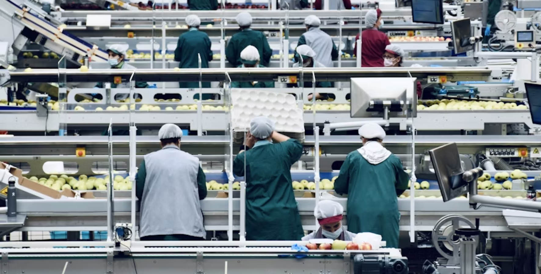
The HRC is a collective of brands, manufacturers and retailers voluntarily working together in line with competition rules to ensure that human rights are protected and respected across the entire length of international value chains. The Coalition’s members, who act individually and independently in their business operations, represent an estimated $1.7 trillion in combined annual revenue and supply chains that reach millions of suppliers and workers worldwide.
The report, published five years after the Coalition was first formed and coinciding with the UN’s Human Rights Day, shows that members have individually put in place strong governance, clear policies, structured risk assessments and mitigation plans that integrate worker input; 91% of member companies have now reached maturity through their own individual efforts.
At the heart of the report is the full CGF ‘Maturity Journey Framework for Human Rights Due Diligence (HRDD) Systems Focused on Forced Labour in Own Operations’. A six-step pathway for companies to individually evaluate and strengthen their individual due diligence systems. The framework offers companies an option for a transparent self-assessment to understand their own progress and identify what comes next:
John Ross, CEO of IGA and a CGF board member and Coalition sponsor, said:
“This report underscores a simple truth: tackling forced labour starts with strong governance, clear expectations, and leaders willing to hold their own organisations accountable. By making this assessment public, the Human Rights Coalition shows how important it is to keep due diligence front and centre.”
HRC Co-Chairs, Virginie Mahin, Senior Director Global Social Sustainability & Stakeholder Engagement, Mondelēz International and Rachel Elliott, General Manager Sustainability – Human Rights, Woolworths Group, and outgoing Co-Chair Jessica Rivas, Director, Climate and Nature Sourcing Transformation, McDonald’s Corporation, said:
“We’re proud to see that 91% of Coalition members have reached maturity in embedding due diligence in their operations. Our recommended Framework is an effective way for the wide consumer goods industry to approach human rights due diligence and ensure we’re all delivering best practices for people.”
The report contains a range of practical examples illustrating how member companies are applying due diligence best practices, including:
- APP Group launching a full due diligence process including training over 5000 employees and managers;
- Danone connecting assessment action and monitoring through an integrated governance system;
- Ferrero operationalising a new forced labour prevention policy;
- Jerónimo Martins embedding Human and Labour Rights Through Training, Audits and Worker Integration
- Mondelēz International expanding risk assessment across its operations;
- McDonald’s updating its Human Rights Policy, Supplier Code of Conduct and supporting guidance;
- Neste extending access to grievance mechanisms for third-party workers;
- Unilever evaluating the impact of fee remediation on migrant workers in Malaysia and Thailand.
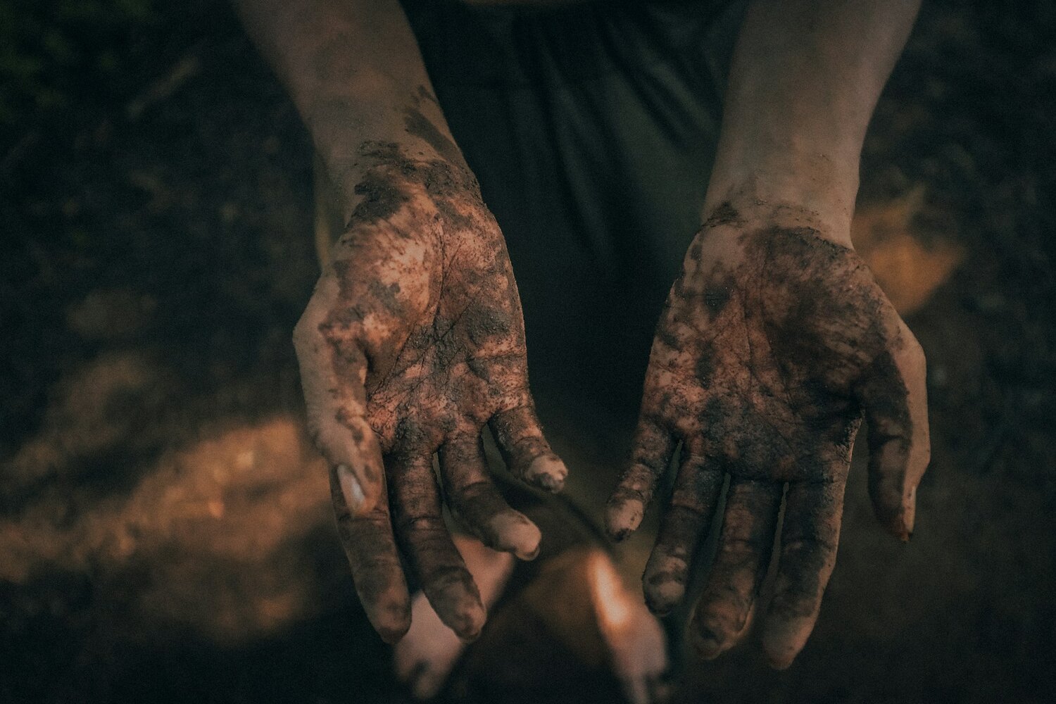
While marking important headway, the report also points to significant steps that remain. For example, remedy systems – designed to correct harm and restore the rights of impacted workers – are being developed and rolled out by many companies but have not yet reached the scale required by the size of the challenge. Structured assessment practices allow companies to collect data, but consistency still needs to be strengthened. Companies are individually, tracking and monitoring outcomes of their programs, but have yet to embed a feedback cycle that feeds directly into company decision making.
Wai-Chan Chan, Managing Director of The Consumer Goods Forum, said:
“I’m proud that the Human Rights Coalition members have individually strengthened governance, clarified responsibilities and taken steps to identify and act on risk, following collaborative action through the Coalition. I look forward to seeing how the wider consumer goods sector can pick up these recommended best practices, supporting not only workers across the globe, but also helping deliver against strategic business priorities and meet key company commitments.”
The members of the Human Rights Coalition are: Ahold Delhaize, APP Group, The Coca Cola Company, Colgate-Palmolive, Danone, Ferrero, Flora Food Group, Haleon, Heineken, IGA, Jerónimo Martins, L’Oréal, The Lindt & Sprüngli Group, Lipton Teas & Infusions, Mars, Inc., McDonald’s Corporation, Mondelēz International, Neste, Nestlé, PepsiCo, Tesco Unilever, Walmart and Woolworths Group.
Explore the findings and access the full report