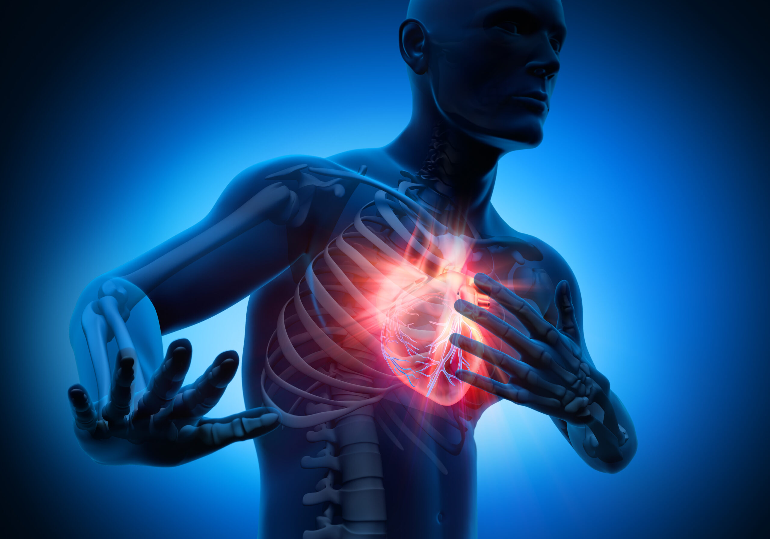
- U.S. researchers develop AI model to improve sudden cardiac death prediction news.cgtn.com
- Multimodal AI to forecast arrhythmic death in hypertrophic cardiomyopathy Nature
- This Model Beats Docs at Predicting Sudden Cardiac Arrest Medscape
- US researchers develop AI to better predict sudden cardiac death tripuratimes.com
- AI predicts patients likely to die of sudden cardiac arrest Johns Hopkins University
Author: admin
-
U.S. researchers develop AI model to improve sudden cardiac death prediction – news.cgtn.com
-
Staffordshire’s Lichfield Festival announces this year’s headline acts
Former Spandau Ballet frontman Tony Hadley and folk group The Unthanks are among headline acts at this year’s Lichfield Festival, organisers have announced.
The festival, which runs from 8 to 20 July, takes place across venues such as Lichfield Cathedral, St Michael’s Church, the Guildhall and Beacon Park.
Organisers said the event, which it dubbed one of the country’s “most eclectic multi-arts festivals” would feature world-class artists and local voices from music, theatre, dance and comedy.
The festival also included a world premiere of Gingerland, a new dance show created by Strictly Come Dancing dancer Neil Jones, a spokesperson added.
On for two nights, the family-friendly comedy features a “dazzling cast of top professional dancers, with glamorous costumes and original music,” organisers added.
Other artists headlining this year’s event include brass ensemble Black Dyke Band, Sam Every (Little) Big Band, choir Tenebrae and jazz artist Jacqui Dankworth.
The 11-day festival will close with a candlelit concert by Jeneba Kanneh-Mason in Lichfield Cathedral.
A new discounted ticket initiative was also launched this year, offering discounted prices for under 30s, carers, children and eligible community groups,
Tickets and the full event programme have been listed on the festival’s website.
Continue Reading
-
Inflammatory Markers as Predictors of Diabetes Mellitus in Patients wi
Introduction
As an ancient disease, tuberculosis (TB) has existed for thousands of years since the origin and evolution of mankind.1 Pulmonary tuberculosis (PTB) is caused by the infection with Mycobacterium tuberculosis (Mtb), which primarily spreads among people through the air and affects the lung.2 PTB was classified as a Global Health Emergency by the World Health Organization (WHO) in 1993, and it was the world’s second leading cause of death from a single infectious agent, after Coronavirus disease 2019 (COVID-19) in 2022. According to the statistics of the WHO, the number of people who developed TB was approximately 10.6 million and the number of people newly diagnosed with TB was 7.5 million in 2022, of which TB patients newly diagnosed in China were approximately 748,000 (accounted for 7.1%),3 ranking third among the 30 countries with a high TB burden.4 Although the global incidence of TB has been well controlled, it still poses a severe challenge to global public health because of the poor prognosis caused by such as rising resistance rates and the severe complications. Currently, the epidemic situation of TB epidemics in China remains very serious. The risk factors for tuberculosis include overcrowding, poverty, malnutrition, and immunosuppression including human immunodeficiency virus (HIV)/acquired immunodeficiency syndrome (AIDS).5 Diabetes mellitus (DM) is increasingly being recognized as an independent risk factor for tuberculosis.6,7 DM is a chronic metabolic disease resulting from a combination of genetic and environmental factors.8,9 The main pathogenesis of DM is an absolute or relative reduction in insulin secretion, which affects the metabolism of carbohydrates, proteins, fats, electrolytes, and water, resulting in chronic organ injury and dysfunction.10,11 DM epidemic has grown worldwide and is associated with high morbidity and mortality.12 During recent decades, the prevalence of DM has been sharply increased owing to an aging population, urbanization, physical inactivity and obesity caused by lifestyle changes.13 According to International Diabetes Federation (IDF) reports in 2019, the number of patients with DM worldwide was as high as 463 million, with the most rapid increase occurring in low- and middle-income countries (LMICs).14 Simultaneously, these countries face serious TB situations. The rising prevalence of diabetes may be contributed to the persistently high incidence of TB in countries with a high TB burden.
The bidirectional association between PTB and DM is well established, and the relationship between them is bidirectional. Studies have shown that the overall risk of PTB in patients with DM is three times higher than in the general population,15,16 and the prevalence of DM among PTB patients ranges from 1.9% to as high as 35%.17 Nearly 80% of adult DM cases are expected to occur in developing countries, and the convergence of these two epidemics may lead to an increased incidence of PTB.18 The patients with PTB and DM lead to treatment failure, longer sputum conversion time to normal, relapse, increased risk of developing multidrug-resistant tuberculosis (MDR-TB), and high mortality.19 According to the WHO PTB screening guidelines, uncontrolled diabetes doubles the risk of TB treatment failure, relapse, and death.20 There are significant challenges in the treatment and care of patients with DM and TB. Systematic evaluation of Asian countries showed that the prevalence of diabetes among PTB patients is between 5% and 50%, while the prevalence among DM patients in developing Asian countries is 1.8–9.5 times the general population.21 China has experienced the largest dual DM and TB epidemic globally, and DM combined with PTB poses a major public health problem. The incidence rates of DM and PTB comorbidity (PTB-DM) among Chinese individuals increased from 19.3% to 24.1%.22 Therefore, clarifying the diagnostic value of clinical laboratory indices for PTB-DM is of great clinical significance.
Inflammation has long been identified as an essential component of both DM and TB.23,24 DM increases the risk of TB infection by inducing chronic inflammation and immune deficiency. TB infection aggravates abnormal blood glucose through inflammatory responses, forming a bidirectional worsening cycle of “DM-tuberculosis”. Inflammation is the core mechanism connecting diabetes and tuberculosis, running through the entire process of disease occurrence and development. The neutrophil-to-lymphocyte ratio (NLR), platelet-to-lymphocyte ratio (PLR), and monocyte-to-lymphocyte ratio (MLR) have been found to be useful markers for the diagnosis and differential diagnosis of TB,25,26 and DM related disease and prognosis.27–29 In addition, system immune inflammation index (SII) and system inflammation response index (SIRI) are two markers of system immune inflammation, and their links to DM are being revealed.30,31 However, the association between immunoinflammatory markers and PTB-DM remains unclear. In the present study, we aimed to investigate whether these immunoinflammatory markers and clinical features are associated with the risk of DM in patients with PTB. It would provide a scientific basis for the prevention and control of PTB in patients with DM.
Materials and Methods
Study Population
A total of 1106 patients with PTB were selected as the case group at Meizhou People’s Hospital between April 2016 and December 2020 were retrospectively. During the study period, 326 cases with PTB (observation group) of DM patients with PTB were randomly selected, and compared with 780 PTB patients without DM during the same period (control group). PTB patients were diagnosed according to the criteria of “WS 288–2017 Pulmonary Tuberculosis Diagnosis”32 by microbiological diagnosis. The diagnostic criteria for T2DM were as follows: (1) There were typical clinical symptoms of DM (polydipsia, polydipsia, polyuria, polydipsia, and unexplained weight loss), and random intravenous plasma glucose ≥11.1mmol/L; or fasting blood glucose (FBG) ≥7mmol/L; or blood glucose level at the 2-hour oral glucose tolerance test ≥11.1mmol/L.33 Patients with leukemia, HIV infection, septic shock, organ failure, malignancy, or mental disorders; those with diseases that can affect immune function, such as AIDS, malignant tumor, chronic hepatitis, cirrhosis, primary kidney disease, renal failure, blood disease, renal transplantation, gastrectomy, or use of hormones and immunosuppressants within four months were also excluded. Clinical data, including age, sex, cough, fever, respiratory symptoms, expectoration, and extrapulmonary tuberculosis, were collected from all study subjects. This study was approved by the Human Ethics Committee of Meizhou People’s Hospital.
Data Collection
Data on clinical characteristics, laboratory outcomes, and inflammation indices were systematically collected from the medical record system of Meizhou People’s Hospital. Clinical symptoms recorded included fever (defined as a body temperature ≥38°C, measured using a standard clinical thermometer), sputum production (assessed based on the presence and quantity of sputum, categorized as mild, moderate, or severe), shortness of breath/difficulty breathing (evaluated using clinical assessment tools such as the Respiratory Distress Observation Scale or the Modified Borg Dyspnea Scale), and extrapulmonary tuberculosis (diagnosed based on clinical presentation, imaging studies, and laboratory confirmation). Laboratory outcomes included erythrocyte sedimentation rate (ESR), measured using the Westergren method and reported in millimeters per hour (mm/hr); C-reactive protein (CRP), quantified using high-sensitivity CRP assays and reported in milligrams per liter (mg/L); and complete blood count (CBC), analyzed using automated hematology analyzers to record absolute neutrophil count (ANC), absolute lymphocyte count (ALC), absolute monocyte count (AMC), and platelet count (reported as cells per microliter). Inflammation indices were calculated as follows: neutrophil-to-lymphocyte ratio (NLR=ANC/ALC), platelet-to-lymphocyte ratio (PLR=Platelet count/ALC), monocyte-to-lymphocyte ratio (MLR=AMC/ALC), systemic immune-inflammation index (SII=Platelet count × ANC/ALC), and systemic inflammation response index (SIRI = AMC × ANC/ALC). These indices were used to assess systemic inflammation and immune response.
Data Processing and Statistical Analysis
SPSS 26.0 and GraphPad Prism software were used for the statistical analysis of the experimental data. Data with non-normal distributions were described as median and interquartile range (IQR) values, and evaluated using the Mann–Whitney U-test. Categorical variables were represented numerically and as percentages, and were compared using the chi-squared test. Univariate regression analysis (Pearson) and Spearman correlation analysis were used to analyze the relationship between the correlation test indicators. Receiver operating characteristic (ROC) curve analysis was used to determine the optimal cutoff values of ESR, NLR, MLR, PLR, SII, and SIRI for differentiating whether pulmonary tuberculosis patients developed DM or not, and the area under the ROC curve (AUC) was calculated. In addition to the logistic regression model, a 95% confidence interval (95% CI) was used to determine the diagnostic probability of PTB combined with DM. The significance level was set at P < 0.05.
Results
General Characteristics in PTB Patients with or without DM
A total of 1106 patients diagnosed with PTB were enrolled, including 326 (29.5%) PTB patients with DM and 780 (70.5%) without DM. The clinical characteristics of the two patient groups of patients are shown in Table 1. The majority of PTB patients were male (84.6%), and most had no fever (83.7%) or shortness of breath/difficulty breathing (76.9%). There were 39 (3.5%) had concurrent extrapulmonary tuberculosis. The differences in gender distribution, age distribution, and clinical manifestations including fever, shortness of breath/difficulty breathing, and expectoration, and extrapulmonary tuberculosis between the two groups were not statistically significant. The level of ESR (44.00 (22.00, 80.00) vs 30.00 (12.00, 54.00), p<0.001) was higher while the levels of NLR (4.61 (2.90, 7.64) vs 6.43 (3.62, 11.20), p<0.001), MLR (0.50 (0.31, 0.75) vs 0.64 (0.38, 1.00), p<0.001), PLR (197.38 (135.53, 299.16) vs 248.44 (149.74, 396.43), p<0.001), SII (1333.06 (712.37, 2289.35) vs 1603.72 (844.73, 3224.20), p<0.001), and SIRI (3.13 (1.73, 6.42) vs 3.93 (2.00, 8.79), p<0.001) were lower in PTB-DM patients than those in non-DM PTB patients.
Table 1 Comparison of Clinical Features and Peripheral Blood Inflammatory Markers Between Non-DM PTB Group and PTB-DM Group
Logistic Regression Analysis of Related Factors for DM in Patients with PTB
Logistic regression analyses of the association between PTB-DM and related factors were performed (Table 2). Univariate logistic regression analysis showed that PTB patients with DM were more likely to have a higher ESR (odds ratio (OR): 1.024, 95% CI: 1.018–1.30, p<0.001), lower levels of NLR (OR: 0.964, 95% CI 0.945–0.983, p<0.001), MLR (OR: 0.440, 95% CI 0.319–0.607, p<0.001), PLR (OR: 0.998, 95% CI: 0.998–0.999, p<0.001), and SIRI (OR: 0.965, 95% CI: 0.944–0.987, p=0.002). Clinical features such as gender, age, fever, expectoration, shortness of breath/difficulty breathing, extrapulmonary tuberculosis, and other blood indicators were not associated with DM in PTB patients. Multivariable logistic regression analyses indicated that a high ESR (OR: 1.024, 95% CI: 1.018–1.030, p<0.001), low levels of MLR (OR: 0.352, 95% CI 0.145–0.856, p=0.021), and PLR (OR: 0.997, 95% CI: 0.995–0.999, p=0.003) were independent risk factors for DM in patients with PTB.
Table 2 Logistic Regression Analysis of Related Factors for DM in Patients with PTB
The Value of Different Indexes and Their Combined Detection in the Differential Diagnosis of PTB-DM
To analyze the discriminating ability of these inflammatory parameters in the PTB-DM versus PTB groups, ROC curves for the related parameters were plotted (Figure 1). Results revealed the AUC value of ESR was 0.619 (95% CI: 0.590–0.648, cut-off value: 45.5), MLR was 0.600 (95% CI 0.570–0.629, cut-off value: 0.765), PLR was 0.584 (95% CI: 0.554–0.613, cut-off value: 239.615), ESR+MLR was 0.689 (95% CI: 0.661–0.716), ESR+PLR was 0.694 (95% CI: 0.666–0.721), MLR+PLR was 0.610 (95% CI: 0.574–0.645), and ESR+MLR+PLR was 0.712 (95% CI 0.685–0.739), respectively. The PTB-DM and PTB groups could be well discriminated by the combination of indicators ESR, MLR and PLR, with sensitivity and specificity of 63.8% and 70.6%, respectively. Table 3 presents the comprehensive features of ESR, MLR, and PLR for the diagnosis.
Table 3 The Diagnostic Efficacy of ESR, MLR, PLR, and Their Combination on PTB-DM
Figure 1 The ROC curve of ESR, MLR, PLR, and their combination on PTB-DM.
Discussion
This study compared the characteristics of the PTB patients with and without DM. Among the patients diagnosed with PTB, 29.5% had DM. The results showed that there were no significant differences in clinical manifestations including gender distribution, age distribution, fever, shortness of breath/difficulty breathing, expectoration, and extrapulmonary tuberculosis. ESR was higher, while NLR, MLR, PLR, SII, and SIRI were lower in PTB-DM patients than in non-DM PTB patients. In addition, high ESR and low MLR and PLR were independent risk factors for PTB-DM.
The high prevalence of DM creates more pressure on the PTB burden. DM increases the risk of PTB, posing a significant threat to the public health, particularly, in countries with a high burden of both diseases.34 Thus, experts have raised concerns regarding the co-prevalence of PTB and DM. PTB patients with DM often have nutritional deficiency, leading to body injury and disease recurrence, which ultimately affects prognosis and increases the risk of mortality.22,35 In many studies on the Chinese population, male sex and advanced age were identified as factors associated with PTB with DM;36–38 however, in this study, age and gender were not statistically different. In addition, the presence of symptoms such as fever, cough, sputum, shortness of breath, difficulty breathing, or extrapulmonary tuberculosis was similar between patients with and without DM. Therefore, we cannot estimate whether TB patients are at risk for diabetes based on simple clinical manifestations.
Chronic infection with Mtb can induce hematopoietic stem cell proliferation and immune changes, which in turn cause changes in the proportion of lymphocyte and other cells.39 There is a correlation between the immune status (including ESR, NLR, MLR, PLR, SII, and SIRI) and clinicopathological features of PTB patients,40 which are some of the more novel inflammatory markers currently available.41 ESR is a sensitive marker of the inflammatory response, and is often used to obtain information regarding disease progression and retrogression.42 The ESR value was significantly higher in tuberculosis patients with tuberculosis, and was even elevated in 98% of the patients.43,44 MLR has been proven to be associated with the diagnosis of PTB and the predictive value of MLR in patients with tuberculosis, and higher MLR levels are associated with more severe disease and poorer prognosis.45,46 The importance of PLR has been emphasized as a marker in some disorders such as non-small-cell lung cancer, acute coronary syndrome, end-stage renal disease, and so on.47,48 PLR could be developed as a valuable maker for identifying tuberculosis infection in chronic obstructive pulmonary disease (COPD) patients,40 indicating that PLR is a convenient, and easily measured prognostic indicator. In this study, the inflammation index of ESR was significantly increased, MLR, PLR, SII, and SIRI were significantly decreased in the PTB patients with DM compared to those in PTB patients alone. Further regression analysis indicated that the ESR, MLR, and PLR were relevant factors for PTB-DM. It indicates that a higher ESR and lower MLR and PLR may indicate PTB-DM.
However, these indicators fluctuate to a certain extent and do not have the significance of an independent diagnosis in patients with PTB-DM. Hence, these factors need to be combined to improve the diagnostic value of PTB complicated by DM. Thus, we analyzed the diagnostic efficacy of ESR, MLR, and PLR in PTB patients with DM, and found that ESR has low sensitivity and MLR has low specificity, while PLR has slightly higher sensitivity and specificity. In addition, we also analyzed the sensitivity and specificity of ESR, MLR, and PLR combined tests, and found that the combined tests of these indicators were superior to the single indicator in both sensitivity and specificity. Therefore, the combined detection of ESR, MLR, and PLR is helpful in the differential diagnosis of PTB with DM and non-DM PTB. The results of this study provide a convenient method for clinicians to assess the risk of developing DM in patients with PTB.
This study offers valuable insights into the relationship between hematological markers and DM in patients with PTB, though there are opportunities for further exploration. Firstly, the relationship between these indicators and the severity of DM has not been studied. Future research could investigate the association between inflammation markers (ESR, MLR, and PLR) and the severity of DM. Secondly, the research subjects included in this study were from a single medical structure. Due to the incomplete representativeness of the research subjects, the application of the results of this study in other populations was limited. So, expanding the study to multiple centers would provide a more diverse sample, enhancing the generalizability of the results. Thirdly, this study only analyzed the differences in ESR, NLR, MLR, PLR, SII, and SIRI levels, and did not investigate the role of other factors in the occurrence of DM in patients with PTB, especially some confounding factors. Lastly, collecting data at multiple time points, rather than a single pre-treatment measure, would allow for a more comprehensive analysis of the dynamic changes in these hematological indicators and their clinical significance throughout the treatment process. Addressing these factors would provide a more complete understanding of the role of these markers in DM and PTB, which depends on more research in the future.
Conclusion
ESR, MLR, and PLR were associated with the risk of DM in patients with PTB. In particular, combined tests of these indicators were superior to the single indicator in both sensitivity and specificity in the diagnosis of DM among patients with PTB. It provides a convenient method for clinicians to assess the risk of developing DM in patients with PTB. Specifically, during the treatment of tuberculosis, it is necessary to closely monitor the changes in the patient’s blood sugar, adjust the diabetes treatment plan in a timely manner, and reduce the fluctuations in blood sugar caused by inflammation. Secondly, for pulmonary tuberculosis patients with abnormal inflammatory indicators, their association with diabetes should be emphasized. Through anti-inflammatory treatment or immunomodulatory measures, insulin resistance can be improved, immune balance can be regulated, and the risk of disease progression can be reduced.
Data Sharing Statement
The data that support the findings of this study are available from the corresponding author upon reasonable request.
Ethics Approval and Consent to Participate
The study was approved by the Ethics Committee of Medicine, Meizhou People’s Hospital number. All participants signed informed consent in accordance with the Declaration of Helsinki.
Acknowledgments
The author would like to thank other colleagues whom were not listed in the authorship of Meizhou People’s Hospital for their helpful comments on the manuscript.
Author Contributions
All authors made a significant contribution to the work reported, whether that is in the conception, study design, execution, acquisition of data, analysis and interpretation, or in all these areas; took part in drafting, revising or critically reviewing the article; gave final approval of the version to be published; have agreed on the journal to which the article has been submitted; and agree to be accountable for all aspects of the work.
Funding
This study was supported by the Science and Technology Program of Meizhou (Grant No.: 2019B0202001).
Disclosure
The authors declare that they have no competing interests.
References
1. Sabin S, Herbig A, Vågene ÅJ, et al. A seventeenth-century Mycobacterium tuberculosis genome supports a Neolithic emergence of the Mycobacterium tuberculosis complex. Genome Biol. 2020;21(1):201. doi:10.1186/s13059-020-02112-1
2. Tsareva A, Shelyakin PV, Shagina IA. Aberrant adaptive immune response underlies genetic susceptibility to tuberculosis. Front Immunol. 2024;15:1380971. doi:10.3389/fimmu.2024.1380971
3. Feng Q, Zhang G, Chen L, et al. Roadmap for ending TB in China by 2035: the challenges and strategies. Biosci Trends. 2024;18(1):11–20. doi:10.5582/bst.2023.01325
4. Organization WH. Global tuberculosis report 2023. Available from: https://www.who.int/publications/i/item/9789240083851.
Accessed .July 3 , 20255. Chakaya J, Khan M, Ntoumi F, et al. Global Tuberculosis report 2020 – reflections on the global TB burden, treatment and prevention efforts. Int J Infect Dis. 2021;113 Suppl 1(Suppl 1):S7–s12. doi:10.1016/j.ijid.2021.02.107
6. Lachmandas E, Vrieling F, Wilson LG, et al. The effect of hyperglycaemia on in vitro cytokine production and macrophage infection with Mycobacterium tuberculosis. PLoS One. 2015;10(2):e0117941. doi:10.1371/journal.pone.0117941
7. Lu CL, Perera R, Farrah H, Waring J. Diabetes screening among active tuberculosis patients in Western Australia tuberculosis control program using HbA1c. Intern Med J. 2019;49(5):630–633. doi:10.1111/imj.14143
8. Lovic D, Piperidou A, Zografou I, Grassos H, Pittaras A, Manolis A. The growing epidemic of diabetes mellitus. Curr Vasc Pharmacol. 2020;18(2):104–109. doi:10.2174/1570161117666190405165911
9. Aloke C, Egwu CO. Current Advances in the Management of Diabetes Mellitus. Biomedicines. 2022;10(10):2436. doi:10.3390/biomedicines10102436
10. Seo WD, Lee JH, Jia Y, et al. Saponarin activates AMPK in a calcium-dependent manner and suppresses gluconeogenesis and increases glucose uptake via phosphorylation of CRTC2 and HDAC5. Bioorg Med Chem Lett. 2015;25(22):5237–5242. doi:10.1016/j.bmcl.2015.09.057
11. Ahmed S, Adnan H, Khawaja MA, Butler AE. Novel micro-ribonucleic acid biomarkers for early detection of type 2 diabetes mellitus and associated complications-A literature review. Int J Mol Sci. 2025;26(2):753. doi:10.3390/ijms26020753
12. Moosaie F, Mohammadi S, Saghazadeh A, Dehghani Firouzabadi F, Rezaei N. Brain-derived neurotrophic factor in diabetes mellitus: a systematic review and meta-analysis. PLoS One. 2023;18(2):e0268816. doi:10.1371/journal.pone.0268816
13. Saeedi P, Salpea P, Karuranga S, et al. Mortality attributable to diabetes in 20-79 years old adults, 2019 estimates: results from the International Diabetes Federation Diabetes Atlas, 9(th) edition. Diabet Res Clin Pract. 2020;162:108086. doi:10.1016/j.diabres.2020.108086
14. Yue Y, Ye K, Lu J, et al. Probiotic strain Lactobacillus plantarum YYC-3 prevents colon cancer in mice by regulating the tumour microenvironment. Biomed Pharmacother. 2020;127:110159. doi:10.1016/j.biopha.2020.110159
15. Nyirenda JLZ, Wagner D, Ngwira B, Lange B. Bidirectional screening and treatment outcomes of diabetes mellitus (DM) and Tuberculosis (TB) patients in hospitals with measures to integrate care of DM and TB and those without integration measures in Malawi. BMC Infect Dis. 2022;22(1):28. doi:10.1186/s12879-021-07017-3
16. Quist-Therson R, Kuupiel D, Hlongwana K. Mapping evidence on the implementation of the WHO’s collaborative framework for the management of tuberculosis and diabetes: a scoping review protocol. BMJ Open. 2020;10(1):e033341. doi:10.1136/bmjopen-2019-033341
17. Noubiap JJ, Nansseu JR, Nyaga UF, et al. Global prevalence of diabetes in active tuberculosis: a systematic review and meta-analysis of data from 2·3 million patients with tuberculosis. Lancet Glob Health. 2019;7(4):e448–e460. doi:10.1016/S2214-109X(18)30487-X
18. Chan JCN, Yang A, Chu N, Chow E. Current type 2 diabetes guidelines: individualized treatment and how to make the most of metformin. Diabetes Obes Metab. 2024;26 Suppl 3:55–74. doi:10.1111/dom.15700
19. Yorke E, Atiase Y, Akpalu J, Sarfo-Kantanka O, Boima V, Dey ID. The bidirectional relationship between tuberculosis and diabetes. Tuberc Res Treat. 2017;2017:1702578. doi:10.1155/2017/1702578
20. WHO. Guidelines Approved by the Guidelines Review Committee. In: Systematic Screening for Active Tuberculosis: Principles and Recommendations. Vol. 2013. Geneva: World Health Organization Copyright © World Health Organization; 2013.
21. Zheng C, Hu M, Gao F. Diabetes and pulmonary tuberculosis: a global overview with special focus on the situation in Asian countries with high TB-DM burden. Glob Health Action. 2017;10(1):1–11. doi:10.1080/16549716.2016.1264702
22. Shi H, Yuan Y, Li X, Li YF, Fan L, Yang XM. Analysis of the influencing factors and clinical related characteristics of pulmonary tuberculosis in patients with type 2 diabetes mellitus. World J Diabetes. 2024;15(2):196–208. doi:10.4239/wjd.v15.i2.196
23. Korniluk A, Koper-Lenkiewicz OM. Mean platelet volume (MPV): new perspectives for an old marker in the course and prognosis of inflammatory conditions. Mediators Inflamm. 2019;2019:9213074. doi:10.1155/2019/9213074
24. López-González JA, Martínez-Soto JM, Avila-Cervantes C, et al. Evaluation of systemic inflammation before and after standard anti-tuberculosis treatment in patients with active pulmonary tuberculosis and diabetes mellitus. Cureus. 2024;16(3):e55391. doi:10.7759/cureus.55391
25. Liu QX, Tang DY, Xiang X, He JQ. Associations between nutritional and immune status and clinicopathologic factors in patients with tuberculosis: a comprehensive analysis. Front Cell Infect Microbiol. 2022;12:1013751. doi:10.3389/fcimb.2022.1013751
26. Tu HZ, Lai TJ, Chen YS, Lee HS, Chen JS. Hematological parameters as potential markers for distinguishing pulmonary tuberculosis from genitourinary tuberculosis. Pathogens. 2023;12(1):84. doi:10.3390/pathogens12010084
27. Regassa DA, Kiya GT, Kebede RA, Beyene W. Assessment of hematological profiles and prognostic role of hemogram-derived novel markers for diabetes mellitus and its complications among type 2 diabetes mellitus adult patients attending Bishoftu General Hospital, central, Ethiopia: a comparative cross-sectional study. J Blood Med. 2023;14:681–699. doi:10.2147/JBM.S435452
28. Ning P, Yang F, Kang J, et al. Predictive value of novel inflammatory markers platelet-to-lymphocyte ratio, neutrophil-to-lymphocyte ratio, and monocyte-to-lymphocyte ratio in arterial stiffness in patients with diabetes: a propensity score-matched analysis. Front Endocrinol. 2022;13:1039700. doi:10.3389/fendo.2022.1039700
29. Mureșan AV, Tomac A, Opriș DR, et al. Inflammatory markers used as predictors of subclinical atherosclerosis in patients with diabetic polyneuropathy. Life. 2023;13(9):1861. doi:10.3390/life13091861
30. Liu W, Zheng S, Du X. Association of systemic immune-inflammation index and systemic inflammation response index with diabetic kidney disease in patients with type 2 diabetes mellitus. diabetes, metabolic syndrome and obesity: targets and therapy. Diabetes Metab Syndr Obes. 2024;17:517–531. doi:10.2147/DMSO.S447026
31. Song Y, Shu Y, Zhao Y, et al. Combination model of neutrophil to high-density lipoprotein ratio and system inflammation response index is more valuable for predicting peripheral arterial disease in type 2 diabetic patients: a cross-sectional study. Front Endocrinol. 2023;14:1100453. doi:10.3389/fendo.2023.1100453
32. Tendolkar MS, Tyagi R, Handa A. Review of advances in diagnosis and treatment of pulmonary tuberculosis. Indian J Tuberc. 2021;68(4):510–515. doi:10.1016/j.ijtb.2021.07.002
33. Benhalima K, Van Crombrugge P, Moyson C, et al. Risk factor screening for gestational diabetes mellitus based on the 2013 WHO criteria. Eur J Endocrinol. 2019;180(6):353–363. doi:10.1530/EJE-19-0117
34. Gadallah M, Amin W, Fawzy M, Mokhtar A, Mohsen A. Screening for diabetes among tuberculosis patients: a nationwide population-based study in Egypt. Afr Health Sci. 2018;18(4):884–890. doi:10.4314/ahs.v18i4.6
35. Traub J, Reiss L, Aliwa B, Stadlbauer V. Malnutrition in patients with liver cirrhosis. Nutrients. 2021;13(2):540. doi:10.3390/nu13020540
36. Wu Q, Wang M, Zhang Y, et al. Epidemiological characteristics and their influencing factors among pulmonary tuberculosis patients with and without diabetes mellitus: a survey study from drug resistance surveillance in East China. Front Public Health. 2021;9:777000. doi:10.3389/fpubh.2021.777000
37. Du Q, Wang L, Long Q, et al. Systematic review and meta-analysis: prevalence of diabetes among patients with tuberculosis in China. Trop Med Int Health. 2021;26(12):1553–1559. doi:10.1111/tmi.13686
38. Ling Y, Chen X, Zhou M, et al. The effect of diabetes mellitus on tuberculosis in eastern China: a decision-tree analysis based on a real-world study. J Diabetes. 2023;15(11):920–930. doi:10.1111/1753-0407.13444
39. Chen L, Liu C, Liang T, et al. Monocyte-to-lymphocyte ratio was an independent factor of the severity of spinal tuberculosis. Oxid Med Cell Longev. 2022;2022:7340330. doi:10.1155/2022/7340330
40. Chen G, Wu C, Luo Z, Teng Y, Mao S. Platelet-lymphocyte ratios: a potential marker for pulmonary tuberculosis diagnosis in COPD patients. Int J Chron Obstruct Pulmon Dis. 2016;11:2737–2740. doi:10.2147/COPD.S111254
41. Xu H, Xie J, Zhang S. Potential blood biomarkers for diagnosing periprosthetic joint infection: a single-center, retrospective study. Antibiotics. 2022;11(4):505. doi:10.3390/antibiotics11040505
42. Zeng H, Zhang P, Shen X, et al. One-stage posterior-only approach in surgical treatment of single-segment thoracic spinal tuberculosis with neurological deficits in adults: a retrospective study of 34 cases. BMC Musculoskelet Disord. 2015;16:186. doi:10.1186/s12891-015-0640-0
43. Shah AR, Desai KN, Maru AM. Evaluation of hematological parameters in pulmonary tuberculosis patients. J Family Med Prim Care. 2022;11(8):4424–4428. doi:10.4103/jfmpc.jfmpc_2451_21
44. Batool Y, Pervaiz G, Arooj A, Fatima S. Hematological manifestations in patients newly diagnosed with pulmonary tuberculosis. Pak J Med Sci. 2022;38(7):1968–1972. doi:10.12669/pjms.38.7.5911
45. Mayito J, Meya DB, Rhein J, Sekaggya-Wiltshire C. Utility of the monocyte to lymphocyte ratio in diagnosing latent tuberculosis among HIV-infected individuals with a negative tuberculosis symptom screen. PLoS One. 2020;15(11):e0241786. doi:10.1371/journal.pone.0241786
46. Buttle TS, Hummerstone CY, Billahalli T, et al. The monocyte-to-lymphocyte ratio: sex-specific differences in the tuberculosis disease spectrum, diagnostic indices and defining normal ranges. PLoS One. 2021;16(8):e0247745. doi:10.1371/journal.pone.0247745
47. Cao W, Yu H, Zhu S, et al. Clinical significance of preoperative neutrophil-lymphocyte ratio and platelet-lymphocyte ratio in the prognosis of resected early-stage patients with non-small cell lung cancer: a meta-analysis. Cancer Med. 2023;12(6):7065–7076. doi:10.1002/cam4.5505
48. Li P, Xia C, Liu P, et al. Neutrophil-to-lymphocyte ratio and platelet-to-lymphocyte ratio in evaluation of inflammation in non-dialysis patients with end-stage renal disease (ESRD). BMC Nephrol. 2020;21(1):511. doi:10.1186/s12882-020-02174-0
Continue Reading
-
Knowledge, Attitudes, and Practices Among Patients Undergoing Gastroin
Introduction
Globally, diseases of the digestive system, including functional gastrointestinal disorders, gastritis, and peptic ulcers, are common and contribute significantly to both morbidity and mortality.1 Chronic gastritis affects over half of the world’s population, underscoring its widespread impact.2 Epidemiological studies suggest that the morbidity rate for peptic ulcers and associated disorders could be as high as 10%; however, recent years have shown a declining trend in incidence, particularly due to improved H. pylori eradication strategies and proton pump inhibitor use.3,4 The burden of gastrointestinal diseases remains high due to the prevalence of both benign conditions, such as acute and chronic gastroenteritis, and malignant diseases like colorectal and gastric cancers, which, while distinct, also represent major global public health concerns. These cancers rank as the third and fourth most common cancers globally, each causing approximately 800,000 deaths annually.5,6 These conditions lead to considerable pain and disability, while also placing a heavy financial strain on both families and society.
Gastrointestinal endoscopy represents the gold standard for diagnosing and monitoring digestive diseases. While primarily performed under conscious sedation, this procedure can provoke significant anxiety and psychological distress in patients, potentially affecting both procedure outcomes and patient compliance with future screening recommendations.7 Despite its clinical benefits, gastrointestinal endoscopy can provoke anxiety, fear, and even stress reactions among patients, as reported in numerous studies.8,9 Endoscopy is primarily utilized in outpatient settings and is well-known for its effectiveness in diagnosing and treating gastrointestinal disorders.10 Given the increasing global incidence of digestive system diseases, the role of endoscopy is more critical than ever.11,12
The KAP framework provides a structured approach to understanding how patients’ knowledge influences their attitudes and subsequent health behaviors. In the context of gastrointestinal endoscopy, this understanding is particularly crucial as patient preparation and cooperation directly impact procedure quality and diagnostic yield. It is based on the idea that knowledge has a beneficial effect on attitudes, which in turn influence behaviors.13,14 Inadequate knowledge and negative attitudes may lead to poor bowel preparation, increased anxiety, refusal to undergo the procedure, or reduced compliance with follow-up recommendations-factors that can significantly compromise diagnostic accuracy and therapeutic outcomes.9,15 With the increasing prevalence of gastrointestinal disorders, ranging from benign conditions such as gastritis to life-threatening cancers, there is a critical need to understand patient experiences and behaviors to enhance clinical outcomes. Gastrointestinal endoscopy, an essential diagnostic and therapeutic tool, often triggers anxiety and fear among patients, potentially affecting their willingness to undergo necessary procedures. This study is pivotal as it focuses on a patient group whose insights are crucial for the efficacy of endoscopic procedures and overall treatment success. By examining patients’ understanding, reactions, and adjustments post-endoscopy, healthcare providers can develop targeted communication strategies and support mechanisms that enhance patient comfort, increase engagement, and promote adherence. This approach ultimately leading to optimized healthcare resource utilization and improved health outcomes.
Despite extensive research on the technology and effectiveness of endoscopy,16–18 studies delving into patient feedback post-examination remain limited. A recent narrative review by Minciullo et al (2022) summarized available tools for assessing patient satisfaction in digestive endoscopy, emphasizing its importance as a quality indicator.19 While that review primarily focused on satisfaction measurement, our study explores patients’ KAP, offering a broader behavioral and cognitive perspective that complements satisfaction-based evaluations. This study aimed to quantify patients’ knowledge levels regarding gastrointestinal endoscopy procedures; assess attitudes and psychological barriers towards endoscopic examination; evaluate adherence to pre- and post-procedure care instructions; and examine the connections between knowledge, attitudes, and practices to identify targeted intervention opportunities. These specific objectives will help address the current gap in understanding patient perspectives and guide the development of evidence-based educational programs. Therefore, herein, we sought to explore the KAP of patients toward gastrointestinal endoscopy in the Endoscopy Unit.
Materials and Methods
Study Design and Participants
The required sample size was estimated using the formula for cross-sectional studies:20 n = Z² × P × (1 – P) / d², where n is the sample size, Z is the standard normal deviate at 95% confidence level (Z = 1.96), P is the expected proportion (assumed to be 50% for maximum variability), and d is the margin of error (set at 4%). Based on this formula, a minimum sample size of 384 was required. Considering potential non-response or invalid submissions, we distributed a larger number of questionnaires. Ultimately, 612 valid responses were obtained, meeting the minimum statistical power requirement.
This cross-sectional study used a survey to collect data at the author’s Hospital from December 2023 to February 2024. Consecutive adult patients who were referred for diagnostic gastrointestinal endoscopy were enrolled using convenience sampling. To minimize heterogeneity, patients with a confirmed inflammatory bowel disease diagnosis or those undergoing therapeutic procedures were excluded. The research received approval from the Medical Ethics Committee of the author’s Hospital, and all the study participants provided informed consent.
Inclusion criteria were the following: 1) those who underwent gastrointestinal endoscopy (gastroscopy or colonoscopy); 2) those who voluntarily participated; 3) those who were conscious and able to cooperate.
Exclusion criteria were the following: 1) those with critical condition; 2) those who underwent gastrointestinal endoscopy for gastrointestinal bleeding; 3) those unable to complete the questionnaire for other reasons.
Questionnaire
The questionnaire was developed through a systematic review of relevant literature and current clinical practice guidelines. Content validity was established through an expert panel review by two gastroenterologists and two endoscopy nurses.21,22 Following the questionnaire’s design, a pilot study was carried out involving 76 participants, generating a Cronbach’s α coefficient = 0.912, indicative of good internal consistency. The final questionnaire included 4 dimensions: knowledge, attitude, practice, and demographic characteristics, comprising basic patient demographics, reasons for seeking medical care, daily routines, and dietary habits. The knowledge section consisted of 9 questions, with responses scored assigned 2 points for “complete understanding”, 1 point for “partial understanding”, and 0 for “lack of understanding”, generating a total score of 0 to 18. The attitude section consisted of 7 questions, rated on a five-point Likert scale from “strongly agree” to “strongly disagree”, with scores ranging from 5 points to 1 point and a total score range of 7 to 35. The practice section included 8 items, also rated on a five-point Likert scale from “always” to “never”, with values ranging from 5 points to 1 point and a total score of 8 to 40. According to Bloom’s cutoff, participants who scored > 80% of the total were classified as possessing satisfactory knowledge, constructive attitudes, and active practices, while those scoring between 60% and 80% were classified as possessing average levels in these dimensions. Scoring < 60% of the total suggested inadequate knowledge, negative attitudes, and passive behaviors.23
The questionnaire was generated using the “Questionnaire Star” platform, after which a QR code was obtained, printed, and placed in the endoscopy room. After patients completed their endoscopy and fully regained consciousness, two specially trained nurses invited them to the endoscopy room to scan the QR code and complete the questionnaire. Patients scanned the QR code and completed the questionnaire using their phone, with each phone allowing only one submission of responses. Researchers will only provide clarifications about the questions without offering any hints for the answers. For elderly patients who cannot use a mobile phone, the nurses will record the patients’ answers and fill out the questionnaire on their behalf.
To further evaluate the validity of the questionnaire, both content validity and construct validity were assessed. Content validity was established through expert review by two gastroenterologists and two endoscopy nurses. Construct validity was examined using confirmatory factor analysis (CFA) (Supplementary Table 1). The Kaiser-Meyer-Olkin (KMO) test yielded a value of 0.913 (P < 0.001), indicating sampling adequacy. Model fit indices demonstrated good construct validity (CMIN/DF = 4.487; RMSEA = 0.076; IFI = 0.937; TLI = 0.929; CFI = 0.937), and all standardized factor loadings were statistically significant (P < 0.001). A CFA path diagram is presented in Supplementary Figure 1 to illustrate the measurement structure.
Statistical Analysis
Statistical analysis was conducted using SPSS 22.0 (IBM, Armonk, NY, USA). Continuous data are reported as mean ± standard deviation (SD), and categorical data are expressed as n (%). Continuous data that were confirmed to follow a normal distribution were analyzed using independent-sample t-tests or one-way ANOVA. The Wilcoxon Mann–Whitney test or Kruskal–Wallis test was used to compare continuous data with skewed distribution. The correlation among KAP was analyzed using Pearson correlation analysis, and interactions among KAP were explored using structural equation modeling (SEM) performed with AMOS version 26.0. A two-sided P-value less than 0.05 was considered statistically significant.
Results
Patients’ Characteristics
Among a total of 744 collected questionnaires, 20 refused to participate in the study, 9 had a short response time (< 90 seconds), 13 had a logical conflict, and 80 were incomplete, resulting in 612 valid questionnaires, with a validity rate of 82.26%.
Out of these 612 participants, 308 (50.33%) were filled out by males; the mean age of participants was 51.11 ± 12.61 years; 363 (62.42%) had a BMI in the normal range; 152 (24.84%) had poor dietary habits, 250 (40.85%) had heavy taste preference in their diets and 147 (24.02%) were frequently constipated. Meanwhile, 355 (58.01%) had their first gastroscopy, 561 (91.67%) opted for a painless procedure, and 308 (50.33%) had gastrointestinal polyps (Table 1). Among the 308 patients with polyps, the majority (225 cases, 73.1%) underwent painless enteroscopy alone, followed by 33 cases (10.7%) who received both painless enteroscopy and painless gastroscopy, and 29 cases (9.4%) with painless gastroscopy alone. This distribution, as visualized in the UpSet plot (Figure 1), indicates that painless colonoscopy was the predominant modality associated with polyp detection in this study. The reason for the endoscopy examination and results are shown in Figure 2. “Life stage screening” refers to routine health check-ups commonly recommended based on age or risk factors, such as colorectal cancer screening in adults over 50.
Table 1 Demographic Characteristics, Knowledge, Attitude, and Practice
Figure 1 UpSet plot showing the distribution of gastrointestinal endoscopic procedures among the 308 patients diagnosed with polyps.
Figure 2 (A) Reasons for undergoing digestive endoscopy (including upper and lower GI procedures); “Life stage screening” refers to routine health check-ups. (B) Results of endoscopic examinations.
Knowledge, Attitude, and Practice
The mean scores for knowledge, attitude, and practice were 11.66 ± 3.95 (possible range: 0–18), 29.79 ± 3.27 (possible range: 7–35), and 36.69 ± 4.99 (possible range: 8–40), separately. Knowledge scores varied depending on education (P < 0.001), monthly income (P = 0.010), alcohol consumption (P = 0.047), family history of gastric or colorectal cancer (immediate family) (P = 0.008), unexplained changes in bowel habits or fecal abnormalities (P = 0.002), number of gastroscopies (P < 0.001), and the number of polyps (P = 0.015). Attitude scores were more likely to vary depending on unexplained changes in bowel habits or fecal abnormalities (P = 0.049), number of gastroscopies (P = 0.047), presence of polyps (P = 0.015), and whether the polyp was >5 mm (P = 0.030). Practice scores varied depending on education (P = 0.020), monthly income (P < 0.010), number of gastroscopies (P = 0.015), and type of the current gastroenteroscopy (P < 0.001) (Table 1).
For all the knowledge items, no more than 40% of the participants answered “Very well known”, while more than 60% chose “Heard of it”, indicating that while progress has been made, there remains ample opportunity for further improvement. Specifically, for “ Endoscopic examinations can assess different areas of the gastrointestinal tract, such as the esophagus, stomach, duodenum, and colon.”, 70.1% chose “Heard of it” (K3). As for “This examination is typically used to diagnose gastrointestinal diseases such as ulcers, tumors, and inflammation.”, 69.77% chose “Heard of it” (K2) (Table 2).
Table 2 Responses to the Knowledge Section
For the attitude dimension, more than 90% of participants chose either “strongly agree” or “agree”, except for A1 and A3. Specifically, 23.37% and 23.53% were neutral on whether they felt nervous or anxious before the examination (A1) and whether they were worried about the safety and side effects of endoscopy (A3), respectively (Table 3).
Table 3 Responses to the Attitude Section
Responses on the practice revealed that > 70% of the participants chose “always” for all items except P2 and P3. Specifically, 68.63% of the participants were always fully aware of the endoscopy procedure and possible discomfort before the examination (P2), and 61.44% of the participants always raised concerns and questions about the procedure with the doctor prior to the examination (P3) (Table 4).
Table 4 Responses to the Practice Section
The Correlation and Interaction Among KAP
The correlation analyses revealed statistically significant, weak to moderate positive correlations: knowledge was weakly correlated with attitude (r = 0.281, P < 0.001) and with practice (r = 0.148, P < 0.001), while attitude demonstrated a moderate correlation with practice (r = 0.370, P < 0.001) (Table 5).
Table 5 Correlation Analysis
The SEM model showed that the questionnaire fit the KAP model well (Figure 3 and Supplementary Table 2), and the analysis of direct and indirect effects showed that knowledge directly affected attitude (β = 0.397, P = 0.026) and attitude directly affected practice (β = 0.402, P = 0.007). Although the direct effect of knowledge on practice is not significant (β = 0.032, P = 0.568), knowledge has an indirect effect on practice through attitude (β = 0.159, P = 0.014) (Table 6).
Table 6 Direct and Indirect Effects in SEM
Figure 3 Structural Equation Model.
Discussion
Our findings reveal important knowledge gaps among patients undergoing diagnostic endoscopy, despite their generally positive attitudes and adherence to recommended practices. Notably, over 60% of participants reported only partially understanding basic endoscopic concepts, highlighting a critical need for enhanced pre-procedure education. Healthcare providers in the Endoscopy Unit should prioritize patient education to enhance their understanding of gastrointestinal endoscopy, ultimately improving their overall experience and outcomes.
This study investigates the KAP of patients undergoing gastrointestinal endoscopy, unveiling a paradoxical scenario in which patients display inadequate knowledge yet exhibit positive attitudes and proactive practices towards the procedure. Previous studies have explored patient involvement in gastrointestinal endoscopy from the patients’ viewpoints, revealing that patient participation typically varied from minimal to basic levels, it occasionally reached higher levels when staff actively involved patients in decision-making processes.24 These findings underscore the significant responsibility of endoscopy staff to recognize individual patient needs and enhance patient engagement. The results of the current study resonate with these observations, suggesting a consistent pattern across different settings.
Significant disparities in KAP scores were noted across demographic and clinical variables. Notably, education level emerged as a pivotal factor, aligning with existing literature suggesting a positive association between higher education and health literacy.25,26 Individuals with higher levels of education typically exhibited better knowledge and practice scores, highlighting the essential part that education has in enhancing patient understanding and involvement in medical procedures. This highlights the necessity for focused educational initiatives designed for different backgrounds to effectively bridge knowledge gaps. Similarly, income level emerged as a significant determinant of KAP, corroborating prior research linking socioeconomic status with health outcomes.27,28 Higher-income groups exhibited better knowledge and practices, possibly because of enhanced access to healthcare resources and information dissemination channels. This highlights the significance of addressing socioeconomic disparities in healthcare delivery and designing interventions that are accessible and affordable for socioeconomically disadvantaged groups.
Patients with a family member diagnosed with stomach or colon cancer exhibited higher knowledge scores, likely due to several interconnected factors. Familial experiences of cancer diagnosis and treatment may have heightened awareness and understanding of gastrointestinal health conditions among these individuals. Additionally, the perceived susceptibility to similar health issues within the family may have motivated proactive information-seeking behaviors. Open communication patterns within families about health concerns, including genetic predispositions and disease experiences, likely facilitated the exchange of knowledge and support, contributing to greater awareness among patients. Moreover, the familial context may have increased exposure to healthcare services and screening programs, encouraging individuals to actively engage in preventive healthcare practices.29,30 Additionally, the positive relationship between the number of gastroscopies a patient underwent and KAP scores highlights how previous experiences influence patient engagement and empowerment. Patients undergoing repeated procedures demonstrated superior knowledge and practices, possibly attributed to familiarity with the process and ongoing education through healthcare interactions.31 This highlights the importance of continuity of care and patient-provider communication in fostering health literacy and promoting proactive healthcare behaviors over time.
In the correlation analyses and SEM, the interaction among knowledge, attitudes, and practices was elucidated, revealing a pathway through which knowledge influences attitude, which in turn impacts practice. Although the direct impact of knowledge on practice was not significant, the indirect effect mediated by attitude highlights the crucial influence of patient perceptions and beliefs in shaping health-related behaviors. This underscores the importance of addressing not only factual knowledge but also attitudinal barriers in promoting behavior change and adherence to medical recommendations.32
The results from the knowledge section reveal a substantial awareness of gastrointestinal endoscopy but also highlight specific gaps in understanding. Remarkably, the highest familiarity is with the requirement for patients to fast before the examination, which reflects a basic understanding that might stem from general pre-procedure instructions given in various medical contexts. Conversely, the concept of endoscopic examinations covering various parts of the gastrointestinal tract was among the least recognized. This could indicate a lack of detailed communication about the procedure’s scope, which is often not elaborated on unless directly relevant to the patient’s condition. Recommendations to improve patient knowledge could include providing comprehensive pre-procedural education materials covering all aspects of the endoscopic process, including detailed instructions on preparatory measures and post-examination care. Interactive educational sessions led by healthcare providers could also address patient queries and concerns in real-time, enhancing understanding and adherence to pre-procedural instructions.15,33
The attitudes section illustrates a strong recognition of the importance of gastrointestinal endoscopy in detecting potential health issues, with significant agreement observed. This positive attitude is pivotal for patient compliance and procedural success. However, the results also show a notable percentage of respondents feeling nervous or anxious before the examination. This anxiety can adversely affect the preparation and cooperation needed during the procedure. To address these concerns, healthcare providers could implement pre-procedural counseling sessions focusing on anxiety management techniques and addressing patient-specific fears. Creating a supportive and reassuring environment during the examination through clear communication and empathetic care practices can also alleviate patient anxiety and enhance overall satisfaction with the procedure.33
In the practice section, adherence to pre-examination preparations like dietary restrictions is notably high, indicating effective communication of these requirements. However, the least compliance was observed in discussing personal concerns and questions about the examination with doctors, which could hinder personalized care and lead to increased anxiety. These findings highlight the crucial need to enhance patient education and enable individuals to actively participate in their healthcare journey. Implementing personalized care plans and follow-up protocols tailored to individual patient needs can promote sustained engagement in recommended practices and enhance long-term health outcomes. Moreover, incorporating technology-based solutions like mobile apps or telehealth platforms can enhance communication between patients and healthcare providers, enabling ongoing support and monitoring outside of the clinical environment.34,35
This study has important practical implications for routine clinical practice. Incorporating KAP assessments into standard pre-endoscopy evaluations may help identify patients with limited understanding or elevated anxiety, allowing for targeted educational interventions. Tailored communication strategies, such as illustrated booklets or video-based tools, could be developed to address specific knowledge gaps and improve psychological preparedness. To maximize impact, these efforts should go beyond conveying factual information and also aim to reinforce positive attitudes and encourage proactive health behaviors. Enhancing public awareness of the indications, diagnostic and therapeutic roles of endoscopy—and its value in the early detection of malignancies—may ultimately support more timely diagnoses and improved long-term outcomes in gastrointestinal health. Future research should focus on evaluating the effectiveness of these approaches in improving patient comprehension, satisfaction, and procedural outcomes.
Several limitations warrant consideration. First, our post-procedure questionnaire timing may have introduced recall bias and potentially overestimated patient knowledge due to pre-procedure education. Second, the heterogeneous study population, including both diagnostic and screening patients, limits the generalizability of our findings to specific patient subgroups. Third, our single-center design and convenience sampling method may not fully represent the broader patient population. Finally, self-reported data collection could introduce social desirability bias. Furthermore, the cross-sectional design of the study prevents the establishment of causality and temporal relationships between variables. Notwithstanding these limitations, the strengths of this paper are found in its thorough evaluation of knowledge, attitudes, and practices regarding gastrointestinal endoscopy among patients, as well as its utilization of both correlation and structural equation modeling analyses to explore the relationships between these variables, providing valuable insights for enhancing patient care and education within Endoscopy Units. In addition, while we analyzed the relationship between KAP and the number of gastroscopies, we did not assess colonoscopy frequency separately. Considering the high proportion of patients with polyps, this may have limited our ability to fully explore associations related to lower gastrointestinal endoscopy. Additionally, indications and findings for upper and lower gastrointestinal endoscopies were not collected separately. This may have limited the interpretability of certain result distributions.
Conclusion
To conclude, patients in the Endoscopy Unit demonstrated limited knowledge, favorable attitudes, and proactive practices regarding gastrointestinal endoscopy. Specifically, knowledge gaps were most prominent in understanding the scope of endoscopic examination-including the anatomical regions visualized (eg, esophagus, stomach, duodenum, and colon) and its diagnostic capabilities for conditions such as ulcers, tumors, and inflammation. These findings highlight the need for targeted educational interventions that address these fundamental concepts to enhance patient comprehension, informed consent, and overall procedural cooperation, thereby improving clinical outcomes and satisfaction.
Data Sharing Statement
All data generated or analysed during this study are included in this published article.
Ethics Approval and Consent to Participate
All procedures were performed in accordance with the ethical standards laid down in the 1964 Declaration of Helsinki and its later amendments. The study was approved by the Medical Ethics Committee of Dongying People’s Hospital (DYYX-2023-182) All participants provided written informed consent prior to enrollment. For elderly participants requiring assistance with questionnaire completion, trained research nurses provided support while ensuring participant privacy and autonomy. The study was carried out in accordance with the applicable guidelines and regulations.
Author Contributions
All authors made a significant contribution to the work reported, whether that is in the conception, study design, execution, acquisition of data, analysis and interpretation, or in all these areas; took part in drafting, revising or critically reviewing the article; gave final approval of the version to be published; have agreed on the journal to which the article has been submitted; and agree to be accountable for all aspects of the work.
Disclosure
Lianmin Wei and Qing Niu are co-first authors for this study. The authors report no other conflicts of interest in this work.
References
1. Wen Z, Li X, Lu Q, et al. Health related quality of life in patients with chronic gastritis and peptic ulcer and factors with impact: a longitudinal study. BMC Gastroenterol. 2014;14(1):149. doi:10.1186/1471-230X-14-149
2. Sipponen P, Maaroos HI. Chronic gastritis. Scand J Gastroenterol. 2015;50(6):657–667. doi:10.3109/00365521.2015.1019918
3. Kuna L, Jakab J, Smolic R, et al. Peptic ulcer disease: a brief review of conventional therapy and herbal treatment options. J Clin Med. 2019;8(2):179. doi:10.3390/jcm8020179
4. Lanas A, Chan FKL. Peptic ulcer disease. Lancet. 2017;390(10094):613–624. doi:10.1016/S0140-6736(16)32404-7
5. Burisch J, Jess T, Martinato M, et al. The burden of inflammatory bowel disease in Europe. J Crohns Colitis. 2013;7(4):322–337. doi:10.1016/j.crohns.2013.01.010
6. Siegel RL, Miller KD, Jemal A. Cancer statistics, 2020. CA Cancer J Clin. 2020;70(1):7–30. doi:10.3322/caac.21590
7. Triantafillidis JK, Merikas E, Nikolakis D, et al. Sedation in gastrointestinal endoscopy: current issues. World J Gastroenterol. 2013;19(4):463–481. doi:10.3748/wjg.v19.i4.463
8. Liu T, Deng H, Yu X, et al. Compilation and evaluation of gastrointestinal endoscopy satisfaction scale. Zhong Nan Da Xue Xue Bao Yi Xue Ban. 2023;48(6):859–867. doi:10.11817/j.issn.1672-7347.2023.220615
9. Yang C, Sriranjan V, Abou-Setta AM, et al. Anxiety associated with colonoscopy and flexible sigmoidoscopy: a systematic review. Am J Gastroenterol. 2018;113(12):1810–1818. doi:10.1038/s41395-018-0398-8
10. Loftus R, Nugent Z, Graff LA, et al. Patient satisfaction with the endoscopy experience and willingness to return in a central Canadian health region. Can J Gastroenterol. 2013;27(5):259–266. doi:10.1155/2013/615206
11. Pourshams A, Sepanlou SG, Ikuta KS. The global, regional, and national burden of pancreatic cancer and its attributable risk factors in 195 countries and territories, 1990-2017: a systematic analysis for the global burden of disease study 2017. Lancet Gastroenterol Hepatol. 2019;4(12):934–947. doi:10.1016/S2468-1253(19)30347-4
12. Alatab S, Sepanlou SG, Ikuta K. The global, regional, and national burden of inflammatory bowel disease in 195 countries and territories, 1990-2017: a systematic analysis for the global burden of disease study 2017. Lancet Gastroenterol Hepatol. 2020;5(1):17–30. doi:10.1016/S2468-1253(19)30333-4
13. Koni A, Taha S, Daifallah A, et al. A cross-sectional evaluation of knowledge, attitudes, practices, and perceived challenges among Palestinian pharmacists regarding COVID-19. SAGE Open Med. 2022;10:20503121211069278. doi:10.1177/20503121211069278
14. Shubayr MA, Kruger E, Tennant M. Oral health providers’ views of oral health promotion in Jazan, Saudi Arabia: a qualitative study. BMC Health Serv Res. 2023;23(1):214. doi:10.1186/s12913-023-09170-8
15. Berry P, Kotha S, Tritto G, et al. A three-tiered approach to investigating patient safety incidents in endoscopy: 4-year experience in a teaching hospital. Endosc Int Open. 2021;9(8):E1188–e1195. doi:10.1055/a-1479-2556
16. Bazerbachi F, Chahal P, Shaukat A. Improving upper gastrointestinal endoscopy quality. Clin Gastroenterol Hepatol. 2023;21(10):2457–2461. doi:10.1016/j.cgh.2023.04.002
17. Libânio D, Pimentel-Nunes P, Bastiaansen B, et al. Endoscopic submucosal dissection techniques and technology: European society of gastrointestinal endoscopy (ESGE. Technical Rev Endoscopy. 2023;55(4):361–389. doi:10.1055/a-2031-0874
18. Nassani N, Alsheikh M, Carroll B, et al. Theranostic gastrointestinal endoscopy: bringing healing light to the lumen. Clin Transl Gastroenterol. 2020;11(3):e00119. doi:10.14309/ctg.0000000000000119
19. Minciullo A, Filomeno L. Assessment of patient satisfaction in digestive endoscopy: a narrative review of the literature. Gastroenterol Nurs. 2022;45(1):43–51. doi:10.1097/SGA.0000000000000605
20. Charan J, Biswas T. How to calculate sample size for different study designs in medical research? Indian J Psychol Med. 2013;35(2):121–126. doi:10.4103/0253-7176.116232
21. Kuzhively J, Pandit JJ. Anesthesia and airway management for gastrointestinal endoscopic procedures outside the operating room. Curr Opin Anaesthesiol. 2019;32(4):517–522. doi:10.1097/ACO.0000000000000745
22. Sidhu R, Turnbull D, Haboubi H, et al. British society of gastroenterology guidelines on sedation in gastrointestinal endoscopy. Gut. 2024;73(2):219–245. doi:10.1136/gutjnl-2023-330396
23. Bloom BS. Learning for mastery. instruction and curriculum. regional education laboratory for the Carolinas and Virginia, topical papers and reprints Number 1. Eval Comment. 1968;1(2):n2.
24. Dubois H, Creutzfeldt J, Törnqvist M, et al. Patient participation in gastrointestinal endoscopy – from patients’ perspectives. Health Expect. 2020;23(4):893–903. doi:10.1111/hex.13066
25. Abheiden H, Teut M, Berghöfer A. Predictors of the use and approval of CAM: results from the German general social survey (ALLBUS). BMC Complement Med Ther. 2020;20(1):183. doi:10.1186/s12906-020-02966-9
26. Irigoyen-Camacho ME, Velazquez-Alva MC, Zepeda-Zepeda MA, et al. Effect of income level and perception of susceptibility and severity of COVID-19 on stay-at-home preventive behavior in a group of older adults in Mexico City. Int J Environ Res Public Health. 2020;17(20):7418. doi:10.3390/ijerph17207418
27. Atri SB, Sahebihagh M, Jafarabadi M, et al. The relationship between health literacy and stages of change in smoking behavior among employees of educational health centers of tabriz university of medical sciences (2016). Int J Prev Med. 2018;9(1):91. doi:10.4103/ijpvm.IJPVM_259_17
28. Mularczyk-Tomczewska P, Zarnowski A, Gujski M, et al. Barriers to accessing health services during the COVID-19 pandemic in Poland: a nationwide cross-sectional survey among 109,928 adults in Poland. Front Public Health. 2022;10:986996. doi:10.3389/fpubh.2022.986996
29. Bai Y, Cho Lee W, Li G, et al. Development and feasibility of an evidence-based and theory-driven tailored mHealth communication intervention to increase colonoscopy screening rate in first-degree relatives of people with colorectal cancer. Eur J Oncol Nurs. 2022;56:102063. doi:10.1016/j.ejon.2021.102063
30. Bisschops R, Rutter MD, Areia M, et al. Overcoming the barriers to dissemination and implementation of quality measures for gastrointestinal endoscopy: European society of gastrointestinal endoscopy (ESGE) and United European gastroenterology (UEG) position statement. United Europ Gastroenterol J. 2021;9(1):120–126. doi:10.1177/2050640620981366
31. Sato S, Shiozawa M, Uchiyama M, et al. Correlation between endoscopic features of the anastomosis after low anterior resection and postsurgical anorectal function. J Gastrointest Oncol. 2019;10(2):188–193. doi:10.21037/jgo.2018.11.09
32. Hashemiparast M, Sharma M, Asghari Jafarabadi M, et al. Still careless: findings from a cross-sectional study of young pedestrians’ risky road crossing behaviors. Arch Public Health. 2020;78(1):44. doi:10.1186/s13690-020-00421-2
33. Broder E, Davies A, Alrubaiy L. Using information videos to improve patient satisfaction in endoscopy: a prospective service improvement project. Cureus. 2022;14(4):e24108. doi:10.7759/cureus.24108
34. Kutyla MJ, Gray MA, von Hippel C, et al. Improving the quality of bowel preparation: rewarding patients for success or intensive patient education? Dig Dis. 2021;39(2):113–118. doi:10.1159/000510461
35. Walter B, Klare P, Strehle K, et al. Improving the quality and acceptance of colonoscopy preparation by reinforced patient education with short message service: results from a randomized, multicenter study (PERICLES-II). Gastrointest Endosc. 2019;89(3):506–13.e4. doi:10.1016/j.gie.2018.08.014
Continue Reading
-
Band to play gig in Doctor Who village East Hagbourne
Dave GilyeatBBC News Online / BBC Introducing
BBC
Tom Baker’s appearance in East Hagbourne brought out a crowd of young autograph hunters “I suspect my childhood love of Doctor Who has had a huge bearing on where I’ve ended up living.”
Life-long Whovian Tim Masters lives just down the road from the picturesque Oxfordshire village of East Hagbourne, where Tom Baker’s Time Lord once battled villainous aliens.
Musician Tim is now organising a special live gig to commemorate 50 years since the filming of the four-part story The Android Invasion.
He tells the BBC: “I thought, well, I’m in a space rock band, I live in the area, and it would be almost rude not to mark it in some way.”
His band Lunar Kites, whose influences include Hawkwind, Muse, and Pink Floyd, will play Hagbourne Village Hall on Sunday.
Tim, 60, formed the band in 2023, with other members hailing from Lewknor, Witney, Didcot, and Oxford.
“After I finished working I thought what am I going to do with my life now?” Tim explains.
“I thought I’m going to go back to what I loved doing as a teenager and form a rock band, and that’s exactly what happened.”
Lunar Kites
Lunar Kites consists of (l-r) guitarist Tim Masters, drummer Andrew Findlay, guitarist Jason Foster, singer Antonio Serrano, and bassist Roger Bowley Tim’s love of Doctor Who goes much further back. His “first proper memory” of the programme is of evil mannequins gunning down innocent shoppers in Jon Pertwee’s Doctor Who debut.
“I was a very imaginative child and I think it absolutely clicked with me,” he says.
“I just love the endless invention of the show, the way it can refresh itself and it can literally do anything, go anywhere, at any time.
“That is a format which is just gold, and there’s no other show that can do that.”
The monstrous Kraals touched down in the Oxfordshire village In July 1975 the human race was targeted again, as the monstrous Kraals touched down in East Hagbourne – named Devesham in the show – with a devious plan to replace all the villagers with robots.
Filming took place around the distinctive Upper Cross monument, the Fleur de Lys pub, and outside the Post Office.
The story featured a particular scene that terrified youngsters, as the Doctor’s companion Sarah Jane took a tumble, revealing her true android face.
Filming took place around the distinctive Upper Cross monument Tim, who lives in neighbouring village West Hagbourne, calls the filming location “beautiful”.
He adds: “I’m always struck by how gorgeous it is… it’s almost unchanged from when it appeared in the episode, it’s almost identical.
“I think that’s part of the beauty of it, it’s a very timeless, archetypical English village.”
The site has since become a place of pilgrimage for dedicated Doctor Who fans.
“You will often see people walking around in long scarves, posing on the village monument, and hanging out in the local pub,” Tim explains.
“If you go into the Fleur de Lys today they’ve got photos up on the wall of Tom Baker meeting all the local kids.”
Google
The picturesque village has barely changed since The Android Invasion Despite his fond memories of The Android Invasion, which averaged 11.6 million viewers half a century ago, Tim concedes quietly: “It’s actually not that good.”
“The stories are all absolute bangers in that season so The Android Invasion does actually look a bit weak.
“But its first episode is amazing. I’d happily show that to any non-fan as an example of a really good Doctor Who story because it’s full of mystery.”
Continue Reading
-
Tasanapol Inthraphuvasak earns maiden Formula 3 victory to become first winner from Thailand
Tasanapol Inthraphuvasak claimed his maiden victory in FIA Formula 3, becoming the first driver from Thailand to win in the Championship.
The Campos Racing driver started from reverse grid pole and fended off his rivals at lights out in the opening corners.
An early Safety Car gave Van Amersfoort Racing’s Théophile Nael the chance to close back up, but an overtake outside of track limits meant the Frenchman eventually ceded the place back to the Campos driver.
From there, Inthraphuvasak established a comfortable gap over Martinius Stenshorne, who went from sixth on the grid up to P2 for Hitech TGR.
Mari Boya also climbed up the order onto the podium, going from P9 on the grid to third in an impressive display. It included a memorable pass by the Campos driver at the final corner on Nael and ART Grand Prix’s Laurens van Hoepen.
Elsewhere, title contender Nikola Tsolov was involved in a Lap 1 collision with PREMA Racing’s Brando Badoer and wound up 29th.
Championship leader Rafael Câmara extended his advantage in the Drivers’ Championship by finishing in P8, with closest rival in the points Tim Tramnitz ending up outside the top 10 in 11th.
For an in-depth report of the FIA Formula 3 Sprint Race, visit the official website here.
Continue Reading
-
Mechanisms and Targeted Therapeutic Strategies in Sepsis-Induced Myoca
Introduction
Sepsis is widely recognized as a critical condition triggered by severe infections, and its definition has evolved from Sepsis 1.0 in 1991 to Sepsis 3.0 in 2016.1,2 Sepsis is currently understood as a systemic inflammatory response to infection, where an excessive immune response can lead to multi-organ failure and shock. It remains one of the most common and life-threatening conditions in clinical practice. Due to its high mortality rate and the frequent occurrence of complex complications, sepsis imposes a significant economic and social burden worldwide.2–4 According to data from 409 hospitals in the United States, approximately 1.7 million patients develop sepsis annually, with this number steadily rising.5 A cross-sectional study conducted across 44 hospitals in China found that the 90-day mortality rate for hospitalized sepsis patients was around 35.5%.6 The heart, which is rich in mitochondria, is one of the primary target organs affected by sepsis. Sepsis-induced myocardial dysfunction (SIMD) is a poor prognostic indicator in sepsis patients, characterized by adverse outcomes and an increased mortality rate.7 Epidemiological studies suggest that myocardial injury or heart failure is commonly observed in sepsis patients, with an incidence ranging from 10% to 70%.8–10
During sepsis, myocardial hypoxia, coupled with mitochondrial dysfunction and oxidative stress, leads to cardiac dysfunction and hemodynamic instability. This is primarily manifested by left ventricular dilation, normal or reduced filling pressures, decreased ventricular contractility, and right or left ventricular dysfunction, resulting in a diminished response to volume infusion.11,12 Current treatment strategies for SIMD focus on two main approaches: one involves traditional symptomatic management such as fluid resuscitation and antimicrobial therapy, which often show limited efficacy. The other emerging approach includes advanced technologies like extracorporeal membrane oxygenation (ECMO) and remote ischemic conditioning (RIC), which offer potential benefits for cardiac and pulmonary support in sepsis patients. However, these advanced interventions are costly and increase the financial burden on patients. Furthermore, they are predominantly available in large, tertiary hospitals with specialized intensive care units, making their routine use impractical. Thus, exploring the molecular mechanisms underlying SIMD is critical to developing targeted therapies. Several factors contribute to the pathogenesis of SIMD, including the activation of inflammatory responses, dysregulation of calcium homeostasis, mitochondrial dysfunction, oxidative stress, and cell death.13–16 Recent studies have shown that when the host encounters injury, infection, or viral invasion, pathogen-associated molecular patterns (PAMPs) and damage-associated molecular patterns (DAMPs) are activated. The body’s pattern recognition receptors (PRRs) recognize specific pathogen structures, triggering the release of pro-inflammatory mediators and initiating an inflammatory cascade. A controlled inflammatory response can facilitate immune activation, enabling pathogen clearance and defending against external threats.17 Nevertheless, when the inflammatory response becomes dysregulated, it can trigger immune dysfunction, contributing to sepsis and damage to target organs, including the heart.
NLRP3 (Nucleotide-binding oligomerization domain-like receptor family pyrin domain containing 3) is a well-established cytosolic pattern recognition receptor that plays a pivotal role in cellular responses to stress signals.18 It is predominantly activated during host infections or inflammatory responses, leading to the assembly of the NLRP3 inflammasome, which subsequently induces apoptosis or pyroptosis.18 As a crucial sensor of the innate immune system, NLRP3 detects various DAMPs and initiates inflammatory responses.19 Recent studies have highlighted that NLRP3 modulates several pathophysiological processes, including pyroptosis, oxidative stress, autophagy, and mitochondrial dysfunction. Inhibition of NLRP3 has been shown to mitigate sepsis-induced myocardial injury and improve survival outcomes.20 For example, Zhang et al demonstrated that in a cecal ligation and puncture (CLP) mouse model of sepsis and lipopolysaccharide (LPS)-stimulated cardiac fibroblasts, corticosteroid treatment effectively suppressed the formation of the NLRP3 inflammasome, caspase-1 activation, and IL-1β secretion, thereby offering protection against myocardial damage.21 Likewise, Qiu et al showed that high-dose ulinastatin (UTI) attenuated NLRP3 inflammasome activation, resulting in myocardial protection and enhanced survival rates in septic rats.22
Recent research has increasingly underscored the critical role of aberrant NLRP3 inflammasome activation in driving a variety of inflammatory responses, including SIMD. The NLRP3 inflammasome is integral to a range of pathological processes such as pyroptosis, oxidative stress, autophagy, and mitochondrial dysfunction, and it is also involved in modulating the cardiac impairment associated with sepsis. Despite these advances, the current body of research remains dispersed, and a systematic review that consolidates these findings is lacking. Therefore, there is a pressing need for a comprehensive synthesis of the existing literature to enhance our understanding of SIMD. This review aims to provide a thorough analysis of the specific role of the NLRP3 inflammasome and the inflammatory pathways it orchestrates in the pathogenesis of septic myocardial dysfunction. We will focus on the NLRP3 inflammasome’s involvement in various mechanistic pathways, including pyroptosis, oxidative stress, autophagy, mitochondrial injury, exosome secretion, and endoplasmic reticulum stress. Additionally, we will explore how these processes may contribute to the pathophysiological development of SIMD. Finally, the review will summarize the principal signaling pathways implicated in SIMD and briefly discuss current therapeutic strategies and their potential molecular targets for mitigating SIMD.
NLRP3 Inflammasome
Composition of NLRP3 Inflammasome
The NLRP3 inflammasome is a multi-protein complex composed of various intracellular components that recognize and respond to activation signals through cytosolic sensors.23 These sensors include nucleotide-binding oligomerization domain (NBD), nucleotide-binding oligomerization domain-like receptors (NLRs), adaptor proteins, and effector molecules.23 The assembly of the inflammasome typically involves PRRs, apoptosis-associated speck-like protein (ASC), and caspase-1. PRRs involved in pathogen recognition are classified into two categories: membrane-bound PRRs, such as Toll-like receptors (TLRs) and C-type lectin receptors, and cytosolic PRRs, such as NLRs and retinoic acid-inducible gene I-like receptors (RIG-I-like receptors).24 Some PRRs are capable of recognizing conserved microbial components or PAMPs, including peptidoglycan.24 They can also detect DAMPs, which are released from cells or tissues undergoing injury, such as adenosine triphosphate (ATP).24 Notably, not all of the aforementioned PRRs are involved in inflammasome formation. For instance, RIG-I-like receptors primarily detect viral RNA in the cytoplasm of infected cells, triggering the synthesis of type I interferons to initiate an antiviral response, but they are not directly related to inflammasome formation.25 To date, five PRRs have been identified that are capable of forming inflammasomes: NLRP1, NLRP3, NLRC4, Pyrin, and AIM2.26–29 These PRRs are considered to play important roles in pathological conditions, such as myocarditis.26–29 The activated inflammasome detects DAMPs released from damaged cells and PAMPs derived from pathogens in the gut-liver axis. The assembly of these complexes induces the activation of caspase-1, which subsequently participates in the caspase-1-dependent pyroptotic pathway. Current research has demonstrated that the NLRP3 inflammasome plays a critical role in the inflammatory response in cardiomyocytes, immune cell activation, and myocardial injury.29 Therefore, the NLRP3 inflammasome is regarded as a key player in the inflammatory response associated with SIMD.30 Targeting the NLRP3 inflammasome for therapeutic intervention in SIMD holds great promise for the future.
The NLRP3 inflammasome is a large multimeric protein complex with an approximate molecular mass of 700,000 Da, composed of NLRP3, the adaptor protein ASC, and the effector protein caspase-1.31 The assembly of the NLRP3 inflammasome requires interactions between the NLRP3 receptor, the adaptor protein ASC, and pro-caspase-1.31
NLRP3 is a member of the NLR (nucleotide-binding oligomerization domain-like receptor) family, which share a conserved structural framework. NLRP3 itself consists of three main structural domains. Leucine-rich repeat (LRR) domain at the C-terminus, which is primarily responsible for recognizing and binding PAMPs or DAMPs.32 This domain engages with microbial or host-derived signals that trigger immune responses.32 It is vital to note that the activation of the NLRP3 inflammasome does not always occur through direct interaction with PAMPs or DAMPs. It can also be triggered by secondary mechanisms, such as disruption of the mitochondrial membrane potential or potassium efflux.33–35 Nucleotide-binding oligomerization domain (NACHT) in the central region, which facilitates self-oligomerization and is involved in mediating the formation of inflammasome complexes. This domain shares similarities with other proteins such as NAIP, CIITA, HET-E, and TP1. Caspase recruitment domain (CARD), pyrin domain (PYD), and baculovirus inhibitor of apoptosis protein repeat (BIR) domains at the N-terminus, which are involved in downstream protein-protein interactions. These domains facilitate the recruitment of other proteins necessary for inflammasome assembly and subsequent activation of caspase-1, leading to pyroptosis and inflammatory responses. Through the coordinated function of these domains, NLRP3 detects a wide range of PAMPs and DAMPs, triggering the assembly of the inflammasome complex and activating caspase-1, which plays a crucial role in the inflammatory response and cellular damage.
Under basal conditions, the NACHT domain of NLRP3 interacts with the LRR domain, thereby maintaining the protein in a self-inhibited conformation. The NACHT domain, which possesses ATPase activity, represents the central structural and functional unit of NLRs. Upon the detection of PAMPs or DAMPs, NLRP3 undergoes a conformational shift that disrupts its autoinhibition, resulting in the exposure of the NACHT domain and its subsequent oligomerization. This process enables NLRP3 to function as a scaffold for inflammasome assembly.36 The N-terminal PYD of NLRP3 recruits the adaptor protein ASC, which also contains a PYD domain. The CARD of ASC then recruits pro-caspase-1, which contains a CARD domain, facilitating the assembly of the inflammasome complex.36 In addition to these interactions, the domains of NLRP3 and its associated proteins are capable of engaging with other ligands, thereby activating downstream signaling pathways that regulate cellular responses and contribute to the inflammatory response.
ASC is recognized as a crucial adaptor protein closely involved in the formation of the NLRP3 inflammasome and its associated cell death mechanisms. Current studies on its structure and function reveal that ASC comprises two key domains: the PYD at the C-terminus and the CARD at the N-terminus.37 Under conditions of cell damage or infection, activation of pattern recognition receptors, such as NLRP3, triggers the binding of its PYD domain to ASC, which in turn facilitates the interaction between ASC’s CARD domain and caspase-1, leading to the activation of caspase-1. Activated caspase-1 then cleaves pro-inflammatory cytokines, such as IL-1β and IL-18, thereby initiating their secretion and triggering an inflammatory response.37 Furthermore, during ASC activation, visible intracellular aggregates known as ASC specks are formed. These specks are a result of ASC aggregation and indicate the process of inflammasome assembly. ASC specks are considered a marker of inflammasome activity, and their formation is essential for the detection and study of inflammasome activation.38
Caspase-1, alternatively referred to as interleukin-1β converting enzyme (ICE), functions as the effector protease within the NLRP3 inflammasome complex. Initially present as an inactive zymogen, caspase-1 is activated through interaction with upstream signals, leading to the formation of a highly conserved protease complex. Caspase-1 is involved in a variety of physiological processes, including signal transduction and transcriptional regulation.39 Its primary role is to cleave precursor forms of interleukins (pro-IL-1β and pro-IL-18) into their mature, biologically active forms, IL-1β and IL-18.39 These pro-inflammatory cytokines are critical for the regulation of innate immune responses and play key roles in the pathogenesis of numerous inflammatory and autoimmune disorders.40,41
Activation of the NLRP3 Inflammasome
The activation of the NLRP3 inflammasome facilitates the activation of pro-caspase-1 and the release of key inflammatory cytokines, which is crucial for the onset and progression of septic cardiomyopathy. The mechanism of NLRP3 inflammasome activation is complex, involving various inflammatory pathways and processes.42 The prevailing hypothesis for NLRP3 inflammasome activation is the “two-signal model”, which includes both the “priming” signal and the “activation” signal.43,44 First, the “priming” signal provided by microbial or endogenous molecules is transduced via the TLR signaling pathway, leading to the activation of the NF-κB pathway. The transcriptional activity of NF-κB is tightly regulated by both intracellular and extracellular mechanisms. NF-κB remains inactive in the cytoplasm in complex with IκB.45 Post-translational modifications or ubiquitination of IκB, in response to extracellular signaling, leads to its degradation, enabling NF-κB to translocate to the nucleus and become activated.45 Bacterial components bind to TLRs and activate NF-κB transcriptional activity through the MyD88, IRAK, and TRAF6 signaling cascade.46 As a result, the baseline expression of pro-IL-1β and NLRP3 proteins is significantly increased. Notably, the priming signal also induces post-translational modifications of NLRP3, such as deubiquitination and phosphorylation, to promote subsequent inflammasome activation.47 For instance, NLRP3 can be considered a substrate of the BRISC complex containing the cytoplasmic BRCC3, which deubiquitinates NLRP3 and activates the inflammasome.48 Once the priming signal is complete, various DAMPs and PAMPs trigger the assembly of the NLRP3 inflammasome through homologous interactions within its NACHT domain.
The activation signal in the second step can occur via three main pathways: the first involves extracellular ATP, which stimulates ion channels, promoting K+ efflux and the formation of membrane channels, directly facilitating the assembly and activation of the NLRP3 inflammasome.33 The P2X7 receptor acts as a cation channel activated by ATP, allowing K+ efflux.49 K+ efflux is widely recognized as a key mechanism in NLRP3 inflammasome activation.50 The second pathway involves the internalization of extracellular crystals or specific particles, such as calcium or chloride ions, leading to lysosomal rupture and facilitating the aggregation and activation of the NLRP3 inflammasome.34 The third pathway involves PAMPs and DAMPs, which, through ROS-dependent signaling, enhance intracellular ROS production and promote NLRP3 inflammasome assembly and activation.35 Studies have shown that NLRP3 activators can initiate the production of mitochondrial ROS (mtROS), which further oxidize mtDNA. mtDNA, a potent inducer of IL-1β production, can co-localize with NLRP3 and promote inflammasome activation.51–53
In addition to the aforementioned factors, some non-degradable substances can activate the NLRP3 inflammasome through “frustrated phagocytosis”. Many non-digestible particles are taken up by macrophages into intracellular phagolysosomes, leading to the release of stress-related substances and lysosomal proteases into the cytoplasm. For example, Cathepsin B, a representative lysosomal protease, can activate the NLRP3 inflammasome.54,55
The Role of NLRP3 Inflammasome in SIMD
Multiple studies have demonstrated that the activation of the NLRP3 inflammasome regulates myocardial inflammation in sepsis-induced myocardial injury through various intracellular pathways, including oxidative stress, pyroptosis, autophagy, mitochondrial dysfunction, exosome response, and endoplasmic reticulum (ER) stress. In the following sections, we will elaborate on the role of the NLRP3 inflammasome in septic cardiomyopathy from these perspectives.
Role of the NLRP3 Inflammasome in Pyroptosis-Mediated Pathogenesis of SIMD
Pyroptosis is a form of programmed cell death that functions as a defensive response to cellular injury or infection; however, when dysregulated, it can contribute to extensive tissue damage and the onset of sepsis. This process is largely mediated by members of the gasdermin (GSMD) protein family, which form pores in the plasma membrane, leading to the release of pro-inflammatory cytokines such as IL-1β and IL-18.56 Key features of pyroptosis include the formation of membrane pores, cellular swelling, membrane rupture, and the subsequent release of inflammatory mediators and cellular contents into the extracellular space.57,58 The excessive release of these cytokines plays a central role in driving the inflammatory cascade seen in sepsis. As such, pyroptosis is closely associated with both the systemic inflammatory response and the resultant organ dysfunction in sepsis, particularly in the context of SIMD. In a study by Kalbitz et al, it was observed that in a CLP-induced sepsis model, the expression levels of NLRP3 and IL-1β were markedly elevated in the left ventricular myocardium.59 Notably, in mice with NLRP3 gene knockdown, both cardiovascular damage and plasma levels of IL-1β and IL-6 were significantly reduced compared to wild-type controls. These findings suggest that the NLRP3 inflammasome plays a critical role in the pathogenesis of SIMD by driving pyroptosis-mediated myocardial injury59 (Figure 1).
Figure 1 Role of the NLRP3 Inflammasome in Pyroptosis-Mediated Pathogenesis of SIMD. NLRP3 inflammasome-mediated pyroptosis can be classified into two distinct types based on the dependence on caspase-1. In caspase-1-dependent pyroptosis, the process is initiated by the assembly of the inflammasome. In contrast, caspase-1-independent pyroptosis is triggered by the interaction between caspase-4, caspase-5, or caspase-11 (depending on the species) and LPS.
Classic Pathways of Pyroptosis in SIMD
The prevailing view in the field of SIMD suggests that the classical pyroptosis pathway can be classified into two types based on whether or not it depends on caspase-1.50 In the classical caspase-1-dependent pyroptosis pathway, when the body recognizes DAMPs and PAMPs in response to various endogenous and exogenous stimuli, the NLRP3 inflammasome is activated by these signals. The inflammasome then interacts with the adaptor protein ASC, leading to the activation of pro-caspase-1, which is subsequently cleaved into active caspase-1.60 On one hand, active caspase-1 cleaves Gasdermin D (GSDMD), a protein belonging to the GSDM family, which is a key player in pyroptosis. The structure of GSDMD consists of a toxic N-terminal domain and a C-terminal inhibitory domain connected by a flexible linker. Upon cleavage of the C-terminal domain, the N-terminal domain of GSDMD is recruited to the cell membrane, where it interacts with lipids, forming intermediate structures known as pre-pores.61 These pre-pores undergo conformational rearrangement, forming oligomeric arcs that further transition into ring-like structures, which ultimately form membrane pores.61 This pore formation leads to the release of cellular contents and triggers pyroptosis.61 Electron microscopy reveals that the inner diameter of the GSDMD-N pore is 10–15 nm, allowing the passage of pro-inflammatory cytokines such as IL-1β and IL-18, thereby enhancing the inflammatory response.62,63 Additionally, the transcription of GSDMD is regulated by multiple molecules.64 For example, in adipocytes, NF-κB can activate the transcription of GSDMD, while in endothelial or macrophage cells, activation of IRF1/2 can enhance GSDMD expression.65–67 As a key effector molecule of the inflammasome, inhibiting the cleavage and oligomerization of GSDMD can block its role in pyroptosis, potentially providing a therapeutic strategy for disease treatment. On the other hand, activated caspase-1 cleaves and activates the precursor forms of IL-1β and IL-18. The mature cytokines are then released extracellularly, further amplifying the inflammatory response.
In the caspase-1-independent pyroptosis pathway, following LPS stimulation, caspase-4, caspase-5, and caspase-11 can directly bind to LPS and become activated, leading to the cleavage of GSDMD and the exposure of its N-terminal domain, initiating pyroptosis. Furthermore, the activation of caspase-4/5/11 also activates the Pannexin-1 channel and facilitates the release of K+ ions, which in turn activates the NLRP3 inflammasome, leading to the activation of caspase-1 and further pyroptosis via the caspase-1-dependent pathway.68 Notably, the NLRP3 inflammasome/caspase-1/IL-1β pathway is implicated in the development of SIMD due to excessive inflammation.69,70 In a mouse model of SIMD, Busch et al observed that compared to wild-type septic mice, NLRP3 knockout mice exhibited lower serum levels of IL-1β, reduced cardiac and cardiomyocyte atrophy, improved cardiac diastolic and systolic functions, and increased survival rates.71 Furthermore, Intermedin1-53 (IMD1-53) suppressed NLRP3 activity through the NLRP3/caspase-1/IL-1β pathway in septic cardiomyocytes, thereby alleviating SIMD.72 In conclusion, targeting NLRP3 inflammasome-mediated pyroptosis to mitigate SIMD presents a promising new therapeutic target for the prevention and treatment of septic cardiomyopathy.
Classic Signaling Pathways of Pyroptosis in SIMD
Studies have shown that the ER/SIRT1/NLRP3/GSDMD signaling pathway, mediated by the NLRP3 inflammasome, is one of the classical pathways involved in SIMD, regulating pyroptosis and contributing to the pathogenesis of SIMD.73 Inhibiting this signaling pathway effectively suppresses pyroptosis and alleviates the symptoms of SIMD.73 Additionally, the STING-IRF3 pathway can activate the NLRP3 inflammasome, further participating in the progression of SIMD. In an endotoxemic model mimicking Gram (-) bacterial sepsis via LPS, Li et al found that after LPS treatment, STING undergoes perinuclear translocation, interacts with interferon regulatory factor 3 (IRF3), and phosphorylates IRF3.74 The phosphorylated IRF3 is subsequently transported to the nucleus, where it increases NLRP3 expression and activates the NLRP3 inflammasome, triggering myocardial cell apoptosis and pyroptosis, ultimately leading to heart dysfunction. Knockout of the STING gene, inhibition of IRF3 phosphorylation, and blocking its nuclear translocation significantly reduced NLRP3-mediated myocardial inflammation and improved sepsis-induced myocardial injury.74 Similarly, the SMC4/NEMO signaling pathway has been identified as a promoter of NLRP3 vesicle activation, inducing myocardial cell pyroptosis and contributing to SIMD development.75
Moreover, transcription factors play a crucial role in the activation of the NLRP3 inflammasome, influencing the onset and progression of sepsis-induced myocardial disease. NF-κB facilitates the activation and assembly of the NLRP3 inflammasome by upregulating the transcription of NLRP3 and pro-IL-1β.76 The p65 subunit directly binds to the NLRP3 gene promoter, regulating LPS-induced NLRP3 expression in brain microvascular endothelial cells (BMECs).77 Interestingly, the expression of nuclear factor erythroid 2-related factor 2 (Nrf2) is negatively correlated with the activation of pyroptosis and the development of sepsis-induced myocardial dysfunction.78,79 Based on these findings, some researchers propose that melatonin, by activating the Nrf2 pathway and inhibiting NLRP3 inflammasome formation, could alleviate sepsis-induced myocardial injury.80
In summary, when the NLRP3 inflammasome is activated by a range of danger signals, including hypoxia, PAMPs, DAMPs, and molecules associated with metabolic disturbances (such as ATP and K⁺), the GSDMD pores open, and IL-1β and IL-18 are released into the bloodstream. This leads to widespread inflammatory responses and immune dysregulation, ultimately triggering sepsis and sepsis-induced myocardial injury. Therefore, targeting the inhibition of the NLRP3 inflammasome and its associated pyroptosis pathway may represent a potential therapeutic strategy for SIMD.
The Role of NLRP3 Inflammasome via Oxidative Stress in SIMD
ROS are by-products of oxygen metabolism and possess highly reactive properties. They primarily include peroxides, superoxides, hydroxyl radicals, and singlet oxygen.81 ROS participate in various physiological processes such as differentiation, proliferation, necrosis, autophagy, and apoptosis by acting as signaling molecules or regulatory factors, often functioning as transcriptional activators.82 In this context, maintaining appropriate cellular ROS levels is essential for redox homeostasis.83 Furthermore, ROS serve as antimicrobial agents, capable of directly destroying microbial pathogens.84 However, an excess of ROS can have detrimental effects. For instance, oxidative stress arises from an imbalance between ROS production and antioxidant defense mechanisms.85 Studies have shown that during sepsis, stressors such as hypoxia led to an overproduction of ROS, which induces significant cellular apoptosis and organ dysfunction, contributing to sepsis and target organ damage.86 Additionally, ROS are known to promote apoptosis, mitochondrial oxidation, and alterations in cellular signaling pathways. It is well-established that the onset of sepsis is associated with a range of dysregulated inflammatory responses, and ROS have been found to be closely linked to the NLRP3 inflammasome in inflammation. Therefore, the roles of ROS and the NLRP3 inflammasome in sepsis and related organ damage are of great interest. Research on the interaction between ROS and the NLRP3 inflammasome in the pathophysiological processes of septic cardiomyopathy primarily focuses on the following areas (Figure 2).
Figure 2 The Role of NLRP3 Inflammasome via Oxidative Stress in SIMD. Sepsis-induced oxidative stress promotes the generation of ROS through mitochondrial oxidation. The activation of P2X7 receptors by PAMPs and DAMPs triggers the influx of Ca2+ and the efflux of K+. The resulting calcium overload and ROS disrupt mitochondrial integrity, leading to the release of cytochrome C into the cytoplasm. This process facilitates the assembly of NLRP3 inflammasomes and induces apoptosis. Additionally, LPS activates the NF-κB signaling pathway, which promotes the expression of pro-IL-1β and pro-IL-18. Subsequently, activated caspase-1 cleaves pro-IL-1β and pro-IL-18 into their active forms.
The Production of ROS Is Partially Dependent on the Activation of NLRP3 in SIMD
NLRP3 is a widely recognized cellular stress sensor, whose activation is closely associated with the generation of ROS and subsequent inflammasome activation.87,88 Studies have shown that LPS triggers ROS production through the activation of TLR4, a key event in the initial activation of NLRP3.89,90 The P2X7 receptor, a trimeric ATP-gated cation channel, facilitates increased membrane permeability, resulting in potassium (K⁺) efflux. During sepsis and its associated renal and myocardial injuries, ATP stimulates NLRP3 inflammasome activation via P2X7 receptor-mediated feedback mechanisms, leading to the processing and release of IL-1β.91,92 The production of ROS is often linked to K⁺ efflux, suggesting that a decrease in intracellular K⁺ concentrations may play a role in inducing ROS generation during NLRP3 inflammasome activation.93 Therefore, it can be hypothesized that ROS production in sepsis-induced myocardial injury may be dependent on the synergistic activation of the P2X7 receptor and the NLRP3 inflammasome.94 Based on this, targeting the activation of the NLRP3 inflammasome and inhibiting excessive ROS production and oxidative stress may offer a novel therapeutic strategy for sepsis-related cardiomyopathy.
ROS Promotes the Activation of the NLRP3 Inflammasome in SIMD
Recent studies have indicated that ROS are potential signals for the activation of the NLRP3 inflammasome. Two main hypotheses have been proposed regarding the role of ROS in promoting NLRP3 inflammasome activation. The first hypothesis involves thioredoxin-interacting protein (TXNIP). It has been demonstrated that ROS are sensed by a complex consisting of thioredoxin (TXN) and TXNIP.94 The TXNIP-TRX system, along with NADPH and thioredoxin reductase (TRX-R), forms a redox system.94 TRX exists in different isoforms, including TRX1 (12 kDa) in the cytoplasm and TRX2 (15.5 kDa) in the mitochondria.95 TRX functions to reduce oxidized proteins, leading to the oxidation of its two cysteine residues, and alternates between oxidized (inactive) and reduced (active) states.96 Oxidized TRX is converted to its reduced form via NADPH-dependent TRX-R activity, which catalyzes the transfer of electrons from NADPH to oxidized TRX, thereby regulating the cellular redox balance.97 The primary physiological function of the TRX system is to remove ROS and protect cells from oxidative damage while maintaining a reducing intracellular environment. TRX1 and TRX2 regulate ROS levels in the cytoplasm and mitochondria, respectively. However, TXNIP inhibits the antioxidant activity of TRX.98 Under physiological conditions, TXNIP is localized to the nucleus, preventing its translocation to the cytoplasm. Under ROS-overproducing conditions, TXNIP upregulates its expression by inhibiting the phosphorylation of AMP-activated protein kinase (AMPK), which triggers the nuclear-to-cytoplasmic translocation of TXNIP, leading to endoplasmic reticulum and mitochondrial stress.99 On one hand, the translocation of TXNIP from the nucleus to the cytoplasm promotes its interaction with TRX1, inhibiting TRX1 activity.100 On the other hand, TXNIP translocated to the mitochondria, where it binds to TRX2 via disulfide bonds, inhibiting the reducing function of TRX2 and oxidizing it, forming the TXNIP/TRX2 complex.96 TXNIP dissociates TRX2 from apoptosis signal-regulating kinase 1 (ASK1), triggering mitochondrial ROS generation and inducing ASK1 phosphorylation.101 Phosphorylated ASK1 stimulates cytochrome c (Cyto c) release, leading to caspase 3 activation and mitochondrial apoptosis.101 As mentioned earlier, NF-κB activation is considered the first step in NLRP3 inflammasome activation.102 The second step involves the direct interaction between TXNIP and NLRP3, which is redox-state-dependent. The activation of pro-inflammatory pathways promotes the nuclear-to-mitochondrial translocation of TXNIP, where it forms a complex with TRX2. This process promotes mitochondrial ROS accumulation, oxidizing TRX2 and releasing TXNIP in the mitochondria.101,103 TXNIP binds to the pyrin domain of NLRP3, followed by the recruitment of ASC’s CARD domain, which interacts with the pro-caspase-1 precursor.104 These interactions result in the formation of the NLRP3 inflammasome and the cleavage of pro-caspase-1, leading to the activation of caspase-1 and triggering a widespread inflammatory response. Therefore, the association between TXNIP and NLRP3 is not a direct ROS-sensing mechanism but rather a secondary effect under oxidative stress conditions.105 Li et al found that LPS stimulation promotes ROS generation, further inducing the translocation of NLRP3 from the nucleus. Isolated TXNIP can directly interact with NLRP3 and form the inflammasome, ultimately causing myocardial cell damage.74 Yang et al discovered that knockdown of TXNIP expression inhibited NLRP3 inflammasome activation, accompanied by ROS production and increased activity of catalase and manganese superoxide dismutase (MnSOD), which alleviated SIMD.106
The second hypothesis is related to mtROS and mtDNA. Mitochondria are the primary sites for ROS production and are also the key cellular organelles targeted by ROS. NLRP3 activation factors can initiate the generation of mtROS, which can further oxidize mtDNA.107,108 Ultimately, mtDNA acts as a potent inducer of IL-1β production and can co-localize with NLRP3, promoting the activation of the NLRP3 inflammasome and triggering septic shock and target organ damage.51,53,109,110 Shimada et al observed the co-localization of mtDNA and NLRP3 through microscopy, confirming their interaction.53 Qin et al found that Suhuang antitussive capsule (Suhuang) inhibits the inflammatory response and target organ damage of sepsis by maintaining mitochondrial homeostasis and suppressing ROS production and NLRP3 inflammasome activation.111
ROS-Mediated Activation of the NLRP3 Inflammasome May Trigger Apoptosis in SIMD
Previous studies have indicated that excessive ROS can induce apoptosis through both endogenous and exogenous pathways. In the exogenous pathway, Fas ligand participates in ROS production, subsequently recruiting the Fas-associated death domain and initiating apoptosis. In the endogenous pathway, mitochondrial damage, along with a cascade of caspase activation and oxidative stress, leads to the release of damaged cytochrome c and DNA, triggering apoptosis. In septic cardiomyopathy, excessive ROS can open mitochondrial permeability transition pores (mPTPs), resulting in the release of cytochrome c, apoptosis-inducing factor, mtDNA, and other factors into the extracellular space, which in turn activates the NLRP3 inflammasome and induces septicemia and septic myocardial injury. Additionally, caspase-1 activation following NLRP3 inflammasome stimulation exacerbates mitochondrial damage, increases cell membrane permeability, and enhances endothelial permeability to small molecules, thereby promoting apoptosis through a positive feedback loop.112 Song et al demonstrated that geniposide (GE) activates AMPKα to inhibit myocardial ROS accumulation, thereby blocking NLRP3 inflammasome-mediated cardiomyocyte apoptosis and improving cardiac function in septic mice.113 Similarly, Atractylenolide I was found to downregulate the PARP1/NLRP3 signaling pathway, inhibit LPS-induced M1 polarization in RAW 264.7 cells, and reduce oxidative stress and apoptosis in H9c2 cells, thus alleviating septic myocardial injury.114
Taken together, the prevailing view suggests that ROS and the NLRP3 inflammasome exert a reciprocal enhancing effect. However, some studies have also found that ROS can participate in autophagy to inhibit DAMPs and PAMPs, thereby limiting NLRP3-mediated inflammatory responses. Therefore, the interaction between ROS and the NLRP3 inflammasome in the pathogenesis of septic cardiomyopathy is complex. Further research is needed to elucidate the specific molecular mechanisms underlying this process.
The Role of NLRP3 Inflammasome via Mitochondrial Damage in SIMD
The myocardium, characterized by its high mitochondrial content, plays a crucial role in cellular energy metabolism. In the pathogenesis and progression of sepsis, myocardial hypoxia triggers mitochondrial damage or dysfunction, leading to metabolic disturbances, oxidative stress, immune dysregulation, and energy depletion in cardiomyocytes. Ultimately, this results in myocardial injury and functional failure, severely impacting the prognosis of septic patients. In the pathophysiology of SIMD, mitochondrial dysfunction is primarily characterized by the activation of oxidative stress, increased mitochondrial membrane permeability, mitochondrial uncoupling, disturbances in mitochondrial bioenergetics, and mitochondrial autophagy. Recent studies suggest that cellular stress, induced by factors such as infection or external stimuli, can precipitate mitochondrial dysfunction, which, in turn, triggers the activation of the NLRP3 inflammasome through multiple signaling pathways. This process further exacerbates septic myocardial damage. In SIMD, the interplay between mitochondrial dysfunction and NLRP3 inflammasome activation is primarily manifested in two central mechanisms (Figure 3).
Figure 3 The Role of NLRP3 Inflammasome via mitochondrial damage in SIMD. Mitochondrial damage leads to the accumulation of ROS within the cell. ROS are released into the cytoplasm, where they interact with NLRP3 proteins, thereby triggering the activation of the NLRP3 inflammasome. Impaired mitochondria can also induce a high influx of Ca2+ through the mitochondrial calcium uniporter and produce large amounts of ROS, leading to the release of mtDNA into the cytoplasm, which further activates the NLRP3 inflammasome and induces apoptosis. However, the activation of the kinase 1/Parkin pathway promotes the removal of damaged and dysfunctional mitochondria, reduces the levels of ROS and mtDNA, and inhibits the activity of the NLRP3 inflammasome. After mitochondrial damage, cardiolipin redistributes to the outer mitochondrial membrane and directly interacts with the LRR domain of NLRP3 to activate the inflammasome.
Mitochondrial Damage Activates the NLRP3 Inflammasome in SIMD
Mitochondria represent the primary source of mtROS, which play a critical role in cellular stress responses. The accumulation of ROS within the mitochondria subsequently spills over into the cytoplasm, where it interacts with the NLRP3 protein, thereby initiating the activation of the NLRP3 inflammasome.115 Excessive ROS production not only triggers the inflammasome pathway but also activates downstream inflammatory cascades mediated by TLRs, contributing to the exacerbation of myocardial injury in the context of sepsis.116 In addition to inflammasome activation, ROS accumulation promotes oxidative modifications of cellular macromolecules, such as proteins and DNA, which leads to structural damage to mitochondria.117 This damage increases mitochondrial membrane permeability and activates apoptotic pathways, including the release of cytochrome c, which triggers cardiomyocyte apoptosis.117 Recent studies by Bronner et al have shown that inositol-requiring enzyme 1α (IRE1α) can mediate ROS-dependent translocation of NLRP3 to the mitochondrial-associated endoplasmic reticulum membrane, thereby facilitating the activation of the caspase signaling axis and the pro-apoptotic protein Bid.118 This interaction further enhances the release of mitochondrial DAMPs, which contribute to the amplification of NLRP3 inflammasome formation. In the pathophysiology of SIMD, mitochondrial dysfunction is central to disease progression. Moreover, damaged mitochondria facilitate excessive calcium influx via the mitochondrial calcium uniporter (MCU), further elevating ROS levels and promoting the release of mitochondrial DNA into the cytoplasm. These events converge to activate the NLRP3 inflammasome, driving an inflammatory response and ultimately leading to cardiomyocyte apoptosis, which plays a pivotal role in SIMD.112
It is noteworthy that sepsis and organ dysfunction are primarily characterized by hypoxia and ROS production. Hypoxia-induced mitochondrial dysfunction not only activates the NLRP3 inflammasome but also alters cellular metabolic pathways, inducing metabolic reprogramming. Under normal conditions, cells utilize transport proteins to uptake long-chain fatty acids, facilitating their oxidation within the mitochondria to generate acetyl-CoA, FADH2, and NADH. These metabolites further participate in the tricarboxylic acid (TCA) cycle and enter the electron transport chain to produce ATP. However, during sepsis, cellular hypoxia and metabolic alterations lead to a metabolic shift from fatty acid oxidation (FAO)-driven oxidative phosphorylation (OXPHOS) to glycolysis via the activation of the HIF-α (hypoxia-inducible factor) signaling pathway. Glycolysis provides ATP by generating pyruvate, which is subsequently converted to lactate, in order to meet the energy demands of immune responses.119 Studies have shown that lidocaine significantly inhibits the secretion of inflammatory cytokines induced by LPS, exerting anti-inflammatory effects through the suppression of hypoxia inducible factor-1(HIF-1α)-mediated glycolysis.120 In conclusion, mitochondrial dysfunction induced by cellular hypoxia not only activates the NLRP3 inflammasome but also induces metabolic reprogramming, playing a crucial role in the pathogenesis of septic cardiomyopathy.
Mitochondrial Autophagy Can Suppress the Activation of the NLRP3 Inflammasome in SIMD
As previously discussed, mitochondrial dysfunction can trigger excessive ROS release, leading to the activation of the NLRP3 inflammasome. However, in sepsis-induced cardiomyopathy, mitochondrial damage can also activate mitochondrial autophagy mechanisms. Specifically, this occurs through the activation of the AMPK/Parkin pathway, which facilitates the clearance of damaged and dysfunctional mitochondria, thereby reducing ROS and mtDNA levels, and inhibiting NLRP3 inflammasome activation.121 The application of mitochondrial autophagy inhibitors has been pointed to facilitate NLRP3 inflammasome activation.122
Based on the interaction between mitochondrial dysfunction and NLRP3 inflammasome activation, this mechanism can be considered as a potential novel therapeutic target for SIMD. For instance, peroxisome proliferator-activated receptor gamma coactivator 1-alpha (PGC-1α) and Nrf2 are critical factors regulating mitochondrial biogenesis.123 Studies have shown that Nrf2 modulates NLRP3 inflammasome activity through two pathways. First, Nrf2 suppresses NLRP3 inflammasome activation by upregulating the expression of antioxidant genes, thereby reducing the generation of ROS.124 Second, Nrf2 inhibits the activation of the NF-κB signaling pathway, reducing the expression of inflammatory mediators such as caspase-1, IL-1β, and IL-18, further suppressing NLRP3 inflammasome activity. It is worth mentioning cardiolipin. Cardiolipin is a unique phospholipid that does not form bilayers, with a specific structure consisting of two acylated phosphatidyl groups connected by a glycerol bridge.125 It is localized to the inner mitochondrial membrane and redistributes to the outer mitochondrial membrane upon mitochondrial destabilization.126 Given that mitochondria are endosymbionts of early eukaryotic cells and cardiolipin is exclusively found in mitochondria and bacteria, it is hypothesized that cardiolipin may be revealed as an endogenous PAMP during mitochondrial dysfunction and sensed by NLRP3.127 Currently, it is believed that cardiolipin may play a role in the activation of the NLRP3 inflammasome, either by serving as a docking site for inflammasome assembly and subsequent activation on mitochondria, or as a direct activating ligand for NLRP3.127 Furthermore, experimental studies by Shankar S. Iyer et al have shown that under various stress conditions, cardiolipin redistributes to the outer mitochondrial membrane, where it directly binds to the LRR domain of NLRP3, positioning NLRP3 on the mitochondria and activating the NLRP3 inflammasome.127 Nonetheless, the exact mechanism of cardiolipin translocation to the outer mitochondrial membrane and its role in the pro-inflammatory pathway of NLRP3 inflammasome activation remains unclear and requires further investigation.128
The Role of NLRP3 Inflammasome via Exosome in SIMD
Exosomes are extracellular vesicles derived from endosomes, typically ranging in size from 30 to 150 nanometers, making them one of the smallest types of extracellular vesicles, which may contain a diverse array of complex molecules provided by the parent cell, including proteins, lipids, mRNA, miRNA, and DNA.129,130 Unlike other extracellular vesicles, exosomes are formed by the fusion of intracellular multivesicular bodies with the plasma membrane, thereby releasing their contents into the extracellular space. In contrast, other extracellular vesicles are actively released by the cell. Furthermore, exosomes exhibit greater complexity in terms of both molecular weight and the variety of molecules they contain.129 In recent years, exosomes have been recognized for their significant roles in the pathogenesis and progression of various diseases, including neurodegenerative diseases, cancer, liver diseases, and heart failure. Similar to other extracellular vesicles, exosomes selectively capture their “cargo” rather than passively packaging it. The uptake of this cargo is dependent on the type of cell that produces the exosomes.131 Exosomes have increasingly been identified as key carriers of signaling molecules during inflammation, effectively transferring proteins, lipids, and nucleic acids to regulate the metabolic state of target cells in numerous diseases such as cancer, cardiovascular diseases, and neurodegenerative disorders. In the context of inflammasomes, the interaction between exosomes and the NLRP3 inflammasome is considered to play a critical role in the onset and progression of inflammation-related diseases, particularly in systemic inflammatory responses (eg, systemic inflammatory myocardial injury, SIMD). For instance, Xu et al observed that inhibiting miR-484 in an LPS-induced sepsis cardiomyocyte model effectively reduced the formation of NLRP3 inflammasomes, thereby downregulating the expression of pro-inflammatory cytokines (such as TNF-α, IL-1β, and IL-6), alleviating cardiomyocyte apoptosis, and promoting cardiomyocyte viability recovery.132 Similarly, miR-495 has been shown to improve damage and inflammation in cardiac microvascular endothelial cells by inhibiting the NLRP3 inflammasome signaling pathway.133 Liu et al’s research demonstrated that miR-129-5p, by targeting TRPM7 and inhibiting NLRP3 inflammasome activation, alleviated cardiomyocyte injury.134 Further molecular studies have suggested that miRNAs may regulate the transcriptional expression of NLRP3 by directly binding to its 3’ untranslated region (UTR). For example, Li et al discovered that in sepsis cardiomyopathy patients and in septic cardiomyocyte injury models, long non-coding RNA ZFAS1, acting as a competing endogenous RNA (ceRNA), indirectly modulates the expression of SESN2, thereby reducing sepsis-induced myocardial cell damage.135
Current literature suggests that exosome-mediated interactions with the NLRP3 inflammasome play a significant role in the pathophysiology of SIMD, highlighting their potential as promising therapeutic targets for SIMD. However, the precise molecular mechanisms governing these interactions remain poorly understood. Consequently, further research is warranted to elucidate the underlying mechanisms and their implications for future therapeutic strategies.
The Role of NLRP3 Inflammasome via ER Stress in SIMD
ER stress is a cellular response activated to cope with conditions such as the accumulation of misfolded and unfolded proteins within the ER lumen and dysregulation of calcium homeostasis.136 This response involves pathways such as the unfolded protein response (UPR), the ER overload response, and caspase-12 mediated apoptosis.136 Physiological UPR refers to the process by which cells manage mild ER stress under normal physiological conditions.137 During this process, the ER senses the accumulation of misfolded or unfolded proteins and activates a series of signaling pathways to initiate the stress response.137 The goal of this response is to restore ER function, promote proper protein folding, and adjust protein synthesis to maintain normal cellular function.137 This stress response is typically reversible and helps cells cope with transient stress. However, when the stress load becomes overwhelming and intracellular homeostasis is disrupted, physiological UPR may no longer maintain normal cellular function.137,138 Excessive ER stress triggers inflammatory signals within the cell, activating the NLRP3 inflammasome and leading to widespread inflammatory responses137,138(Figure 4).
Figure 4 The Role of NLRP3 Inflammasome via ER stress in SIMD. Under normal conditions, GRP78 binds to and inhibits three transmembrane UPR signaling factors localized in the ER: IRE1, ATF6, and PERK. However, during ER stress, the UPR is activated through these three ER sensors—PERK, IRE1, and ATF6—which subsequently trigger the activation of the NLRP3 inflammasome. Excessive ROS production can induce ER stress. Moreover, ER stress activates PERK and IRE1, which promote the expression of TXNIP, thereby activating the NLRP3 inflammasome.
Abbreviations: TXNIP, Thioredoxin-interacting protein; XBP1, X-box Binding Protein 1; sXBP1, spliced XBP1; ATF4, Activating transcription factor 4; ATF6, Activating Transcription Factor 6; DDIT3, DNA Damage-Inducible Transcript 3; EIF2α, Eukaryotic Initiation Factor 2α; GRP78, Glucose-Regulated Protein 78.
The UPR is a cellular mechanism that helps mitigate ER stress by enhancing the ER’s protein-folding capacity, repairing mildly misfolded proteins, and ultimately clearing irreversibly misfolded proteins.139 The UPR involves multiple signaling pathways aimed at promoting the proper folding of proteins in the ER, reducing overall protein synthesis, and activating ER-associated degradation (ERAD) pathways to remove accumulated misfolded proteins. In cases of excessive or unresolved ER stress, the UPR also triggers apoptotic cascades. A key protein in the UPR process is the chaperone GRP78, which regulates protein synthesis, folding, and assembly.140 GRP78 acts not only as a sensor of misfolded proteins but also as an initiator of UPR signaling cascades.141 Under normal conditions, GRP78 binds to and inhibits the activity of three ER-resident transmembrane UPR signaling proteins: inositol-requiring enzyme 1 (IRE1), activating transcription factor 6 (ATF6), and protein kinase RNA-like endoplasmic reticulum kinase (PERK). However, upon the accumulation of misfolded proteins during ER stress, GRP78 recognizes the error and releases these signaling factors, allowing them to bind to misfolded proteins, thus activating UPR signaling and downstream cascades.142 These factors cooperate to promote correct protein folding and clear misfolded proteins. Although this is the classical mechanism of recognizing misfolded proteins in the ER, increasing evidence suggests that misfolded proteins may also directly interact with IRE1 or PERK, initiating the UPR.143,144 Moreover despite being a protective response, in cases of severe and prolonged ER stress, the UPR can lead to cellular toxicity.145
Recent studies have revealed a close relationship between ER stress, UPR, and inflammatory responses.146,147 A number of investigations have shown that sepsis and sepsis-induced cardiomyopathy are closely linked to excessive ROS production. Thus, enhancing cellular antioxidant defenses or promoting ROS clearance may help restore redox balance and improve pathological conditions in various disease models.148 Excessive ROS generation can induce ER stress, referred to as ROS-induced ER stress,149 which is one of the mechanisms of cell apoptosis mediated by ROS. ER stress -induced apoptosis in cardiomyocytes has been recognized as a primary mechanism of myocardial injury.150,151 Further research has also highlighted the role of the NLRP3 inflammasome in the development of various inflammatory diseases, including sepsis and sepsis-induced cardiomyopathy. In this context, the interplay between ER stress and the NLRP3 inflammasome and their potential synergistic roles in the pathogenesis of sepsis and SIMD have become important areas of investigation.
The UPR is the most significant and widely studied pathway for ER stress. In mammals, UPRs are mediated by three ER stress sensors: IRE1, PERK, and ATF6. Research suggests that ER stress serves as an endogenous trigger for the NLRP3 inflammasome.152 These three ER stress sensors can activate the NLRP3 inflammasome through complex mechanisms, leading to cellular damage, often involving the TXNIP/NLRP3 pathway.
Upon activation, IRE1α phosphorylates and dimerizes, activating its RNase domain and catalyzing the removal of 26 nucleotides from the XBP1 mRNA sequence, allowing its translation.153 X-box binding protein 1 (XBP1) is a key regulator of genes involved in ER-associated degradation (ERAD) and protein folding.153 Additionally, IRE1α activation triggers the TNF receptor-associated factor 2 (TRAF2) and c-Jun N-terminal kinase (JNK) signaling modules, which initiate inflammatory responses.154 Studies indicate that IRE1α overexpression due to ER stress activates XBP1s or stimulates JNK phosphorylation through ASK1, which in turn activates C/EBP homologous protein (CHOP), inducing TXNIP overexpression. TXNIP is then translocated to the mitochondria, where it forms a complex with TRX2.103 This process promotes ROS accumulation and the oxidation of TRX2, releasing TXNIP, which subsequently activates the NLRP3 inflammasome through interaction with NLRP3, triggering an inflammatory response.104 Second, PERK, a type I transmembrane kinase, is activated under ER stress. Its activation induces the phosphorylation of the eukaryotic initiation factor 2α (eIF2α) subunit, which inhibits protein synthesis.155 Under ER stress, sustained phosphorylation of eIF2α induces ATF4 expression, leading to the activation of CHOP and the subsequent activation of the TXNIP/NLRP3 inflammasome.155,156 Third, upon activation of the UPR, ATF6 directly encodes XBP1, which enhances CHOP expression.157 Similar to the IRE1α and PERK pathways, overexpression of CHOP leads to TXNIP overexpression and activation of the NLRP3 inflammasome.101 Liu et al found that silencing TXNIP inhibited NLRP3 inflammasome activation and reduced cardiomyocyte apoptosis induced by ischemia/reperfusion.158 Similarly, studies in a SIMD rat model observed increased expression of TXNIP, NLRP3, IL-1β, and IL-18.106
In addition to the TXNIP/NLRP3 pathway, Yang et al found that inhibiting the IRE1α pathway alleviates NLRP3 activity and IL-1β production, thereby reducing inflammation and ROS in sepsis and organ injury models.152 Activated IRE1 also triggers mitochondrial damage through caspase-2 and BID, leading to NLRP3 inflammasome activation.159 The ER is a calcium ion reservoir, and during ER stress, an imbalance in the ER leads to excessive calcium influx into the mitochondria through the ER-mitochondria contact points (MAM), resulting in mitochondrial calcium overload and damage.160 This overload causes excessive mtROS production, mitochondrial DNA damage, and cardiolipin damage, all of which activate the NLRP3 inflammasome.44,128,161 Consequently, calcium dysregulation serves as a secondary effect of ER membrane instability and subsequent inflammatory responses, rather than directly inducing membrane instability.162 Calcium changes are critical signaling events in cellular stress responses, indirectly promoting NLRP3 inflammasome activation.162 In experimental models of various inflammatory diseases, including sepsis, reducing ER stress and the interaction between ER stress and the NLRP3 inflammasome through pharmacological or gene therapy strategies has successfully alleviated pathology associated with inflammation.118,163 Melatonin and liver X receptor agonists have been shown to attenuate sepsis-induced myocardial dysfunction by inhibiting ER stress.164,165
The aforementioned findings highlight the critical role of the interaction between ER stress and the NLRP3 inflammasome in the pathogenesis of septic myocardial injury. However, the precise mechanisms by which ER stress triggers the activation and inflammatory functions of the NLRP3 inflammasome remain unclear. To date, there has been no in-depth investigation directly exploring the role of ERS markers, such as GRP78, IRE1α, PERK, or ATF6α, within the NLRP3 inflammasome signaling cascade. Future research is required to further elucidate the specific molecular mechanisms underlying the interplay between ER stress and the NLRP3 inflammasome in septic cardiomyopathy. This will be crucial for the development of targeted therapeutic strategies for SIMD.
Targeted Therapy for SIMD Focusing on the NLRP3 Inflammasome
As previously highlighted, the activation of the NLRP3 inflammasome is a critical factor in the pathogenesis of sepsis and SIMD, contributing to disease progression through mechanisms such as pyroptosis, ROS, mitochondrial dysfunction, exosome release, and ERS stress. These interconnected pathological processes play a significant role in the onset and advancement of SIMD. Consequently, targeting the NLRP3 inflammasome has emerged as a promising therapeutic strategy for both the prevention and treatment of SIMD (Table 1). For instance, melatonin has been shown to modulate the Nrf2 signaling pathway, thereby inhibiting NLRP3 inflammasome activation and mitigating myocardial injury induced by sepsis.80 In a similar vein, geniposide (GE) exerts its effects by activating AMPKα, which suppresses the accumulation of myocardial ROS and prevents NLRP3 inflammasome-mediated cardiomyocyte apoptosis, leading to improved outcomes in SIMD.113 Given the pivotal role of NLRP3 inflammasome activation and its involvement in multiple pathological processes, including pyroptosis and oxidative stress, targeting the NLRP3 inflammasome represents a promising therapeutic approach and an important area for future research in the treatment of SIMD.
Table 1 Therapeutic Strategies Based on NLRP3 Inflammasome in SIMD
Limitation
In this review, while we have thoroughly explored the role of the NLRP3 inflammasome in septic cardiomyopathy, several limitations remain to be addressed. First, our research lacks clinical trial data, and as a result, our conclusions are primarily based on animal models and in vitro experiments. Although these experimental findings provide important insights into the mechanisms of the NLRP3 inflammasome, their validation in clinical settings remains insufficient. Therefore, future studies should design and conduct more clinical trials to verify the theories and discoveries we have proposed. Secondly, our research identified the involvement of the NLRP3 inflammasome in multiple intracellular signaling pathways, including mitochondrial dysfunction, oxidative stress, and endoplasmic reticulum stress. These pathways may exhibit overlapping and interactive effects to some extent. However, the current study has not fully elucidated the precise relationships and interactions between these pathways. Therefore, future research needs to further investigate the cross-talk between these signaling pathways in order to gain a more comprehensive understanding of the multifaceted role of the NLRP3 inflammasome in septic cardiomyopathy. In conclusion, although this study provides new insights into the role of the NLRP3 inflammasome in septic cardiomyopathy, we acknowledge the limitations of the current research. We look forward to future studies that will complement and refine these findings through more clinical investigations and in-depth mechanistic studies.
Conclusion
In summary, we explored the activation of the NLRP3 inflammasome as a central mechanism in the pathogenesis of SIMD, with a particular focus on its interactions with various pathological processes, including pyroptosis, oxidative stress, mitochondrial damage, exosome release, and endoplasmic reticulum stress. The findings suggest that the NLRP3 inflammasome may serve as a potential therapeutic target or a preventive approach for SIMD. While significant progress has been made in the development of NLRP3 inflammasome-targeted therapies, existing research has predominantly been confined to cell lines and animal models, with a lack of clinical evidence to support these findings. Therefore, there is an urgent need for more clinical studies focusing on the application of NLRP3 inflammasome-based therapies.
Acknowledgments
The authors thank Department of Emergency Medical, General Hospital of Ningxia Medical University, Department of Nuclear Medical, General Hospital of Ningxia Medical University and Department of Pediatrics Emergency Medical, General Hospital of Ningxia Medical University for supporting this work.
Author Contributions
All authors made a significant contribution to the work reported, whether that is in the conception, study design, execution, acquisition of data, analysis and interpretation, or in all these areas; took part in drafting, revising or critically reviewing the article; gave final approval of the version to be published; have agreed on the journal to which the article has been submitted; and agree to be accountable for all aspects of the work.
Disclosure
All authors declare that there is no conflict of interest in this work.
References
1. American College of Chest Physicians/Society of Critical Care Medicine Consensus Conference. Definitions for sepsis and organ failure and guidelines for the use of innovative therapies in sepsis. Crit Care Med. 1992;20:864–874. doi:10.1097/00003246-199206000-00025
2. Singer M, Deutschman CS, Seymour CW, et al. The third international consensus definitions for sepsis and septic shock (Sepsis-3). JAMA. 2016;315(8):801–810. doi:10.1001/jama.2016.0287
3. Rudd KE, Johnson SC, Agesa KM. Global, regional, and national sepsis incidence and mortality, 1990-2017: analysis for the global burden of disease study. Lancet. 2020;395:200–211. doi:10.1016/S0140-6736(19)32989-7
4. Sakr Y, Jaschinski U, Wittebole X. Sepsis in intensive care unit patients: worldwide data from the intensive care over nations audit. Open Forum Infect Dis. 2018;5(12):ofy313. doi:10.1093/ofid/ofy313
5. Rhee C, Dantes R, Epstein L, et al. Incidence and trends of sepsis in US hospitals using clinical vs claims data, 2009-2014. JAMA. 2017;318:1241–1249. doi:10.1001/jama.2017.13836
6. Xie J, Wang H, Kang Y. The epidemiology of sepsis in Chinese ICUs: a national cross-sectional survey. Crit Care Med. 2020;48:e209–e218. doi:10.1097/CCM.0000000000004155
7. Zeng X-M, Liu D-H, Han Y, Huang Z-Q, Zhang J-W, Huang Q. Assessment of inflammatory markers and mitochondrial factors in a rat model of sepsis-induced myocardial dysfunction. Am J Transl Res. 2020;12:901–911.
8. Hollenberg SM, Singer M. Pathophysiology of sepsis-induced cardiomyopathy. Nat Rev Cardiol. 2021;18:424–434. doi:10.1038/s41569-020-00492-2
9. Frencken JF, Donker DW, Spitoni C, et al. Myocardial injury in patients with sepsis and its association with long-term outcome. Circ Cardiovasc Qual Outcomes. 2018;11(2):e004040. doi:10.1161/CIRCOUTCOMES.117.004040
10. Bessière F, Khenifer S, Dubourg J, Durieu I, Lega J-C. Prognostic value of troponins in sepsis: a meta-analysis. Intensive Care Med. 2013;39:1181–1189. doi:10.1007/s00134-013-2902-3
11. Sanfilippo F, Corredor C, Fletcher N. Left ventricular systolic function evaluated by strain echocardiography and relationship with mortality in patients with severe sepsis or septic shock: a systematic review and meta-analysis. Crit Care. 2018;22(1):183. doi:10.1186/s13054-018-2113-y
12. Martin L, Derwall M, Al Zoubi S, et al. the septic heart: current understanding of molecular mechanisms and clinical implications. Chest. 2019;155(2):427–437. doi:10.1016/j.chest.2018.08.1037
13. Feng D, Guo L, Liu J, et al. DDX3X deficiency alleviates LPS-induced H9c2 cardiomyocytes pyroptosis by suppressing activation of NLRP3 inflammasome. Exp Ther Med. 2021;22:1389. doi:10.3892/etm.2021.10825
14. Ruan W, Ji X, Qin Y, et al. Harmine alleviated sepsis-induced cardiac dysfunction by modulating macrophage polarization via the STAT/MAPK/NF-κB pathway. Front Cell Dev Biol. 2021;9:792257. doi:10.3389/fcell.2021.792257
15. Ehrman RR, Sullivan AN, Favot MJ. Pathophysiology, echocardiographic evaluation, biomarker findings, and prognostic implications of septic cardiomyopathy: a review of the literature. Crit Care. 2018;22(1):112. doi:10.1186/s13054-018-2043-8
16. Song C, Zhang Y, Pei Q, et al. HSP70 alleviates sepsis-induced cardiomyopathy by attenuating mitochondrial dysfunction-initiated NLRP3 inflammasome-mediated pyroptosis in cardiomyocytes. Burns Trauma. 2022;10:tkac043. doi:10.1093/burnst/tkac043
17. Conway-Morris A, Wilson J, Shankar-Hari M. Immune activation in sepsis. Crit Care Clin. 2018;34:29–42. doi:10.1016/j.ccc.2017.08.002
18. Rivera A, Siracusa MC, Yap GS, Gause WC. Innate cell communication kick-starts pathogen-specific immunity. Nat Immunol. 2016;17:356–363. doi:10.1038/ni.3375
19. Chen H, Mao X, Meng X, et al. Hydrogen alleviates mitochondrial dysfunction and organ damage via autophagy‑mediated NLRP3 inflammasome inactivation in sepsis. Int J Mol Med. 2019;44:1309–1324. doi:10.3892/ijmm.2019.4311
20. Fusco R, Siracusa R, Genovese T, Cuzzocrea S, Di Paola R. Focus on the role of NLRP3 inflammasome in diseases. Int J Mol Sci. 2020;21:4223. doi:10.3390/ijms21124223
21. Zhang B, Liu Y, Sui Y-B, et al. Cortistatin inhibits NLRP3 inflammasome activation of cardiac fibroblasts during sepsis. J Card Fail. 2015;21(5):426–433. doi:10.1016/j.cardfail.2015.01.002
22. Qiu J, Xiao X, Gao X, Zhang Y. Ulinastatin protects against sepsis‑induced myocardial injury by inhibiting NLRP3 inflammasome activation. Mol Med Rep. 2021;24:730. doi:10.3892/mmr.2021.12369
23. Luo J, Zhou Y, Wang M, Zhang J, Jiang E. Inflammasomes: potential therapeutic targets in hematopoietic stem cell transplantation. Cell Commun Signal. 2024;22:596. doi:10.1186/s12964-024-01974-3
24. Li X, Hu X, You H, Zheng K, Tang R, Kong F. Regulation of pattern recognition receptor signaling by palmitoylation. iScience. 2025;28(2):111667. doi:10.1016/j.isci.2024.111667
25. Hromić-Jahjefendić A, Aljabali AAA. Analysis of the immune response in COVID-19. Prog Mol Biol Transl Sci. 2025;213:31–71.
26. Liao Y, Liu K, Zhu L. Emerging roles of inflammasomes in cardiovascular diseases. Front Immunol. 2022;13:834289. doi:10.3389/fimmu.2022.834289
27. Calabrese L, Fiocco Z, Mellett M, et al. Role of the NLRP1 inflammasome in skin cancer and inflammatory skin diseases. Br J Dermatol. 2024;190(3):305–315. doi:10.1093/bjd/ljad421
28. Du L, Wang X, Chen S, Guo X. The AIM2 inflammasome: a novel biomarker and target in cardiovascular disease. Pharmacol Res. 2022;186:106533. doi:10.1016/j.phrs.2022.106533
29. Fan X-Y, Liao X-Q, Wang Z-Y, Zhao -J-J, Hu Z-X. Mechanisms of NLRP3 inflammasome-mediated pyroptosis in chronic heart failure and research progress in traditional Chinese medicine. Zhongguo Zhong Yao Za Zhi. 2024;49:2106–2116. doi:10.19540/j.cnki.cjcmm.20231212.708
30. Jin Y, Fleishman J, Ma Y. NLRP3 inflammasome targeting offers a novel therapeutic paradigm for sepsis-induced myocardial injury. DDDT. 2025;19:1025–1041. doi:10.2147/DDDT.S506537
31. Swanson KV, Deng M, Ting JP-Y. The NLRP3 inflammasome: molecular activation and regulation to therapeutics. Nat Rev Immunol. 2019;19:477–489. doi:10.1038/s41577-019-0165-0
32. O’Brien WT, Pham L, Symons GF, et al. The NLRP3 inflammasome in traumatic brain injury: potential as a biomarker and therapeutic target. J Neuroinflammation. 2020;17(1):104. doi:10.1186/s12974-020-01778-5
33. Muñoz-Planillo R, Kuffa P, Martínez-Colón G, Smith B, Rajendiran T, Núñez G. K+ efflux is the common trigger of NLRP3 inflammasome activation by bacterial toxins and particulate matter. Immunity. 2013;38(6):1142–1153. doi:10.1016/j.immuni.2013.05.016
34. He Y, Hara H, Núñez G. Mechanism and regulation of NLRP3 inflammasome activation. Trends Biochem Sci. 2016;41:1012–1021. doi:10.1016/j.tibs.2016.09.002
35. Wu J, Lv T, Liu Y, et al. The role of quercetin in NLRP3-associated inflammation. Inflammopharmacology. 2024;32(6):3585–610. doi:10.1007/s10787-024-01566-0
36. Shen S, Wang Z, Sun H, Ma L. Role of NLRP3 inflammasome in myocardial ischemia-reperfusion injury and ventricular remodeling. Med Sci Monit. 2022;28:e934255. doi:10.12659/MSM.934255
37. Zhang W-J, Li K-Y, Lan Y, Zeng H-Y, Chen S-Q, Wang H. NLRP3 inflammasome: a key contributor to the inflammation formation. Food Chem Toxicol. 2023;174:113683. doi:10.1016/j.fct.2023.113683
38. Lobanova E, Zhang YP, Emin D. ASC specks as a single-molecule fluid biomarker of inflammation in neurodegenerative diseases. Nat Commun. 2024;15(1):9690. doi:10.1038/s41467-024-53547-0
39. Napodano C, Carnazzo V, Basile V, et al. NLRP3 inflammasome involvement in heart, liver, and lung diseases-A Lesson from cytokine storm syndrome. Int J Mol Sci. 2023;24:16556. doi:10.3390/ijms242316556
40. Lu H-F, Zhou Y-C, Hu T-Y. Unraveling the role of NLRP3 inflammasome in allergic inflammation: implications for novel therapies. Front Immunol. 2024;15:1435892. doi:10.3389/fimmu.2024.1435892
41. Gupta S, Cassel SL, Sutterwala FS, Dagvadorj J. Regulation of the NLRP3 inflammasome by autophagy and mitophagy. Immunol Rev. 2025;329(1):e13410. doi:10.1111/imr.13410
42. Danielski LG, Giustina AD, Bonfante S, Barichello T, Petronilho F. The NLRP3 inflammasome and its role in sepsis development. Inflammation. 2020;43:24–31. doi:10.1007/s10753-019-01124-9
43. Gong T, Jiang W, Zhou R. Control of inflammasome activation by phosphorylation. Trends Biochem Sci. 2018;43:685–699. doi:10.1016/j.tibs.2018.06.008
44. Gong T, Yang Y, Jin T, Jiang W, Zhou R. Orchestration of NLRP3 inflammasome activation by ion fluxes. Trend Immunol. 2018;39:393–406. doi:10.1016/j.it.2018.01.009
45. Zhang J, Zhang R, Li W, Ma X-C, Qiu F, Sun C-P. IκB kinase β (IKKβ): structure, transduction mechanism, biological function, and discovery of its inhibitors. Int J Biol Sci. 2023;19(13):4181–4203. doi:10.7150/ijbs.85158
46. Ibrahim A, Saleem N, Naseer F, et al. From cytokines to chemokines: understanding inflammatory signaling in bacterial meningitis. Mol Immunol. 2024;173:117–126. doi:10.1016/j.molimm.2024.07.004
47. Song N, Liu Z-S, Xue W. NLRP3 phosphorylation is an essential priming event for inflammasome activation. Molecular Cell. 2017;68:185–197.e6. doi:10.1016/j.molcel.2017.08.017
48. Py BF, Kim M-S, Vakifahmetoglu-Norberg H, Yuan J. Deubiquitination of NLRP3 by BRCC3 critically regulates inflammasome activity. Molecular Cell. 2013;49:331–338. doi:10.1016/j.molcel.2012.11.009
49. Mishra A, Behura A, Kumar A, et al. P2X7 receptor in multifaceted cellular signalling and its relevance as a potential therapeutic target in different diseases. Eur. J. Pharmacol. 2021;906:174235. doi:10.1016/j.ejphar.2021.174235
50. Jiang H, Gong T, Zhou R. The strategies of targeting the NLRP3 inflammasome to treat inflammatory diseases. Adv Immunol. 2020;145:55–93.
51. Zhong Z, Liang S, Sanchez-Lopez E, et al. New mitochondrial DNA synthesis enables NLRP3 inflammasome activation. Nature. 2018;560:198–203. doi:10.1038/s41586-018-0372-z
52. Zhong Z, Umemura A, Sanchez-Lopez E, et al. NF-κB restricts inflammasome activation via elimination of damaged mitochondria. Cell. 2016;164:896–910. doi:10.1016/j.cell.2015.12.057
53. Shimada K, Crother T, Karlin J, et al. Oxidized mitochondrial DNA activates the NLRP3 inflammasome during apoptosis. Immunity. 2012;36:401–414.
54. Halle A, Hornung V, Petzold GC. The NALP3 inflammasome is involved in the innate immune response to amyloid-β. Nat Immunol. 2008;9:857–865. doi:10.1038/ni.1636
55. Hornung V, Bauernfeind F, Halle A. Silica crystals and aluminum salts activate the NALP3 inflammasome through phagosomal destabilization. Nat Immunol. 2008;9:847–856. doi:10.1038/ni.1631
56. Tang D, Kang R, Berghe TV, Vandenabeele P, Kroemer G. The molecular machinery of regulated cell death. Cell Res. 2019;29:347–364. doi:10.1038/s41422-019-0164-5
57. Cookson BT, Brennan MA. Pro-inflammatory programmed cell death. Trend Microbiol. 2001;9:113–114. doi:10.1016/S0966-842X(00)01936-3
58. Galluzzi L, Vitale I, Aaronson SA, et al. Molecular mechanisms of cell death: recommendations of the nomenclature committee on cell death 2018. Cell Death Differ. 2018;25:486–541. doi:10.1038/s41418-017-0012-4
59. Kalbitz M, Fattahi F, Grailer JJ, et al. Complement‐induced activation of the cardiac NLRP3 inflammasome in sepsis. FASEB J. 2016;30:3997–4006. doi:10.1096/fj.201600728R
60. Kayagaki N, Stowe IB, Lee BL. Caspase-11 cleaves gasdermin D for non-canonical inflammasome signalling. Nature. 2015;526:666–671. doi:10.1038/nature15541
61. Ding J, Wang K, Liu W, et al. Pore-forming activity and structural autoinhibition of the gasdermin family. Nature. 2016;535:111–116. doi:10.1038/nature18590
62. Barnett KC, Ting JP-Y. Mitochondrial GSDMD pores DAMPen pyroptosis. Immunity. 2020;52:424–426. doi:10.1016/j.immuni.2020.02.012
63. Xiaodong L, Xuejun X. GSDMD-mediated pyroptosis in retinal vascular inflammatory diseases: a review. Int Ophthalmol. 2023;43:1405–1411. doi:10.1007/s10792-022-02506-z
64. Li Z, Ji S, Jiang M-L, Xu Y, Zhang C-J. The regulation and modification of GSDMD signaling in diseases. Front Immunol. 2022;13:893912. doi:10.3389/fimmu.2022.893912
65. Jin X, Dong X, Sun Y, Liu Z, Liu L, Gu H. Dietary fatty acid regulation of the NLRP3 inflammasome via the TLR4/NF-κB signaling pathway affects chondrocyte pyroptosis. Oxid Med Cell Longev. 2022;2022(2022):3711371. doi:10.1155/2022/3711371
66. Kayagaki N, Lee BL, Stowe IB, et al. IRF2 transcriptionally induces GSDMD expression for pyroptosis. Sci Signal. 2019;12:eaax4917. doi:10.1126/scisignal.aax4917
67. Benaoudia S, Martin A, Puig Gamez M, et al. A genome‐wide screen identifies IRF2 as a key regulator of caspase‐4 in human cells. EMBO Rep. 2019;20:e48235. doi:10.15252/embr.201948235
68. Liu X, Zhang Z, Ruan J. Inflammasome-activated gasdermin D causes pyroptosis by forming membrane pores. Nature. 2016;535(7610):153–158. doi:10.1038/nature18629
69. Fisher CJ, Slotman GJ, Opal SM, et al. Initial evaluation of human recombinant interleukin-1 receptor antagonist in the treatment of sepsis syndrome: a randomized, open-label, placebo-controlled multicenter trial. Crit Care Med. 1994;22:12–21. doi:10.1097/00003246-199401000-00008
70. Van Tassell BW, Raleigh JMV, Abbate A. Targeting interleukin-1 in heart failure and inflammatory heart disease. Curr Heart Fail Rep. 2015;12:33–41. doi:10.1007/s11897-014-0231-7
71. Busch K, Kny M, Huang N. Inhibition of the NLRP3/IL-1β axis protects against sepsis-induced cardiomyopathy. J Cachexia, Sarcopenia Muscle. 2021;12(6):1653–1668. doi:10.1002/jcsm.12763
72. Wu D, Shi L, Li P. Intermedin1-53 protects cardiac fibroblasts by inhibiting NLRP3 inflammasome activation during sepsis. Inflammation. 2018;41:505–514. doi:10.1007/s10753-017-0706-2
73. Huang W, Wang X, Xie F, Zhang H, Liu D. Serum NLRP3: a biomarker for identifying high-risk septic patients. Cytokine. 2022;149:155725. doi:10.1016/j.cyto.2021.155725
74. Li N, Zhou H, Wu H. STING-IRF3 contributes to lipopolysaccharide-induced cardiac dysfunction, inflammation, apoptosis and pyroptosis by activating NLRP3. Redox Biol. 2019;24:101215.
75. Yang Z, Pan X, Wu X. TREM −1 induces pyroptosis in cardiomyocytes by activating NLRP3 inflammasome through the SMC4 / NEMO pathway. FEBS J. 2023;290:1549–1562. doi:10.1111/febs.16644
76. Wen R, Liu Y-P, Tong -X-X, Zhang T-N, Yang N. Molecular mechanisms and functions of pyroptosis in sepsis and sepsis-associated organ dysfunction. Front Cell Infect Microbiol. 2022;12:962139. doi:10.3389/fcimb.2022.962139
77. Chen S, Tang C, Ding H. Maf1 Ameliorates Sepsis-Associated Encephalopathy by Suppressing the NF-kB/NLRP3 Inflammasome Signaling Pathway. Front Immunol. 2020;11:594071. doi:10.3389/fimmu.2020.594071
78. Pu Q, Gan C, Li R. Atg7 deficiency intensifies inflammasome activation and pyroptosis in Pseudomonas sepsis. J Immunol. 2017;198:3205–3213. doi:10.4049/jimmunol.1601196
79. Li Z, Liu T, Feng Y, et al. PPARγ alleviates sepsis-induced liver injury by inhibiting hepatocyte pyroptosis via inhibition of the ROS/TXNIP/NLRP3 signaling pathway. Oxid Med Cell Longev. 2022;2022:1–15.
80. Rahim I, Sayed RK, Fernández-Ortiz M, et al. Melatonin alleviates sepsis-induced heart injury through activating the Nrf2 pathway and inhibiting the NLRP3 inflammasome. Naunyn Schmiedebergs Arch Pharmacol. 2021;394(2):261–277. doi:10.1007/s00210-020-01972-5
81. Zhao S, Chen F, Yin Q, Wang D, Han W, Zhang Y. Reactive oxygen species interact with NLRP3 inflammasomes and are involved in the inflammation of sepsis: from mechanism to treatment of progression. Front Physiol. 2020;11:571810. doi:10.3389/fphys.2020.571810
82. Fu C, Weng S, Liu D, et al. Review on the role of mitochondrial dysfunction in septic encephalopathy. Cell Biochem Biophys. 2024;83(1):135–145. doi:10.1007/s12013-024-01493-5
83. Ray PD, Huang B-W, Tsuji Y. Reactive oxygen species (ROS) homeostasis and redox regulation in cellular signaling. Cell Signal. 2012;24:981–990. doi:10.1016/j.cellsig.2012.01.008
84. Fang FC. Antimicrobial actions of reactive oxygen species. mBio. 2011;2:e00141–11. doi:10.1128/mBio.00141-11
85. Rauf A, Khalil AA, Awadallah S, et al. Reactive oxygen species in biological systems: pathways, associated diseases, and potential inhibitors—A review. Food sci nutr. 2024;12(2):675–693. doi:10.1002/fsn3.3784
86. Sahoo DK, Wong D, Patani A, et al. Exploring the role of antioxidants in sepsis-associated oxidative stress: a comprehensive review. Front Cell Infect Microbiol. 2024;14:1348713. doi:10.3389/fcimb.2024.1348713
87. Zhao Q, Liu G, Ding Q, et al. The ROS/TXNIP/NLRP3 pathway mediates LPS-induced microglial inflammatory response. Cytokine. 2024;181:156677. doi:10.1016/j.cyto.2024.156677
88. Pétrilli V, Papin S, Dostert C, et al. Activation of the NALP3 inflammasome is triggered by low intracellular potassium concentration. Cell Death Differ. 2007;14(9):1583–1589. doi:10.1038/sj.cdd.4402195
89. Zhou T, Qian H, Zheng N, Lu Q, Han Y. GYY4137 ameliorates sepsis-induced cardiomyopathy via NLRP3 pathway. Biochim Biophys Acta Mol Basis Dis. 2022;1868:166497. doi:10.1016/j.bbadis.2022.166497
90. West AP, Brodsky IE, Rahner C. TLR signalling augments macrophage bactericidal activity through mitochondrial ROS. Nature. 2011;472(7344):476–480. doi:10.1038/nature09973
91. Liu Z, Pan H, Zhang Y. Ginsenoside-Rg1 attenuates sepsis-induced cardiac dysfunction by modulating mitochondrial damage via the P2X7 receptor-mediated Akt/GSK-3β signaling pathway. J Biochem Mol Toxicol. 2022;36(1):e22885. doi:10.1002/jbt.22885
92. Arulkumaran N, Sixma ML, Pollen S, et al. P2X 7 receptor antagonism ameliorates renal dysfunction in a rat model of sepsis. Physiol Rep. 2018;6:e13622. doi:10.14814/phy2.13622
93. Kowaltowski AJ, de Souza-Pinto NC, Castilho RF, Vercesi AE. Mitochondria and reactive oxygen species. Free Radic Biol Med. 2009;47:333–343. doi:10.1016/j.freeradbiomed.2009.05.004
94. Tschopp J, Schroder K. NLRP3 inflammasome activation: the convergence of multiple signalling pathways on ROS production? Nat Rev Immunol. 2010;10:210–215. doi:10.1038/nri2725
95. Spindel ON, World C, Berk BC. Thioredoxin interacting protein: redox dependent and independent regulatory mechanisms. Antioxid Redox Signal. 2012;16:587–596. doi:10.1089/ars.2011.4137
96. Yoshihara E, Masaki S, Matsuo Y, Chen Z, Tian H, Yodoi J. Thioredoxin/Txnip: redoxisome, as a redox switch for the pathogenesis of diseases. Front Immunol. 2014;4:514. doi:10.3389/fimmu.2013.00514
97. Gao P, He -F-F, Tang H. NADPH oxidase-induced NALP3 inflammasome activation is driven by thioredoxin-interacting protein which contributes to podocyte injury in hyperglycemia. J Diabetes Res. 2015;2015(2015):504761. doi:10.1155/2015/504761
98. He L, He T, Farrar S, et al. Antioxidants maintain cellular redox homeostasis by elimination of reactive oxygen species. Cell Physiol Biochem. 2017;44(2):532–553. doi:10.1159/000485089
99. Saxena G, Chen J, Shalev A. Intracellular shuttling and mitochondrial function of thioredoxin-interacting protein. J Biol Chem. 2010;285(6):3997–4005. doi:10.1074/jbc.M109.034421
100. Cao X, He W, Pang Y, Cao Y, Qin A. Redox-dependent and independent effects of thioredoxin interacting protein. Biol Chem. 2020;401:1215–1231.
101. Park S-J, Kim Y, Li C. Blocking CHOP-dependent TXNIP shuttling to mitochondria attenuates albuminuria and mitigates kidney injury in nephrotic syndrome. Proc Natl Acad Sci U S A. 2022;119(35):e2116505119. doi:10.1073/pnas.2116505119
102. Luo B, Huang F, Liu Y, et al. NLRP3 inflammasome as a molecular marker in diabetic cardiomyopathy. Front Physiol. 2017;8:519. doi:10.3389/fphys.2017.00519
103. Zhou R, Tardivel A, Thorens B, Choi I, Tschopp J. Thioredoxin-interacting protein links oxidative stress to inflammasome activation. Nat Immunol. 2010;11:136–140. doi:10.1038/ni.1831
104. Panse M, Kluth O, Lorza-Gil E, et al. Palmitate and insulin counteract glucose-induced thioredoxin interacting protein (TXNIP) expression in insulin secreting cells via distinct mechanisms. PLoS One. 2018;13:e0198016.
105. Choi E-H, Park S-J. TXNIP: a key protein in the cellular stress response pathway and a potential therapeutic target. Exp Mol Med. 2023;55:1348–1356. doi:10.1038/s12276-023-01019-8
106. Yang C, Xia W, Liu X, Lin J, Wu A. Role of TXNIP/NLRP3 in sepsis-induced myocardial dysfunction. Int J Mol Med. 2019;44:417–426.
107. Cortés-Rojo C, Rodríguez-Orozco AR. Importance of oxidative damage on the electron transport chain for the rational use of mitochondria-targeted antioxidants. Mini Rev Med Chem. 2011;11:625–632. doi:10.2174/138955711795906879
108. Guo C, Sun L, Chen X, Zhang D. Oxidative stress, mitochondrial damage and neurodegenerative diseases. Neural Regen Res. 2013;8:2003–2014. doi:10.3969/j.issn.1673-5374.2013.21.009
109. Dominguini D, Michels M, Wessler LB, Streck EL, Barichello T, Dal-Pizzol F. Mitochondrial protective effects caused by the administration of mefenamic acid in sepsis. J Neuroinflammation. 2022;19(1):268. doi:10.1186/s12974-022-02616-6
110. Mohsin M, Tabassum G, Ahmad S, Ali S, Ali Syed M. The role of mitophagy in pulmonary sepsis. Mitochondrion. 2021;59:63–75. doi:10.1016/j.mito.2021.04.009
111. Qin W, Tong X, Liang R. Preservation of mitochondrial homeostasis is responsible for the ameliorative effects of Suhuang antitussive capsule on non-resolving inflammation via inhibition of NF-κB signaling and NLRP3 inflammasome activation. J Ethnopharmacol. 2021;271:113827. doi:10.1016/j.jep.2021.113827
112. Yu J, Nagasu H, Murakami T. Inflammasome activation leads to Caspase-1-dependent mitochondrial damage and block of mitophagy. Proc Natl Acad Sci U S A. 2014;111:15514–15519. doi:10.1073/pnas.1414859111
113. Song P, Shen D-F, Meng -Y-Y. Geniposide protects against sepsis-induced myocardial dysfunction through AMPKα-dependent pathway. Free Radic Biol Med. 2020;152:186–196.
114. Wang D, Lin Z, Zhou Y. Atractylenolide I ameliorates sepsis-induced cardiomyocyte injury by inhibiting macrophage polarization through the modulation of the PARP1/NLRP3 signaling pathway. Tissue Cell. 2024;89:102424. doi:10.1016/j.tice.2024.102424
115. Zhou R, Yazdi AS, Menu P, Tschopp J. A role for mitochondria in NLRP3 inflammasome activation. Nature. 2011;469:221–225. doi:10.1038/nature09663
116. Ren G, Zhou Q, Lu M, Wang H. Rosuvastatin corrects oxidative stress and inflammation induced by LPS to attenuate cardiac injury by inhibiting the NLRP3/TLR4 pathway. Can J Physiol Pharmacol. 2021;99:964–973. doi:10.1139/cjpp-2020-0321
117. Xu P, Zhang W-Q, Xie J, et al. Shenfu injection prevents sepsis-induced myocardial injury by inhibiting mitochondrial apoptosis. J Ethnopharmacol. 2020;261:113068. doi:10.1016/j.jep.2020.113068
118. Bronner DN, Abuaita B, Chen X, et al. Endoplasmic reticulum stress activates the inflammasome via NLRP3- and Caspase-2-driven mitochondrial damage. Immunity. 2015;43(3):451–462. doi:10.1016/j.immuni.2015.08.008
119. Piñeros Alvarez AR, Glosson-Byers N, Brandt S. SOCS1 is a negative regulator of metabolic reprogramming during sepsis. JCI Insight. 2017;2(13):e92530. doi:10.1172/jci.insight.92530
120. Lin S, Jin P, Shao C. Lidocaine attenuates lipopolysaccharide-induced inflammatory responses and protects against endotoxemia in mice by suppressing HIF1α-induced glycolysis. Int Immunopharmacol. 2020;80:106150. doi:10.1016/j.intimp.2019.106150
121. Lin Q, Li S, Jiang N, et al. PINK1-parkin pathway of mitophagy protects against contrast-induced acute kidney injury via decreasing mitochondrial ROS and NLRP3 inflammasome activation. Redox Biol. 2019;26:101254. doi:10.1016/j.redox.2019.101254
122. Su S-H, Wu Y-F, Lin Q, Wang D-P, Hai J. URB597 protects against NLRP3 inflammasome activation by inhibiting autophagy dysfunction in a rat model of chronic cerebral hypoperfusion. J Neuroinflammation. 2019;16:260. doi:10.1186/s12974-019-1668-0
123. Li PA, Hou X, Hao S. Mitochondrial biogenesis in neurodegeneration. J Neurosci Res. 2017;95:2025–2029. doi:10.1002/jnr.24042
124. Liu X, Zhang X, Ding Y. Nuclear factor E2-related factor-2 negatively regulates NLRP3 inflammasome activity by inhibiting reactive oxygen species-induced NLRP3 priming. Antioxid Redox Signal. 2017;26(1):28–43. doi:10.1089/ars.2015.6615
125. O’Neill LAJ. Cardiolipin and the Nlrp3 inflammasome. Cell Metab. 2013;18:610–612. doi:10.1016/j.cmet.2013.10.013
126. Chu CT, Bayır H, Kagan VE. LC3 binds externalized cardiolipin on injured mitochondria to signal mitophagy in neurons: implications for Parkinson disease. Autophagy. 2014;10:376–378. doi:10.4161/auto.27191
127. Liu J, Wang T, He K, Xu M, Gong J-P. Cardiolipin inhibitor ameliorates the non-alcoholic steatohepatitis through suppressing NLRP3 inflammasome activation. Eur Rev Med Pharmacol Sci. 2019;23:8158–8167. doi:10.26355/eurrev_201909_19036
128. Liu Q, Zhang D, Hu D, Zhou X, Zhou Y. The role of mitochondria in NLRP3 inflammasome activation. Mol Immunol. 2018;103:115–124. doi:10.1016/j.molimm.2018.09.010
129. Brennan K, Martin K, FitzGerald SP, et al. A comparison of methods for the isolation and separation of extracellular vesicles from protein and lipid particles in human serum. Sci Rep. 2020;10(1):1039. doi:10.1038/s41598-020-57497-7
130. Ali-Khiavi P, Mohammadi M, Masoumi S, et al. The therapeutic potential of exosome therapy in sepsis management: addressing complications and improving outcomes”. Cell Biochem Biophys. 2024. doi:10.1007/s12013-024-01564-7
131. Muralidharan-Chari V, Clancy JW, Sedgwick A, D’Souza-Schorey C. Microvesicles: mediators of extracellular communication during cancer progression. J Cell Sci. 2010;123:1603–1611. doi:10.1242/jcs.064386
132. Xu M, Li X-Y, Song L, et al. miR-484 targeting of Yap1-induced LPS-inhibited proliferation, and promoted apoptosis and inflammation in cardiomyocyte. Biosci Biotechnol Biochem. 2021;85(2):378–385. doi:10.1093/bbb/zbaa009
133. Zhou T, Xiang D-K, Li S-N, et al. MicroRNA-495 ameliorates cardiac microvascular endothelial cell injury and inflammatory reaction by suppressing the NLRP3 inflammasome signaling pathway. Cell Physiol Biochem. 2018;49(2):798–815. doi:10.1159/000493042
134. Liu S, Liao Q, Xu W, Zhang Z, Yin M, Cao X. MiR-129-5p protects H9c2 cardiac myoblasts from hypoxia/reoxygenation injury by targeting TRPM7 and inhibiting NLRP3 inflammasome activation. J Cardiovasc Pharmacol. 2021;77(5):586–593. doi:10.1097/FJC.0000000000000991
135. An L, Yang T, Zhong Y, et al. Molecular pathways in sepsis-induced cardiomyocyte pyroptosis: novel finding on long non-coding RNA ZFAS1/miR-138-5p/SESN2 axis. Immunol Lett. 2021;238:47–56. doi:10.1016/j.imlet.2021.07.003
136. Zhang X, Xu C, Ji L, Zhang H. Endoplasmic reticulum stress in acute pancreatitis: exploring the molecular mechanisms and therapeutic targets. Cell Stress Chaperones. 2025;30:119–129. doi:10.1016/j.cstres.2025.03.001
137. Zhang Y, Guo S, Fu X, Zhang Q, Wang H. Emerging insights into the role of NLRP3 inflammasome and endoplasmic reticulum stress in renal diseases. Int Immunopharmacol. 2024;136:112342. doi:10.1016/j.intimp.2024.112342
138. Walter P, Ron D. The unfolded protein response: from stress pathway to homeostatic regulation. Science. 2011;334:1081–1086. doi:10.1126/science.1209038
139. Bohnert KR, McMillan JD, Kumar A. Emerging roles of ER stress and unfolded protein response pathways in skeletal muscle health and disease. J Cell Physiol. 2018;233:67–78. doi:10.1002/jcp.25852
140. Zeng H, Liu Y, Liu X, et al. Interplay of α-synuclein oligomers and endoplasmic reticulum stress in parkinson’s disease: insights into cellular dysfunctions. Inflammation. 2024. doi:10.1007/s10753-024-02156-6
141. Dekkers MC, Lambooij JM, Pu X, et al. Extracellular vesicles derived from stressed beta cells mediate monocyte activation and contribute to islet inflammation. Front Immunol. 2024;15:1393248. doi:10.3389/fimmu.2024.1393248
142. Sigurdsson V, Miharada K. Regulation of unfolded protein response in hematopoietic stem cells. Int J Hematol. 2018;107:627–633. doi:10.1007/s12185-018-2458-7
143. Gardner BM, Walter P. Unfolded proteins are Ire1-activating ligands that directly induce the unfolded protein response. Science. 2011;333:1891–1894. doi:10.1126/science.1209126
144. Ron D, Walter P. Signal integration in the endoplasmic reticulum unfolded protein response. Nat Rev Mol Cell Biol. 2007;8:519–529. doi:10.1038/nrm2199
145. Lin JH, Li H, Yasumura D, et al. IRE1 signaling affects cell fate during the unfolded protein response. Science. 2007;318(5852):944–949. doi:10.1126/science.1146361
146. Pathinayake PS, Hsu AC-Y, Waters DW, et al. Understanding the unfolded protein response in the pathogenesis of asthma. Front Immunol. 2018;9:175. doi:10.3389/fimmu.2018.00175
147. Gundamaraju R, Vemuri R, Chong WC, Bulmer AC, Eri R. Bilirubin attenuates ER stress-mediated inflammation, escalates apoptosis and reduces proliferation in the LS174T colonic epithelial cell line. Int J Med Sci. 2019;16:135–144. doi:10.7150/ijms.29134
148. Kakihana Y, Ito T, Nakahara M, Yamaguchi K, Yasuda T. Sepsis-induced myocardial dysfunction: pathophysiology and management. J Intensive Care. 2016;4:22. doi:10.1186/s40560-016-0148-1
149. Ding W, Yang L, Zhang M, Gu Y. Reactive oxygen species-mediated endoplasmic reticulum stress contributes to aldosterone-induced apoptosis in tubular epithelial cells. Biochem Biophys Res Commun. 2012;418:451–456. doi:10.1016/j.bbrc.2012.01.037
150. Li D, Cong Z, Yang C, Zhu X. Inhibition of LPS-induced Nox2 activation by VAS2870 protects alveolar epithelial cells through eliminating ROS and restoring tight junctions. Biochem Biophys Res Commun. 2020;524:575–581. doi:10.1016/j.bbrc.2020.01.134
151. Liu Z-W, Zhu H-T, Chen K-L. Protein kinase RNA-like endoplasmic reticulum kinase (PERK) signaling pathway plays a major role in reactive oxygen species (ROS)-mediated endoplasmic reticulum stress-induced apoptosis in diabetic cardiomyopathy. Cardiovasc Diabetol. 2013;12:158. doi:10.1186/1475-2840-12-158
152. Shao R, Lou X, Xue J. Thioredoxin-1 regulates IRE1α to ameliorate sepsis-induced NLRP3 inflammasome activation and oxidative stress in Raw 264.7 cell. Immunopharmacol Immunotoxicol. 2023;45(3):277–286. doi:10.1080/08923973.2022.2138431
153. Yoshida H, Matsui T, Yamamoto A, Okada T, Mori K. XBP1 mRNA is induced by ATF6 and spliced by IRE1 in response to ER stress to produce a highly active transcription factor. Cell. 2001;107:881–891. doi:10.1016/S0092-8674(01)00611-0
154. Hetz C, Papa FR. The unfolded protein response and cell fate control. Mol Cell. 2018;69:169–181. doi:10.1016/j.molcel.2017.06.017
155. Rozpedek W, Pytel D, Mucha B, Leszczynska H, Diehl JA, Majsterek I. The role of the PERK/eIF2α/ATF4/CHOP signaling pathway in tumor progression during endoplasmic reticulum stress. Curr Mol Med. 2016;16(6):533–544. doi:10.2174/1566524016666160523143937
156. Yan W, Frank CL, Korth MJ. Control of PERK eIF2α kinase activity by the endoplasmic reticulum stress-induced molecular chaperone P58 IPK. Proc Natl Acad Sci U S A. 2002;99:15920–15925. doi:10.1073/pnas.252341799
157. Hu H, Tian M, Ding C, Yu S. The C/EBP homologous protein (CHOP) transcription factor functions in endoplasmic reticulum stress-induced apoptosis and microbial infection. Front Immunol. 2018;9:3083. doi:10.3389/fimmu.2018.03083
158. Liu Y, Lian K, Zhang L. TXNIP mediates NLRP3 inflammasome activation in cardiac microvascular endothelial cells as a novel mechanism in myocardial ischemia/reperfusion injury. Basic Res Cardiol. 2014;109:415. doi:10.1007/s00395-014-0415-z
159. Hise AG, Tomalka J, Ganesan S. An essential role for the NLRP3 inflammasome in host defense against the human fungal pathogen Candida albicans. Cell Host Microbe. 2009;5:487–497. doi:10.1016/j.chom.2009.05.002
160. Gómez-Suaga P, Bravo-San Pedro JM, González-Polo RA, Fuentes JM, Niso-Santano M. ER–mitochondria signaling in Parkinson’s disease. Cell Death Dis. 2018;9:337. doi:10.1038/s41419-017-0079-3
161. Murakami T, Ockinger J, Yu J, et al. Critical role for calcium mobilization in activation of the NLRP3 inflammasome. Proc Natl Acad Sci U S A. 2012;109:11282–11287. doi:10.1073/pnas.1117765109
162. Li W, Cao T, Luo C, et al. Crosstalk between ER stress, NLRP3 inflammasome, and inflammation. Appl Microbiol Biotechnol. 2020;104:6129–6140. doi:10.1007/s00253-020-10614-y
163. Liu L, Wu H, Zang J, et al. 4-phenylbutyric acid reveals good beneficial effects on vital organ function via anti-endoplasmic reticulum stress in septic rats. Crit Care Med. 2016;44:e689–701. doi:10.1097/CCM.0000000000001662
164. Saïd-Sadier N, Padilla E, Langsley G, Ojcius DM. Aspergillus fumigatus stimulates the NLRP3 inflammasome through a pathway requiring ROS production and the Syk tyrosine kinase. PLoS One. 2010;5:e10008. doi:10.1371/journal.pone.0010008
165. Kumar H, Kumagai Y, Tsuchida T, et al. Involvement of the NLRP3 inflammasome in innate and humoral adaptive immune responses to fungal beta-glucan. J Immunol. 2009;183:8061–8067. doi:10.4049/jimmunol.0902477
166. Joshi S, Kundu S, Priya VV, Kulhari U, Mugale MN, Sahu BD. Anti-inflammatory activity of carvacrol protects the heart from lipopolysaccharide-induced cardiac dysfunction by inhibiting pyroptosis via NLRP3/Caspase1/Gasdermin D signaling axis. Life Sci. 2023;324:121743. doi:10.1016/j.lfs.2023.121743
167. Wu C, Chen Y, Zhou P, Hu Z. Recombinant human angiotensin-converting enzyme 2 plays a protective role in mice with sepsis-induced cardiac dysfunction through multiple signaling pathways dependent on converting angiotensin II to angiotensin 1-7. Ann Transl Med. 2023;11:13. doi:10.21037/atm-22-6016
168. Chen P, An Q, Huang Y, Zhang M, Mao S. Prevention of endotoxin-induced cardiomyopathy using sodium tanshinone IIA sulfonate: involvement of augmented autophagy and NLRP3 inflammasome suppression. Eur J Pharmacol. 2021;909:174438. doi:10.1016/j.ejphar.2021.174438
169. Li S, Guo Z, Zhang ZY. Protective effects of NLRP3 inhibitor MCC950 on sepsis-induced myocardial dysfunction. J Biol Regul Homeost Agents. 2021;35:141–150. doi:10.23812/20-662-A
170. Dai S, Ye B, Chen L, Hong G, Zhao G, Lu Z. Emodin alleviates LPS -induced myocardial injury through inhibition of NLRP3 inflammasome activation. Phytother Res. 2021;35:5203–5213. doi:10.1002/ptr.7191
171. Li Q, Zhang M, Zhao Y, Dong M. Irisin protects against LPS-stressed cardiac damage through inhibiting inflammation, apoptosis, and pyroptosis. Shock. 2021;56:1009–1018. doi:10.1097/SHK.0000000000001775
172. Wang J, Zhu Q, Wang Y, Peng J, Shao L, Li X. Irisin protects against sepsis-associated encephalopathy by suppressing ferroptosis via activation of the Nrf2/GPX4 signal axis. Free Radic Biol Med. 2022;187:171–184. doi:10.1016/j.freeradbiomed.2022.05.023
173. Wei A, Liu J, Li D, et al. Syringaresinol attenuates sepsis-induced cardiac dysfunction by inhibiting inflammation and pyroptosis in mice. Eur J Pharmacol. 2021;913:174644. doi:10.1016/j.ejphar.2021.174644
174. Zhang Y, Lv Y, Zhang Q. ALDH2 attenuates myocardial pyroptosis through breaking down Mitochondrion-NLRP3 inflammasome pathway in septic shock. Front Pharmacol. 2023;14:1125866. doi:10.3389/fphar.2023.1125866
175. Rathkey JK, Zhao J, Liu Z, et al. Chemical disruption of the pyroptotic pore-forming protein gasdermin D inhibits inflammatory cell death and sepsis. Sci Immunol. 2018;3(26):eaat2738. doi:10.1126/sciimmunol.aat2738
176. Li H, Li X, Xu G, Zhan F. Minocycline alleviates lipopolysaccharide-induced cardiotoxicity by suppressing the NLRP3/Caspase-1 signaling pathway. Sci Rep. 2024;14:21180. doi:10.1038/s41598-024-72133-4
177. Duan F, Li L, Liu S, et al. Cortistatin protects against septic cardiomyopathy by inhibiting cardiomyocyte pyroptosis through the SSTR2-AMPK-NLRP3 pathway. Int Immunopharmacol. 2024;134:112186. doi:10.1016/j.intimp.2024.112186
178. Kim M-J, Bae SH, Ryu J-C, et al. SESN2/sestrin2 suppresses sepsis by inducing mitophagy and inhibiting NLRP3 activation in macrophages. Autophagy. 2016;12(8):1272–1291. doi:10.1080/15548627.2016.1183081
179. Ji T, Liu Q, Yu L, et al. GAS6 attenuates sepsis-induced cardiac dysfunction through NLRP3 inflammasome-dependent mechanism. Free Radic Biol Med. 2024;210:195–211. doi:10.1016/j.freeradbiomed.2023.11.007
180. Sun H-J, Zheng G-L, Wang Z-C, et al. Chicoric acid ameliorates sepsis-induced cardiomyopathy via regulating macrophage metabolism reprogramming. Phytomedicine. 2024;123:155175. doi:10.1016/j.phymed.2023.155175
181. Zhu -X-X, Meng X-Y, Zhang A-Y, et al. Vaccarin alleviates septic cardiomyopathy by potentiating NLRP3 palmitoylation and inactivation. Phytomedicine. 2024;131:155771. doi:10.1016/j.phymed.2024.155771
182. Deng C, Liu Q, Zhao H, et al. Activation of NR1H3 attenuates the severity of septic myocardial injury by inhibiting NLRP3 inflammasome. Bioeng Transl Med. 2023;8:e10517. doi:10.1002/btm2.10517
183. Wang L, Zhao H, Xu H, et al. Targeting the TXNIP-NLRP3 interaction with PSSM1443 to suppress inflammation in sepsis-induced myocardial dysfunction. J Cell Physiol. 2021;236(6):4625–4639. doi:10.1002/jcp.30186
184. Zhang J, Wang L, Xie W, et al. Melatonin attenuates ER stress and mitochondrial damage in septic cardiomyopathy: a new mechanism involving BAP31 upregulation and MAPK-ERK pathway. J Cell Physiol. 2020;235(3):2847–2856. doi:10.1002/jcp.29190
185. Wang L-X, Ren C, Yao R-Q, et al. Sestrin2 protects against lethal sepsis by suppressing the pyroptosis of dendritic cells. Cell Mol Life Sci. 2021;78(24):8209–8227. doi:10.1007/s00018-021-03970-z
186. Han D, Li X, Li S, et al. Reduced silent information regulator 1 signaling exacerbates sepsis-induced myocardial injury and mitigates the protective effect of a liver X receptor agonist. Free Radic Biol Med. 2017;113:291–303. doi:10.1016/j.freeradbiomed.2017.10.005
187. Wu Q-R, Yang H, Zhang H-D, et al. IP3R2-mediated Ca2+ release promotes LPS-induced cardiomyocyte pyroptosis via the activation of NLRP3/Caspase-1/GSDMD pathway. Cell Death Discov. 2024;10(1):91. doi:10.1038/s41420-024-01840-8
Continue Reading
-
How Coventry’s sewers starred in The Italian Job
Richard Williams & Chloee FrenchBBC CWR
Allen CookBBC News, West Midlands
Listen on BBC Sounds: Minis were filmed in a sewer pipe underneath Coventry for the iconic chase scene in the 1969 movie A stretch of sewage pipe underneath Coventry cemented its place in British film history, thanks to a legendary car chase, a French stuntman and a much-loved motoring icon, the Mini.
In the late 1960s, while producing what would become the classic crime caper, The Italian Job, the filmmakers were stumped.
They could not find a suitable location in Turin, Italy, to film part of the famous escape through the streets of the city and its sewers.
Then, as Oscar-winning producer Michael Deeley recalled, luck intervened: “It was completely by chance we discovered a stretch of pipe in Stoke Aldermoor.”
That find led to the classic scene and, decades later, the exact location, now buried underground, is being tracked down and the story retold by the BBC’s Secret Coventry series.
Silver Screen Collection/Getty Images
Michael Caine starred in the 1969 film about a crime caper and the theft of gold bullion In the heist movie, after stealing a shipment of gold destined for a Turin car factory, the robbers make their escape with the gold in three Minis.
They drive down steps, leap across gaps between buildings and go through the sewers.
But for the latter part, they needed a wide enough section of sewer pipe which, as Mr Deeley said, they found in Coventry.
The 240m (262yd) long pipe was being installed at the time and snaked under part of Stoke Aldermoor, between The Barley Lea and Allard Way.
Photographs from the time show the Minis being lowered down to the pipes which were already being buried underground.
Coventry Telegraph Archive/Mirrorpix/Getty Images
The Minis were winched down to the sewer pipe so they could be filmed Neville Goode was the operator of the crane and still remembers the day clearly – though at the time, he had no idea his work was part of film history.
“It was just putting the cars down the tunnel, no idea why. Nobody told us why it was being done,” he said.
Only later, after seeing the film, did the reality sink in: “We thought, ‘Hang on, I remember working on that film’.”
Kevin Conway, a Mini enthusiast, was the driving force behind the installation of a commemorative plaque at the scene in 2019.
“They arranged for some local cameramen to be able to lower the Minis into the ground and it turned out to be one of the greatest British films ever made,” he said.
Coventry Telegraph Archive/Coventry Telegraph Archive/Mirrorpix/Getty Images
Remy Julienne was among the stunt drivers who undertook the filming Star Michael Caine was not needed for the Coventry filming, but the daredevil behind the wheel in the tunnel was French stunt star Remy Julienne, who orchestrated much of the film’s action.
They attempted to achieve a full 360-degree roll of the car inside the sewer, but Mr Conway said it ended up that Julienne “crashed a few times”.
“[Neville] had to take a smashed Mini out of the tunnel, on its side, drag it out and lift it out,” he added.
Kevin Conway led efforts to get a plaque installed at the scene of the filming in Coventry But the retired crane operator did come to the stuntman’s aid through a pair of gloves lent to the Frenchman.
Mr Goode said: “Julienne came out and said there was too much water, it was making the steering wheel slippy so I said, ‘I’ve got a pair of gloves in my cab if you’d like to borrow them, maybe they would help?’
“So he took those and he kept them.”
The area above the sewer pipe and the plaque at the spot, installed six years ago, has become a surprising landmark among fans of the film, Mr Conway said.
“The amount of people that I meet…it’s popping up on Facebook: ‘Here’s me standing beside it’,” he added.
“Ten feet underneath where that plaque is, was where Remy Julienne sat in the front seat of a Mini and gunned his engine.”
Continue Reading
-
Is your brain 15 seconds behind? Study reveals you are seeing the past, not the present |
Have you ever felt fully present and aware of your surroundings? A groundbreaking 2022 study published in Science Advances suggests that what we perceive as the present moment may actually be an illusion. According to researchers, your brain could be showing you a visual representation that’s up to 15 seconds old. This surprising phenomenon, recently highlighted by Popular Mechanics and , reveals that our brains blend past visual inputs to create a stable, seamless view of the world. In reality, we may constantly be seeing the past carefully edited by the brain to feel like “now.” Explore how your brain does this and why.
Scientists discover why your brain shows you a delayed version of reality
The human brain doesn’t process the visual world in real time. Instead, it delays and blends images from the recent past to create a stable and smooth picture of what’s around us. Scientists call this effect a
“previously unknown visual illusion,”
one that shields us from the chaotic nature of moment-to-moment perception.Rather than a flaw, this delay is a survival feature helping us cope with constant sensory input in a dynamic world. Think about how quickly your environment changes — blinking lights, shifting shadows, moving objects, or your own eyes darting across a room. Processing every single change instantly would overwhelm your brain.To avoid sensory overload, your brain uses a process called serial dependence — it blends what you’re seeing now with what you saw a few moments ago. This technique results in visual smoothing, giving you the impression of a calm, unchanging scene. In other words, your brain sacrifices precision for peace of mind.
Your brain’s visual perception is a 15-second illusion—here’s how it works
The study found that our brains may be relying on visual snapshots from up to 15 seconds in the past. That means what you perceive as the “present moment” is an edited replay of earlier visual input.This delay helps us function in a constantly changing environment by preventing cognitive fatigue. It’s a kind of biological buffering — like your brain is constantly editing a video, always playing back the last few seconds to ensure continuity. Far from being a glitch, this feature offers a massive evolutionary benefit. By focusing on consistency rather than hyper-accurate real-time feedback, the brain allows us to:
- Stay focused on tasks
- Reduce distraction
- Respond more calmly in unpredictable situations
In a fast-moving world, this smoothing effect ensures our attention isn’t hijacked by every minor change around us.
What does it mean to “Live in the Moment”
This discovery challenges a central idea in mindfulness and philosophy — the concept of being fully present. If our visual reality is based on the past, then the “now” we believe we’re living in is not truly present, but rather a curated experience shaped by our brain’s memory and guesswork.It raises intriguing questions:
- Can we ever perceive reality objectively?
- Is consciousness just a story our brain tells us?
- What does “the present” even mean in neuroscience?
You’re seeing the past — and your brain doesn’t want you to know.
Continue Reading
-
Rosters locked in for start of FIBA U18 Women’s EuroBasket
LA PALMA (Spain) – The final rosters at the FIBA U18 Women’s EuroBasket 2025 are now confirmed ahead of Saturday’s tip-off, as the summer’s action of Youth EuroBaskets commence.
There are 16 participating nations looking to take the title in La Palma, Spain, including defending champions France, who claimed their third triumph in 2024.
Click below to see each team’s roster:
Group A: France, Israel, Montenegro, Serbia
Group B: Belgium, Hungary, Latvia, Portugal
Group C: Finland, Greece, Italy, Slovenia
Group D: Czechia, Poland, Spain, TürkiyeThe teams have been split into four groups of four with three days of action ahead of the Round of 16 and Quarter-Finals, which follow the first rest day. The Semi-Finals will be played after the second rest day, before the tournament concludes on Sunday, July 13.
Meanwhile, the FIBA U18 Women’s EuroBasket 2025, Division B in Alytus and Vilnius, Lithuania, is already underway having started on Friday. The competition runs from July 4-13, as 21 nations aim for promotion.
All games at the Youth EuroBaskets this summer are streamed, live and for free, on FIBA’s official YouTube channel.
###
About FIBA
FIBA (fiba.basketball) – the world governing body for basketball, is an independent association formed by 212 National Basketball Federations throughout the world. It is recognized as the sole competent authority in basketball by the International Olympic Committee (IOC).For further information about FIBA, visit fiba.basketball or follow FIBA on facebook.com/fiba, x.com/fiba, instagram.com/fiba and youtube.com/fiba.
Continue Reading