
On the day of the ceremony, accredited journalists will also have the opportunity to interview representatives of the five shortlisted films. The following representatives of the nominated films are expected to attend the ceremony and be…
Author: admin
-
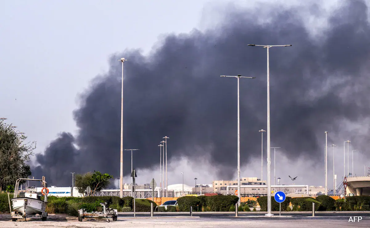
Indian Killed In Abu Dhabi Due To Debris From Intercepted Missile
An Indian national was killed by falling debris after a ballistic missile was intercepted over Abu Dhabi on Thursday….
Continue Reading
-

Atmosphere’s ‘detergent’ may get stronger as Earth warms
Methane may not stay in the air for long, but it still warms the planet quickly. A tiny chemical in the air helps clean it up every day.
Scientists now want to know what will happen to this natural cleaning system as Earth gets hotter.
Continue Reading
Pakistan to play proactive role in regional de-escalation: FO – RADIO PAKISTAN
- Pakistan to play proactive role in regional de-escalation: FO RADIO PAKISTAN
- Pakistan says little, reveals less The Express Tribune
- Pakistan’s SMDA-Led Path to Unity and De-Escalation Daily Times
- foreign office urges media to avoid fake news
Continue Reading
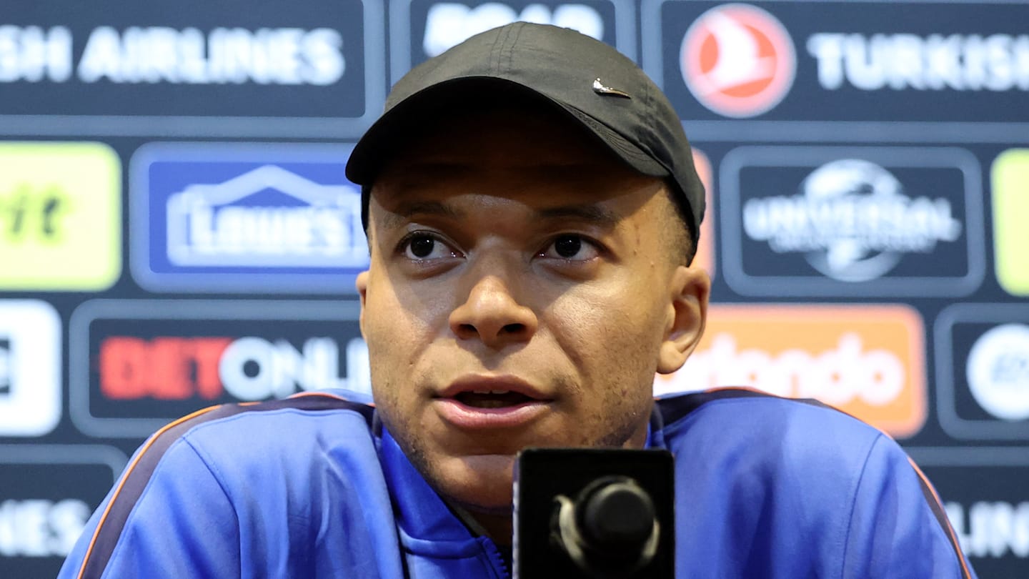
Kylian Mbappe Sets Record Straight on Real Madrid Injury Blunder As Controversy Grows
Kylian Mbappé has disputed claims that Real Madrid misdiagnosed his knee injury because club doctors…
Continue Reading

Hisense Opens UR9 RGB MiniLED TV Pre-Orders with a Complimentary 55-Inch CanvasTV for Early Orders
New Generation of Color Performance Comes Home With the UR9, Built with RGB MiniLED Technology That Generates Red, Green and Blue at the Source for True-to-Life Color That’s Gentler on the Eyes
ALPHARETTA, Ga., March 26,…
Continue Reading
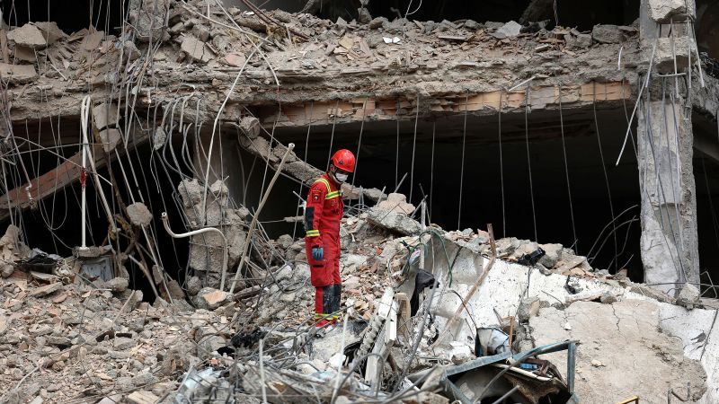
Live updates: Iran war; Trump casts doubt on prospect of an Iran deal
An Israeli man has been killed in a Hezbollah rocket attack on the northern Israeli city of Nahariya, according to Israel’s national emergency service.
It marks the second Israeli…
Continue Reading
The Bus Launches Today on PC, PlayStation®5 and Xbox Series X|S
Paderborn, Germany, 26th March 2026: Today marks the official launch of The Bus, the highly detailed city bus simulation from TML-Studios and Aerosoft, available now on PC (Steam), PlayStation 5, and Xbox Series X|S. After years of…
Continue Reading
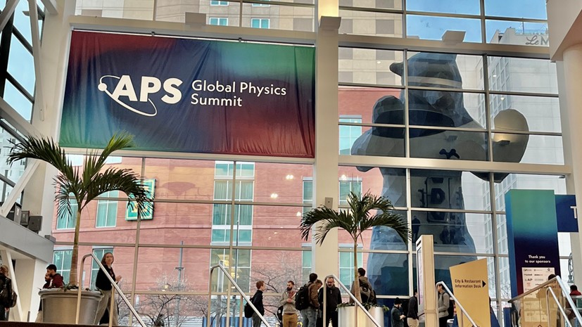
Many-body effects at the world’s largest physics conference – Physics World
Many-body effects at the world’s largest physics conference – Physics World
In 1987, a group of six young Belgian designers put their stamp on fashion. Dubbed the Antwerp Six— playfully referred to as “Twerps” by WWD—Dries Van Noten, Dirk Bikkembergs, Walter Van Saene, Marina Yee (1958-2025), and…
Continue Reading