- Pakistan boosts oil reserves as global supply crisis deepens samaa tv
- Higher education goes online, schools shut for 2 weeks, 4-day work week announced as ‘austerity measures’ Dawn
- Govt approves weekly oil price proposals The Express Tribune
Author: admin
-
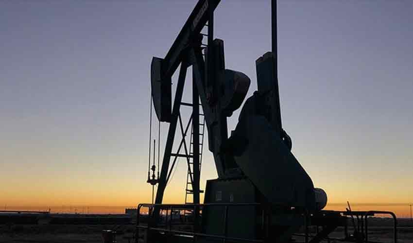
Pakistan boosts oil reserves as global supply crisis deepens – samaa tv
-

Meet the 9 Breakout Models of Fall 2026
Another season has come and gone. While we’re past the upheaval of spring’s 15 designer debuts, fall 2026 proved another lively fashion month. Change was still afoot: Meryll Rogge showed her first collection for Marni, while Pieter Mulier…
Continue Reading
-
Study discovers malaria parasite tricks immune system, paving way for new therapies-Xinhua
JERUSALEM, March 11 (Xinhua) — Israeli researchers have discovered a sophisticated way in which the deadly malaria parasite keeps the host’s immune system jammed, the Weizmann Institute of Science said in a statement on Wednesday.
Their…
Continue Reading
-
How the Middle East conflict has hit daily flight hours – Cirium
- How the Middle East conflict has hit daily flight hours Cirium
- Middle East Conflict Disrupts Commercial Aviation, Fuel Prices Surge, Airspace Closures Hit Gulf Carriers, and Aviation Demand Faces Uncertainty: New Update Travel And Tour World
- ‘Biggest risk for aviation is geopolitics, long-term conflict could hit demand’: Boeing The Economic Times
- Middle East Conflict A ‘Financial Hit’ To Battered Airline Industry, Says Boeing Chief NDTV Profit
- What’s at stake for commercial aviation amid the Middle East conflict Cirium
Continue Reading
-
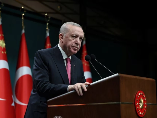
Erdogan says Iran war must stop before whole region dragged in
Turkiye had offered to mediate between the sides before the US-Israeli strikes on Iran began 12 days ago
Turkish President Tayyip Erdogan speaks after a cabinet meeting in Ankara, Turkey on March 9, 2026. PHOTO: REUTERS
…Continue Reading
-

Your Oscars questions answered: ‘The best film of the year hasn’t actually won best picture since 12 Years a Slave’ | Film
Guardian writers have been making their pitches for best picture winner at the 98th Academy Awards in our Oscars hustings series.
Has Chase Infiniti been snubbed? Should Train Dreams win for best cinematography? Who’s the bigger monster,…
Continue Reading
-
Pakistan Navy escorting ships on country’s own trade routes, not Strait of Hormuz— official – Arab News
- Pakistan Navy escorting ships on country’s own trade routes, not Strait of Hormuz— official Arab News
- Pakistan Navy launches Operation Muhafizul Bahr to counter threats to shipping, maritime trade: ISPR Dawn
- Pakistan Sends Navy Escort for Its…
Continue Reading
-

New Materials Shift Crystal Structure with Humidity
NEW YORK, March 11, 2026 — Most solid materials we rely on, from steel, to plastics and ceramics, are designed to have specific properties. Whether a material is soft and flexible, or stiff and tough depends on how molecules within…
Continue Reading
-
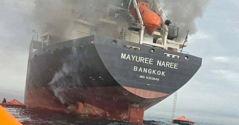
Trump vows to end war soon as Iran hits ships, threatens banks, and toll on U.S. forces emerges
A senior Qatari official, who has worked for years in the Gulf state’s ministry of foreign affairs, told CBS News on Wednesday that the Middle East has, “for all intents and purposes,” been engulfed in a regional war that…
Continue Reading
-
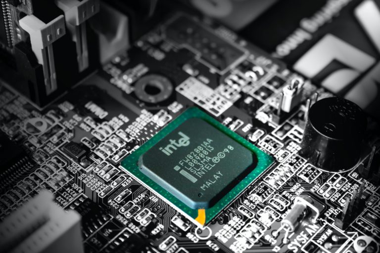
Intel Corporation (INTC) Gains 9% on the Release of Core Series 2 Processors
Intel Corporation (NASDAQ:INTC) is one of the Best Technology Stocks to Buy for the Long Term. Intel Corporation (NASDAQ:INTC) has gained more than 9% since the release of its Core Series 2 processor with P-cores on March 9. The company…
Continue Reading