
Senior Provincial Minister, Marriyum Aurangzeb.
LAHORE:
The Punjab…


China on Tuesday said it will continue its mediation efforts to…

Mr Nobody Against Putin, a primary school teacher’s record of the indoctrination of his pupils to support Russia’s invasion of Ukraine, has won the Oscar for best documentary in Los Angeles.
Pavel Talankin, who is now in exile in Europe,…

Our relationships shape our health in many ways. Friends and family can provide support during difficult times and encourage healthy habits. But not all relationships are positive – some can be a persistent source of stress.
A new study…

By Mike Murphy
Smoke rises after energy installations in the United Arab Emirates were struck by Iran on Saturday.
U.S. stock-index futures reversed early losses on Sunday as the market braced for another surge in oil prices this week, with the conflict with Iran threatening to escalate further.
West Texas Intermediate crude (CL.1) (CLJ26) jumped as high as $102.57 a barrel Sunday, but quickly erased those gains and was last trading around $98 a barrel. Brent crude (BRN00) (BRNK26), the global benchmark, also gave up sharp early gains and was last trading around $103 a barrel. Oil prices crossed the $100-a-barrel level last week for the first time since 2022, and have surged about 40% since the start of the U.S. and Israeli bombing campaign against Iran at the end of February.
After losing more than 100 points early in the session, Dow Jones Industrial Average futures (YM00) were recently up about 172 points, or 0.4%, S&P 500 futures (ES00) and Nasdaq-100 futures (NQ00) also bounced back strongly from early declines.
The turnaround may have been influenced by a Wall Street Journal report late Sunday that the Trump administration is poised to announce that multiple countries have agreed to help escort tanker traffic through the critical Strait of Hormuz, though it was unclear if that might happen before or after the end of hostilities.
Bitcoin (BTCUSD) was above $72,000 after a Friday rally took it to nearly $74,000. Gold (GC00) and silver (SI00) futures fell. The U.S. Dollar Index DXY declined.
Last week, the three major indexes fell for a third straight week, with the Dow DJIA dropping 2%, the S&P 500 SPX sliding 1.6% and the Nasdaq COMP falling 1.3%.
Investors remain worried that oil infrastructure in the Persian Gulf will be targeted, after President Donald Trump announced Friday that the U.S. had bombed military targets on Iran’s Kharg Island, home to Iran’s main terminal for exporting oil. Trump threatened to strike Iran’s vital oil infrastructure sites next if Tehran doesn’t allow tankers to flow freely through the Strait of Hormuz.
On Sunday, Iran warned the United Arab Emirates to evacuate three major ports, suggesting they could be targeted.
Meanwhile, the U.S. is moving about 2,500 Marines to the Middle East, raising the possibility of ground operations in Iran.
The International Energy Agency on Sunday said emergency reserves of oil would soon start flowing, after last week’s approval of the release of 400 million barrels held in strategic reserves. In an effort to relieve “the largest supply disruption in the history of the global oil market,” the agency said “stocks will be made available by IEA member countries in Asia Oceania immediately while stocks from IEA member countries in the Americas and Europe will be made available starting from the end of March.” The U.S. plans to release about 172 million barrels starting this week.
On Sunday, Iranian Foreign Minister Abbas Araghchi claimed on CBS News’ “Face the Nation” that Iran has not closed the strategic waterway and that Iran’s military has decided to let “a group of vessels” belonging to a variety countries to pass the strait safely and securely.
Trump over the weekend called on other countries’ navies to help secure the Strait of Hormuz, but so far none have publicly agreed to.
“The oil market is now flirting with the outer edge of disruption risk,” Stephen Innes, managing partner at SPI Asset Management, said in a weekend note. ” Every uptick in oil tightens financial conditions. Every tightening of conditions feeds back into equities. The market stops behaving like a calm pricing mechanism and starts behaving like a stress test.”
Meanwhile, panic is starting to seep into Wall Street, with technical indicators showing strain on the market, raising new fears of inflation – and potentially a recession.
Read more: The financial sector is sending some spooky technical signals about the stock market
“The economic outlook is increasingly shrouded in the fog of war,” Bob Schwartz, a senior economist at Oxford Economics, said in a Friday note to clients. Skyrocketing gas prices are “delivering an immediate hit to household budgets – particularly for lower-income families that devote a larger share of their spending to energy.”
See: Individual investors are chasing oil’s surge amid Iran conflict; institutions are thinking about what comes next
U.S. gas prices Sunday averaged $3.699 a gallon, according to AAA, about 77 cents higher than a month ago.
Still, most traders seem to be pricing in only a short-term spike in fuel prices, expecting a relatively quick conflict.
Energy Secretary Chris Wright said Sunday on NBC’s “Meet the Press” that the surge in gas prices is likely to last “a few more weeks,” calling the current situation “a short-term disruption to the flow of energy.”
“Depending upon the timing and the manner in which this conflict comes to an end, we’re going to see some elevated pricing until we get there,” Wright said, as he dismissed Iranian threats that a drawn-out conflict will send the price of oil above $200 a barrel. ” I would pay no attention to what Iran says,” he added.
The effects of rising oil and gas prices on transportation and retail spending are expected to be revealed in corporate earnings outlooks this week, as companies including FedEx (FDX), Macy’s (M) and Lululemon (LULU) report quarterly earnings.
Investors will also be closely watching this week’s meeting of the Fed’s rate-setting committee. While the Fed is widely expected to keep interest rates steady Wednesday, any indications of future moves – perhaps even a hike – will be front of mind.
Read more: It was unthinkable a couple of weeks ago, but could the next move by the Fed be a rate hike?
-Mike Murphy
This content was created by MarketWatch, which is operated by Dow Jones & Co. MarketWatch is published independently from Dow Jones Newswires and The Wall Street Journal.
(END) Dow Jones Newswires
03-15-26 2105ET
Copyright (c) 2026 Dow Jones & Company, Inc.
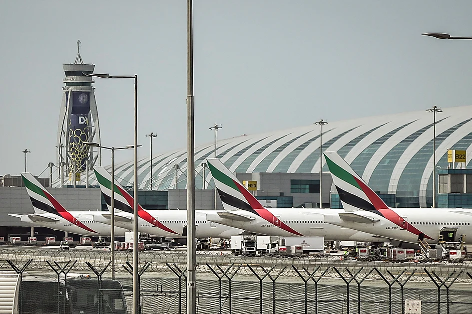
[Editor’s Note: Follow Khaleej Times live blog amid US-Israel-Iran war for the latest regional developments.]
Flights at Dubai International Airport have been temporarily suspended early on Monday after a drone-related incident impacted one of…


RALSTON, NE— The Lincoln Stars (27-25-4-1) skated to a wild 7-6 victory on road over the Omaha Lancers on Sunday…