- PTI formally nominates Mehmood Khan Achakzai as NA opposition leader Dawn
- Speaker sets opp leader appointment process in motion The Express Tribune
- Ayaz Sadiq seeks nominations for opposition leader as PPP stages walkout Dunya News
- Opposition…
Author: admin
-
PTI formally nominates Mehmood Khan Achakzai as NA opposition leader – Dawn
-

Space in 2026
Last year was a busy one for the space industry, and the quest to explore the cosmos is not slowing down any time soon. RICHARD LOWE FRAeS from the RAeS Space Group reports on what is to come in…
Continue Reading
-
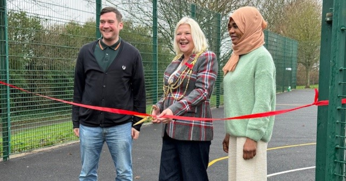
Official opening of ‘good as new’ recycled games area
The innovative recycled multi use games area (MUGA) at Hangleton Park has been officially opened by city Mayor Amanda Grimshaw.
The pitch was re-located from Moulsecoomb last year, completing one of the council’s most innovative recycling…
Continue Reading
-
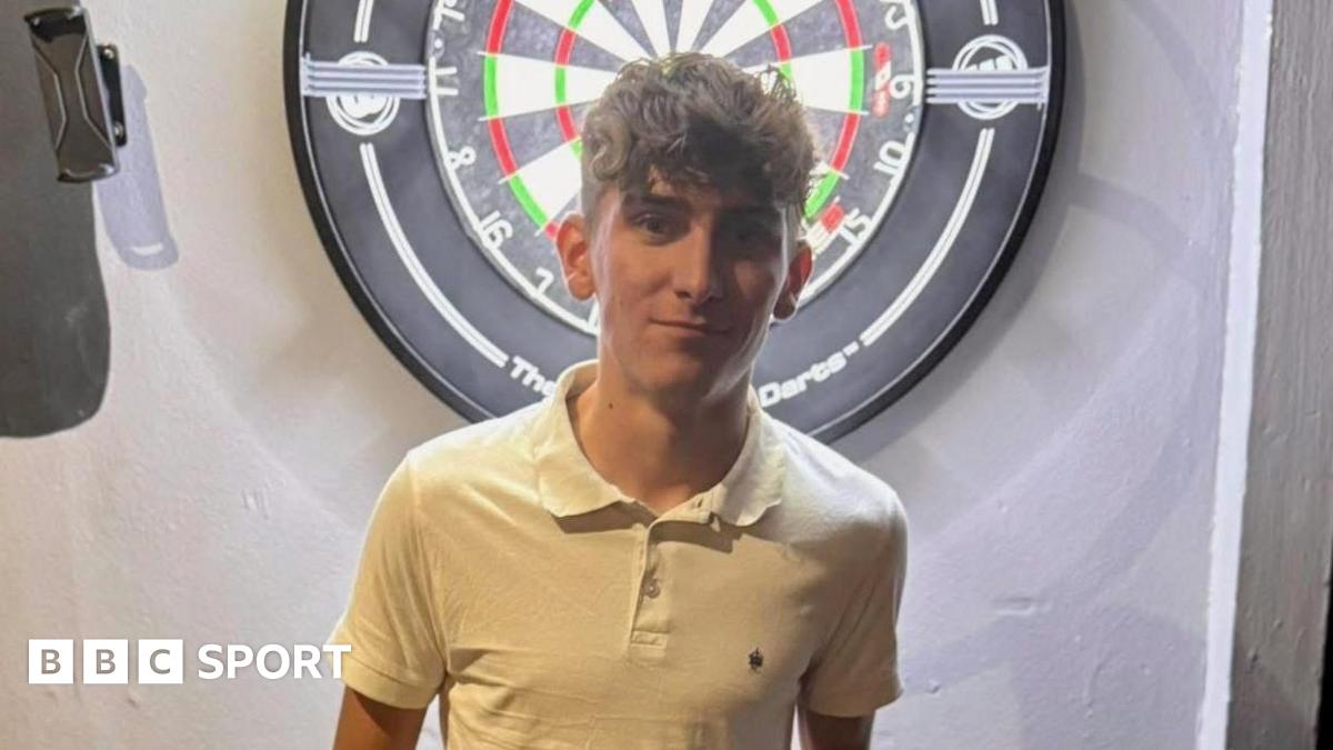
Tyler Thorpe: Norfolk darts player qualifies for PDC world tour
Steve Beaton, the 1996 world champion, was among those who failed to secure a tour card at the Q school with Suffolk’s Stephen Burton and King also making it through on the final day.
But it is 18-year-old Littler, who now sets the standards…
Continue Reading
-
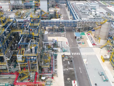
Arkema starts up its new Rilsan® clear transparent polyamide unit in Singapore
This successful start-up represents a major step forward for Arkema, as the new unit triples the Group’s global production capacity of Rilsan® Clear transparent polyamide. This investment of around US$20 million, announced in July 2025, is part of the major growth projects in which Arkema has recently invested to support its strategic roadmap on Specialty Materials.
Designed for operational excellence and reliability, this new unit will enable Arkema to meet the growing demand for sustainable high-performance transparent materials across key markets such as eyewear, AR/VR and smart consumer electronics, industrial filtration, healthcare devices and home appliances.
Continue Reading
-
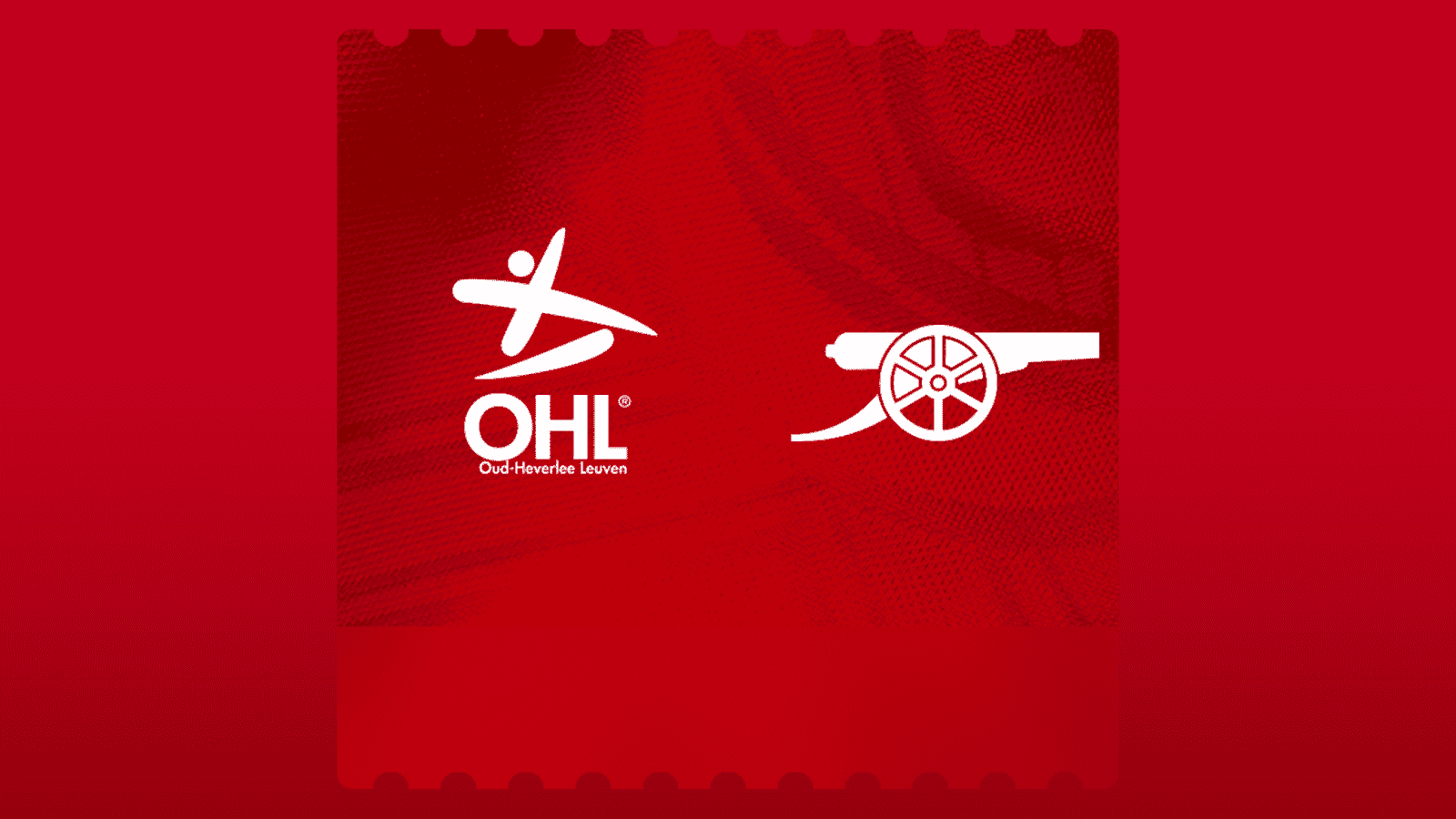
Ticket Info: OH Leuven v Arsenal Women
Arsenal Women will head back to Belgium to face OH Leuven at the King Power at Den Dreef Stadion on Wednesday, February 11 kick off 5:45pm (GMT) for the first leg of the UEFA Women’s Champions League play-offs.
This match will be shown live on…
Continue Reading
-
Ticket info: Arsenal Women v West Ham United
Arsenal Women will take on West Ham United Women in the Barclays Women’s Super League on Saturday, March 21 at Emirates Stadium with kick off at 12:30pm (subject to change due to broadcast).
If you would like to join us at Emirates Stadium…
Continue Reading
-

More Young People are Using AI Chatbots. Is it Safe? – Terms of Service with Clare Duffy
More Young People are Using AI Chatbots. Is it Safe?
Since 2024, multiple families have filed lawsuits against major AI companies, claiming their children were driven to self-harm and even suicide after talking with chatbots. Some AI companies…
Continue Reading
-

A Journalist With 1,800 Phones on Galaxy Z TriFold – Samsung Newsroom India
A Japanese IT journalist with more than 20 years on the smartphone beat shares his Galaxy Z TriFold experience
Yasuhiro Yamane, a Japanese IT journalist who has covered smartphones and…
Continue Reading
-
Synchronous glacier retreats across hemispheres challenge ice age theories: study-Xinhua
SYDNEY, Jan. 13 (Xinhua) — An international research team involving Australian scientists has uncovered evidence that glaciers in the Southern and Northern hemispheres were synchronous during the last ice age.
Published in Nature…
Continue Reading