WASHINGTON — The world economy is experiencing a disorienting flashback to the 1970s.
Oil prices are once again surging in the wake of war in the Middle East, driving up the cost of gasoline, diesel and jet fuel and threatening a return to stagflation – the toxic mix of higher prices and slower growth that made economic life so miserable a half century ago.
But the U.S. and world economies are less vulnerable now than they were when Saudi Arabia and other Middle Eastern petroleum producers withheld oil supplies to punish countries that supported Israel in the 1973 Yom Kippur War.
In response to that shock — and another triggered six years later by the Iranian revolution — countries embarked on a new course to increase their energy efficiency, reduce their dependence on Middle Eastern oil, stockpile fuel against future threats, and find and develop alternative sources of energy.
“We have decades of experience now dealing with these kinds of oil shocks,’’ said Amy Myers Jaffe, research professor at New York University’s Center for Global Affairs.
Of course, the notion that the current Iran energy shock could have been worse is little comfort to frustrated American motorists paying $4 or more for a gallon of gasoline, to European farmers contending with skyrocketing fertilizer prices and to street vendors in India who can’t get enough gas to cook curries and samosas for their customers.
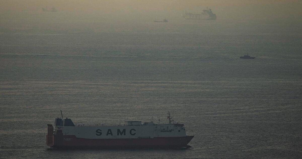
And the sheer scale is unprecedented. In response to attacks by the United States and Israel that began Feb. 28, Iran effectively shut off the Strait of Hormuz, through which 20 million barrels of oil — or one-fifth of global production — flowed daily.
Lutz Kilian, director of the Federal Reserve Bank of Dallas’ Center for Energy and the Economy, figures that 5 million daily barrels can either be rerouted from the Persian Gulf to the Red Sea or continue to transit through the Strait of Hormuz. But that still means that roughly 15 million barrels — or 15% — of daily global oil production is missing, ` with just 6% in the 1973 embargo and after Iraq’s invasion of Kuwait in 1990.
Cushioning the blow
Changes the U.S. and other countries made over the past five decades have limited the economic fallout from the war. In 1973, oil accounted for almost half — 46% — of world energy supplies. By 2023, oil’s share had fallen to 30%, according to the International Energy Agency.
The world still uses more oil than ever: Consumption topped 100 million barrels a day last year, up from fewer than 60 million barrels a day in 1973. But a much bigger share of global energy is coming from other sources — such as natural gas, nuclear, solar — compared with five decades ago.
The U.S., in particular, has weaned itself away from dependence on foreign oil.
When the ’73 oil shock hit, America’s domestic energy production was in decline and its reliance on oil imports was growing alarmingly. But the rise of fracking — pumping high-pressure water deep underground to extract previously hard-to-get oil or gas from rock — rejuvenated U.S. energy production in the 21st century. By 2019, America had become a net petroleum exporter.
“The U.S. economy is much better positioned than it was in the 1970s,” when it was “particularly vulnerable to an oil price shock,” said Sam Ori, executive director of the University of Chicago’s Energy Policy Institute.
In the early ‘70s, for example, the United States got about 20% of its electricity from oil, Ori said. But a law enacted in 1978 prohibited the use of petroleum in power plants. Now the United States gets no electricity from oil — aside from a few generators in, say, the far reaches of Alaska.
Dimming the lights
The 1973 oil embargo was a wake-up call, creating shortages that led to long lines at U.S. gasoline stations.
On Nov. 25, 1973, President Richard Nixon went on television to ask the American people to make sacrifices. To conserve fuel, he urged gasoline stations to shut their pumps from Saturday night through Sunday, hoping to discourage long-distance weekend driving.
He asked Congress to lower the maximum speed limit to 50 miles an hour (lawmakers settled for 55 miles an hour) and to ban ornamental and most commercial lighting (they balked at that). Nixon himself promised to dim the White House Christmas lights.
But while those memories may have left a lasting imprint on some, Jaffe of New York University’s Center for Global Affairs says that today, “a repeat of long gasoline lines, fuel rationing, and outright fuel shortages in the U.S seems highly unlikely.”
Other countries took aggressive action following the 1973 oil embargo as well.
The United Kingdom, contending with a coal strike as well as the energy crisis, cut the work week to three days to slash electricity consumption. France ordered offices to turn off the lights at night.
Japan, almost entirely dependent on imported oil, passed a series of “sho-ene’’ laws — combining the Japanese words for “save’’ or “reduce’’ with “energy’’ — mandating energy efficiency in shipping, buildings, machinery, automobiles and homes.
Japan also encouraged the use of liquefied natural and gas and the rapid growth of nuclear power, an effort set back after a 2011 earthquake and tsunami damaged the Fukushima nuclear plant. Overall, Japan ranks No. 21 in the world in per capita energy consumption, according to International Energy Agency data, as a result of its efficiency drive and widespread use of buses and trains. The United States is No. 9.
More fuel efficient cars, new oil fields
The U.S. government began imposing fuel economy standards in 1975. Fuel economy has risen from 13.1 miles per gallon for model year 1975 vehicles to 27.1 mpg in model year 2023, according to the Environmental Protection Agency. The World Bank, in fact, attributes most of the drop in the global economy’s dependence on oil to stricter fuel efficiency requirements for vehicles around the world.
The ’70s shocks also set off a search for oil outside the Middle East — Prudhoe Bay in Alaska, the North Sea fields off the coasts of the United Kingdom and Norway and Canada’s oil sands deposits.
As fracking boomed, U.S. oil production shot up from 5 million barrels a day in 2008 to 13.6 million barrels a day last year. Over the same period, U.S. natural gas production has more than doubled.
Countries also began stockpiling oil and set up the Paris-based International Energy Agency in 1975 to coordinate responses to energy shocks. Last month, the agency’s 32 member countries agreed to release 400 million barrels of oil in an effort to calm the oil market; included were 172 million barrels from the U.S. Strategic Petroleum Reserve, set up in 1975.
Central banks such as the Federal Reserve also learned lessons. In the ‘70s, they reduced interest rates to protect the economy from the oil shocks. In so doing, they overlooked the threat posed by higher energy costs — and inflation, already elevated, got worse.
In a Feb. 17 commentary – 11 days before the United States and Israel attacked Iran – the Dallas Fed’s Kilian wrote that the Fed erred in cutting rates to boost the economy when the 1970s oil shocks hit: “What we can learn from the 1970s is that a well-intentioned policy of stimulating the economy by lowering interest rates has the potential of inadvertently reigniting inflation.’’
Trump undoes efforts to reduce oil dependence
While much has changed, the University of Chicago’s Ori cautions: “Oil is still king, the No. 1 fuel in the U.S. economy.’’ Cars, planes, trucks and ships get about 90% of their delivered energy from petroleum. “The lifeblood of the economy – the transportation sector —is still overwhelmingly reliant on petroleum fuel, the price of which is set in a global market,’’ Ori said, “and a disruption anywhere affects the price everywhere.’’
He also notes that President Donald Trump is undoing many of the policies meant to reduce America’s dependence on petroleum and to encourage the use of electric vehicles.
Trump’s sweeping tax bill last year ended consumer credits of up to $7,500 for EV purchases. He has announced a proposal to weaken U.S. fuel economy standards and repealed fines on automakers that don’t meet those standards.
“You take all that together, and the fact is, the U.S. is going in the opposite direction of making big changes to further insulate the economy from oil shocks and oil price volatility,’’ Ori said.
Wiseman and Kageyama write for the Associated Press. Kageyama reported from Tokyo.