The Public Service Association of New South Wales and Community and Public Sector Union (SPSF Group) NSW Branch acknowledges the Traditional Custodians of the lands where we work and the places in which we live. We pay respect to Ancestors and Elders, past, present and future. We recognise the unique cultural and spiritual relationship and celebrate the contributions of First Nations peoples to Australia.
For members who are blind or have low levels of vision, a wide range of adaptive technologies and tools are available to assist. Applications such as Jaws Screen Reader can help access union information.
In addition, Vision Australia has a list of resources for the visually impaired HERE.
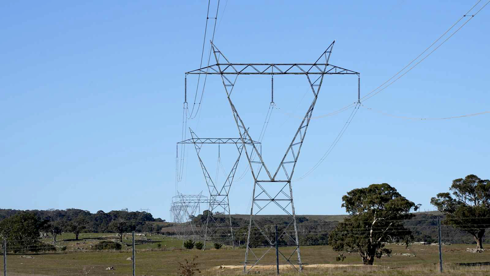
ACCIONA together with our JV partner Genus, has been awarded a contract to construct the Western Renewables Link, an essential transmission project that will assist in facilitating Victoria’s renewable energy future.
The contract is subject to satisfaction of conditions precedent and commencement of construction activities is still subject to the project receiving all necessary state and federal approvals.
Spanning approximately 190 kilometres, the Western Renewables Link will deliver a new 500 kV double-circuit transmission line from Bulgana in western Victoria to the Sydenham terminal station in Melbourne’s North-West.
The project will unlock large-scale renewable energy from the region, enabling more than 3,000 MW of new generation and powering up to one million Victorian homes.
Bede Noonan, ACCIONA CEO, Australia and New Zealand, said ACCIONA is proud to bring its local expertise combined with global experience to deliver this project.
“ACCIONA is proud to be contributing to Australia’s clean energy transformation by playing a leading role in the delivery of transmission networks across Australia.
“Core to our delivery is our collaborative approach, working hand in hand with local communities and stakeholders.” Bede said.
Douglas Shire Council will remove three unsafe Oil Palms on the verge of Port Douglas Road, in front of Oaks Resort Port Douglas, before the end of the year (subject to contractor availability).
Council arborists have been monitoring the palms for the past two months after observing a rapid decline in their health. The trees now appear dead, with failed central fronds and significant basal bracket fungi indicating advanced internal decay.
Given the prominent location and proximity to traffic, Council sought an independent arborist assessment, which confirmed that all three palms are structurally compromised, affected by pathogenic decay, and highly likely to fail.
Based on this expert advice, Council will proceed with their removal as a necessary safety measure.
Council acknowledges the community’s appreciation for this well-known avenue of palms and understands that their removal may be disappointing. This decision follows detailed inspections and independent professional recommendations to ensure public safety.
[TOKYO – 2025/12/15] – NTT DATA, a global leader in AI, digital business and technology services, has been selected as a 2025 CDP Climate Change “A List” company for the fourth consecutive year and CDP Water Security “A List” company for the first time by the international NGO CDP*1. NTT DATA was recognized as a leader for its transparency and performance in climate change and water initiatives.
CDP surveys corporate environmental information at the request of institutional investors who make ESG investments. The companies are assessed by a score of A to D, based on the comprehensiveness of disclosure, awareness and management of environmental risks. CDP also investigates how companies demonstrate environmental leadership best practices, such as setting ambitious and meaningful targets. CDP scores are widely used to drive investment and procurement decisions towards a resilient and Earth-positive economy.
The CDP disclosure cycle 2025 assessed over 22,100 companies around the world. NTT DATA has been recognized as one of the companies achieving an “A” rating, based on its implementation of key criteria emphasized by CDP, such as comprehensive and transparent environmental information disclosure, board-level oversight, ambitious target setting and initiatives across the entire supply chain.
NTT DATA was commended for its approach to water related initiatives and incorporating “efficient water management in data centers” into its environmental policy. NTT DATA’s approach to water security includes conducting water stress assessments, analyzing all data centers for water-related risks and setting improvements of WUE (Water Usage Effectiveness) as a target for efficient water management. As a result of these initiatives, NTT DATA was selected for the A List for the first time.
Notes to editors
About CDP CDP is a global non-profit that runs the world’s only independent environmental disclosure system for companies, capital markets, cities, states and regions to manage their environmental impacts.
About NTT DATA
NTT DATA is a $30+ billion business and technology services leader, serving 75% of the Fortune Global 100. We are committed to accelerating client success and positively impacting society through responsible innovation. We are one of the world’s leading AI and digital infrastructure providers, with unmatched capabilities in enterprise-scale AI, cloud, security, connectivity, data centers and application services. Our consulting and industry solutions help organizations and society move confidently and sustainably into the digital future. As a Global Top Employer, we have experts in more than 70 countries. We also offer clients access to a robust ecosystem of innovation centers as well as established and start-up partners. NTT DATA is part of NTT Group, which invests over $3 billion each year in R&D.
As Australian shares face a challenging start to the week, influenced by global tech sector declines, investors may find themselves exploring alternative opportunities within the market. Penny stocks, often representing smaller or newer companies, continue to capture interest despite their somewhat outdated label. By focusing on those with solid financial foundations and potential for growth, these stocks can offer surprising value and stability in uncertain times.
Name
Share Price
Market Cap
Financial Health Rating
Alfabs Australia (ASX:AAL)
A$0.41
A$117.5M
★★★★★☆
EZZ Life Science Holdings (ASX:EZZ)
A$1.495
A$70.52M
★★★★★★
Dusk Group (ASX:DSK)
A$0.78
A$48.57M
★★★★★★
IVE Group (ASX:IGL)
A$2.88
A$442.63M
★★★★★☆
MotorCycle Holdings (ASX:MTO)
A$3.12
A$230.45M
★★★★★★
Veris (ASX:VRS)
A$0.074
A$39.99M
★★★★★★
West African Resources (ASX:WAF)
A$2.90
A$3.31B
★★★★★★
Service Stream (ASX:SSM)
A$2.24
A$1.37B
★★★★★★
EDU Holdings (ASX:EDU)
A$0.94
A$135.3M
★★★★★☆
GWA Group (ASX:GWA)
A$2.41
A$632.09M
★★★★★☆
Click here to see the full list of 427 stocks from our ASX Penny Stocks screener.
Let’s take a closer look at a couple of our picks from the screened companies.
Simply Wall St Financial Health Rating: ★★★★☆☆
Overview: DUG Technology Ltd is a technology company offering hardware and software solutions to the technology and resource sectors across Australia, the United States, the United Kingdom, Malaysia, and the United Arab Emirates, with a market cap of A$281.66 million.
Operations: The company’s revenue is derived from three main segments: Hpcaas ($27.44 million), Services ($51.87 million), and Software ($10.47 million).
Market Cap: A$281.66M
DUG Technology Ltd, with a market cap of A$281.66 million, provides hardware and software solutions to various sectors globally. The company has three revenue streams: Hpcaas (A$27.44 million), Services (A$51.87 million), and Software (A$10.47 million). Despite being unprofitable, DUG has improved its financial position from negative shareholder equity five years ago to positive now, indicating progress in reducing losses by 51.3% annually over the past five years. It trades at 83.1% below its estimated fair value and maintains more cash than total debt, suggesting potential for future growth despite current challenges in profitability and volatility stability at 7%.
ASX:DUG Debt to Equity History and Analysis as at Dec 2025
Simply Wall St Financial Health Rating: ★★★★★★
Overview: Immutep Limited is a biotechnology company focused on developing novel Lymphocyte Activation Gene-3 related immunotherapies for cancer and autoimmune diseases in Australia, with a market cap of A$552.65 million.
Operations: The company’s revenue is primarily derived from its immunotherapy segment, which generated A$5.03 million.
Market Cap: A$552.65M
Immutep Limited, a biotechnology company with a market cap of A$552.65 million, is focused on developing immunotherapies and has recently entered into a strategic collaboration with Dr. Reddy’s Laboratories for the development and commercialization of its lead product, eftilagimod alfa (efti), in selected markets. The agreement includes an upfront payment of US$20 million and potential milestone payments up to US$349.5 million, alongside double-digit royalties on sales. Despite being pre-revenue with A$5 million in revenue from its immunotherapy segment, Immutep’s strong cash position exceeds both short-term and long-term liabilities, supporting ongoing clinical trials for various cancers.
ASX:IMM Debt to Equity History and Analysis as at Dec 2025
Simply Wall St Financial Health Rating: ★★★★★☆
Overview: Wagners Holding Company Limited produces and sells construction and related building materials across several countries, including Australia, the United States, New Zealand, the United Kingdom, Papua New Guinea, and Malaysia; it has a market cap of A$719.01 million.
Operations: Wagners Holding generates revenue through its Construction Materials segment at A$257.69 million, Project Services at A$105.71 million, Earth Friendly Concrete at A$0.16 million, and Composite Fibre Technology at A$68.45 million.
Market Cap: A$719.01M
Wagners Holding, with a market cap of A$719.01 million, has shown robust financial health and growth potential. Its interest payments are well covered by EBIT, and the net debt to equity ratio of 12.6% is satisfactory. The company has experienced management and board teams with average tenures over eight years. While short-term assets cover short-term liabilities, they fall short for long-term obligations. However, Wagners’ debt reduction over five years and substantial earnings growth of 120.9% last year indicate strong operational performance in its construction materials segment, supported by inclusion in the S&P/ASX Emerging Companies Index recently.
ASX:WGN Financial Position Analysis as at Dec 2025
This article by Simply Wall St is general in nature. We provide commentary based on historical data and analyst forecasts only using an unbiased methodology and our articles are not intended to be financial advice. It does not constitute a recommendation to buy or sell any stock, and does not take account of your objectives, or your financial situation. We aim to bring you long-term focused analysis driven by fundamental data. Note that our analysis may not factor in the latest price-sensitive company announcements or qualitative material. Simply Wall St has no position in any stocks mentioned.
Companies discussed in this article include ASX:DUG ASX:IMM and ASX:WGN.
This article was originally published by Simply Wall St.
Have feedback on this article? Concerned about the content? Get in touch with us directly. Alternatively, email editorial-team@simplywallst.com
You don’t have permission to access “http://www.spglobal.com/energy/en/news-research/latest-news/lng/121425-interview-tokyo-gas-mulls-participation-in-us-gulf-coast-lng-project—ceo” on this server.
Baker McKenzie today announced that leading project finance lawyer Matthias Schemuth has joined the Firm’s Singapore office* as a Principal and Asia Pacific Co-Head of Projects in its Finance & Projects practice, alongside Partner Jon Ornolffson in Tokyo.
Matthias joins the Firm from DLA Piper, bringing more than 20 years of experience in the energy and infrastructure sectors across Asia Pacific. He advises sponsors, developers, commercial banks, multilateral lending agencies, and export credit agencies on the structuring and financing of large-scale projects. His practice also spans international banking, structured commodity and trade finance, with a strong focus on emerging markets. Matthias has been consistently recognised by Chambers Asia Pacific and Who’s Who Legal as a leading project finance practitioner.
James Huang, Managing Principal of Baker McKenzie Wong & Leow in Singapore, said: “We are excited to welcome Matthias to our team. His expertise and proven record in managing teams will be invaluable as we expand our regional and global finance offerings for clients.”
Emmanuel Hadjidakis, Asia Pacific Chair of Baker McKenzie’s Banking & Finance Practice, commented: “Asia Pacific is seeing strong momentum in infrastructure development, energy transition investments, and cross-border project financing, much of it centred in Singapore. Having Matthias on board will further enhance our ability to help clients seize opportunities in the region’s evolving energy and infrastructure markets.”
Steven Sieker, Baker McKenzie’s Asia Chief Executive, added: “Matthias’s appointment underscores Baker McKenzie’s continued commitment to investing in exceptional talent across key markets to support our clients in navigating today’s increasingly complex business and regulatory environment.”
Matthias said: “I’m thrilled to join Baker McKenzie and contribute to its strong growth in Asia Pacific. The Firm’s global reach and local depth provide an unparalleled platform for delivering innovative projects and financing solutions to clients in this dynamic region.”
With more than 2,700 deal practitioners in more than 40 jurisdictions, Baker McKenzie is a transactional powerhouse. The Firm excels in complex, cross-border transactions; over 65% of our deals are multijurisdictional. The teams are a hybrid of ‘local’ and ‘global’, combining money-market sophistication with local excellence. The Firm’s Banking & Finance lawyers are ranked in more jurisdictions than any other firm by Chambers.
Matthias’s hire continues the expansion of Baker McKenzie’s global team. His joining follows the recent arrivals of Carole Turcotte in Toronto; Tom Oslovar in Palo Alto; Jenny Liu in New York and Palo Alto; Helen Johnson, Mark Thompson, Nick Benson, Kevin Heverin, James Wyatt and Michal Berkner in London; Jan Schubert in Frankfurt; Todd Beauchamp and Charles Weinstein in Washington DC; Dan Ouyang, Winfield Lau, and Ke (Ronnie) Li in Beijing, Shanghai, and Hong Kong; and Alexander Stathopoulos in Singapore.
*Baker McKenzie Wong & Leow is the member firm of Baker McKenzie in Singapore
The popular mobile summer rubbish barge service will be back on the water in the Bay of Islands this summer from Monday 29 December.
The staffed barge – a familiar site in the Bay during the summer holiday season – is a joint venture between the Northland Regional, Far North District Council and the Department of Conservation, supported by contractors Northland Waste.
It offers an on-water service for both local and visiting boaties who are encouraged to use it to help keep the Bay of Islands rubbish-free.
The barge service will begin summer operations on Monday 29 December. After then, the weather-dependent service will generally operate Mondays and Fridays until Friday 30 January 2026.
On operational days it will visit Urupukapuka Island campsites from 9am to 10am before mooring close to the south-eastern end of Moturua Island from 10.30am to 1pm (hours will reduce as demand drops off toward the end of January or as capacity of the barge reduces).
A flat $10 fee per rubbish bag applies, regardless of whether pre-paid or plain bags are used. Recyclables are accepted with the cost depending on quantity and cleanliness; typically somewhere between $3 and $5. Additional charges will incur for oversized bags and dirty recycling.
Users are asked to follow the guidance of barge staff. There will be a maximum limit of four people on the barge at any one time and footwear must also be worn for health and safety reasons.
Meanwhile, shore-based facilities are available daily from 8am to 5pm at Ōpua (by the fuel jetty Far North Holdings car park) from Friday 26 December to Thursday 29 January.
A Rāwhiti site (Kaingahoa campground, Kaingahoa Bay, Rāwhiti Rd) will be open daily from 8am – 4pm from Saturday 20 December 2025 to Saturday 28 February 2026. (From 01 March 2026 to 19 December 2026, the site is open daily, 9am – midday.)
Both the Opua and Rāwhiti sites are closed Christmas Day and New Year’s Day.
Meanwhile, a pop-up rubbish and recycling centre will also be installed at Russell Wharf.
The centre will be located at the end of the wharf from 26 December to 11 January, 8am – 11am and 2pm – 5pm. A flat $8 fee per rubbish bag applies, while recyclables are free of charge.
To find out more visit www.nrc.govt.nz/rubbishbarge
Good afternoon and thank you for the opportunity to speak at this conference. The theme – banking
and financial stability – is a topic of deep interest to the RBA given our longstanding mandate to
contribute to financial stability. And I would like to say thank you to all of you, for your work in this
important area and contributing to our understanding of the issues.
Today Im going to explore two broad questions relating to financial stability.
First, why does financial stability matter and what is the RBAs responsibility in relation to it?
Second, how does the RBA deliver on its financial stability responsibility?
Along the way, Ill highlight some of our ongoing work in this space and the issues that are on our
mind. To summarise, our overall assessment is that in this period of elevated global uncertainty, the
Australian financial system is well positioned to weather most shocks. However, we cannot be complacent.
The nature of risks is changing and so there is ongoing work for regulators and industry to remain
prepared in this evolving environment.
Why does financial stability matter and what is the RBAs role?
Financial stability is important because of the role that the financial system plays in the everyday life
of Australians.
The financial system is made up of a range of financial institutions. It includes banks, insurers,
superannuation funds and non-bank lenders. It also includes financial markets, where financial assets
such as shares, bonds and foreign exchange are bought and sold. And it includes market infrastructure,
such as the payments system and other systems that support the trading of financial assets.
It is important that all these elements of the financial system are working well because together they
provide the financial services that Australians depend on in their everyday life. More concretely, a
well-functioning financial system enables households and businesses to:
save, borrow and invest – so that they can prepare for retirement, buy their first home, or
purchase equipment to expand their businesses
make payments – so they can move money efficiently and securely, such as when theyre
tapping their card to pay for groceries, or making payments for their bills online
insure against and manage risk – so they can protect themselves against unexpected costs, such
as damage to or loss of property, or unexpected medical bills.
In other words, the financial system is vital in helping Australians get on in life and plan for the
future. This means that if the financial system overall is not functioning well, it is costly for
everyone.
For example, if banks are unwilling or unable to lend money, businesses might not get the working
capital they need to run their operations, households might reduce spending further than otherwise,
it can become more challenging to purchase a home and economic activity can suffer. Indeed, financial
crises lead to economic hardship for many with long-lasting consequences for economic growth.
Another example is disruptions to payments systems, which can prevent salaries and pensions from
being paid, households from buying the things they need and transactions in financial markets from
settling, causing widespread difficulties.
Maintaining financial stability is about ensuring we have a financial system that is strong, operationally
resilient and prepared for adverse conditions, so we can avoid these types of disruptions and their
broader costs. This doesnt mean reducing risk to the point that we compromise innovation,
competition and efficiency. But it does mean ensuring resilience alongside these other features. Indeed,
innovation and competition can lead to better ways to achieve resilience. We need a financial system that
can support the economy by reliably providing the financial services that households and businesses
depend on, both in good times and bad. A stable financial system promotes saving and investment,
supporting lower funding costs, productive investment and thus longer term growth. So financial stability
is a necessary underpinning to support the economic prosperity and welfare of Australians – which
is the RBAs overarching legislative objective.
Given the alignment between financial stability and the RBAs other functions and objectives, the RBA
– like other central banks – has long had a mandate to contribute to financial stability.
This is a role that has evolved and changed over time. Initially it spanned all the way from liquidity
provision to banking supervision. But in the late 1990s, this changed following the Financial System
Inquiry (Wallis Review). Responsibility for banking supervision was transferred
to a new separate agency – the Australian Prudential Regulation Authority (APRA), which was set up
to bring the prudential functions of the RBA and Insurance and Superannuation Commission together into a
dedicated agency. So unlike in some other countries, in Australia, the RBA, as the central bank, is
not responsible for prudential supervision of these entities – that is APRAs
job.
Although the Australian Governments response to the Wallis Review meant that supervision of these
individual financial institutions was transferred to APRA, the RBA still retained a general (albeit
non-legislated) responsibility to safeguard stability of the financial system as a whole. This reflects
that the RBA, as the central bank, is well positioned to both assess financial system stability –
given the breadth of our system-wide responsibilities – and to support financial system stability
– given our balance sheet capacity to provide liquidity. We also cannot achieve our monetary policy
objectives without financial stability. Relatedly, following the Wallis Review the RBA also gained
enhanced powers to regulate the payments system to ensure it is secure, stable and efficient. The
RBAs responsibility to promote the stability of the Australian financial system was recognised by
the Government at that time, and in subsequent agreements between the RBA and
Treasury.
More recently, following the independent Review of the RBA in 2023, the RBAs financial stability
role was enshrined in legislation – making contributing to financial stability a core
part of the RBAs legislative functions. The Review highlighted the importance of this for
reinforcing accountabilities and strengthening the foundation of cooperation arrangements with other
agencies that share a mandate for promoting financial stability.
How does the RBA deliver on its financial stability responsibility?
So how does the RBA deliver on this high-level responsibility to contribute to the stability of the
Australian financial system? The answer is in a number of ways, and in conjunction with other agencies.
In Australia, delivering on financial stability is a team effort. Responsibilities are shared across
several agencies, each with complementary mandates and policy tools (Figure 1).
Figure 1: The Australian Financial Regulatory Framework
As the central bank, the RBA has a range of responsibilities which Ill cover in more detail shortly.
As Ive already mentioned, APRA is the prudential regulation authority, responsible for regulating
and supervising banks, insurers and superannuation funds, so that Australians financial interests
are protected and the financial system is stable, competitive and efficient. This includes responsibility
for macroprudential policy and bank resolution.
The Australian Securities and Investments Commission (ASIC) and the Treasury also have important financial
stability roles. ASIC is responsible for market integrity and consumer protection across the financial
services sector, and regulates clearing and settlement facilities (complementing the RBAs
supervision of those same facilities from a financial stability perspective). And the Treasury has an
important role in advising the government on financial stability, including the financial regulatory
framework. Treasury also has important policy tools that can help alleviate the economic impacts of
financial crises.
These institutional arrangements and mix of responsibilities make cross-agency collaboration essential in
Australias financial regulatory framework. And this is where the Council of Financial Regulators
(CFR) comes in.
The role of the CFR
The CFR plays a crucial role in bringing together the RBA, APRA, ASIC and Treasury to coordinate and
collaborate on financial stability issues. Its ultimate aim is to promote the stability of the Australian
financial system and support effective and efficient regulation.
The CFR agencies work together to promote financial stability in three key ways:
We analyse vulnerabilities in the financial system that could amplify shocks, and work together to
understand their potential impacts and coordinate policy and other actions to address them.
We support coordination of financial regulation, so that it is effective, efficient and promotes
competition in the financial sector.
We maintain crisis readiness so the CFR agencies are as ready as can be to work together to respond
to support financial stability in the event of future shocks.
Earlier this month, the CFR published its first annual update on its initiatives to address risks and
vulnerabilities in the financial system – covering progress over 2025 and focus areas for the year
ahead. These focus areas for 2026 are geopolitical vulnerabilities, operational vulnerabilities, systemic
liquidity risk and high household leverage. Given the international environment of elevated
uncertainty and geopolitical tensions, strengthening crisis readiness is a common theme running through
much of this work.
While the CFR does not have formal regulatory or decision-making powers separate from those of its
individual members, it has a strong track record of effective collaboration – including in response
to the COVID-19 pandemic and the global financial crisis. The RBA Review in
2023 highlighted the importance of reinforcing cooperation arrangements among the CFR agencies for
promoting financial stability. Consistent with this, the CFR Charter and Memorandums of Understanding
between the agencies were updated earlier this year to clarify roles and responsibilities, and how
agencies work together to promote financial stability.
Having talked about the CFR and its agencies, let me now turn to the specifics of the RBAs role and
how exactly we fulfil our financial stability responsibilities.
The RBAs contribution to financial stability
The RBA contributes to financial stability in several distinct ways. Some contributions aim to be
preventative – focused on promoting resilience and mitigating vulnerabilities, so that
the financial system can withstand a wide range of adverse conditions. Others are curative
– designed to contain disruptions and restore confidence during periods of financial stress.
Weve recently set out the RBAs framework for contributing to financial stability on our
website as part of our broader commitment to enhance transparency and public understanding of the work we
do.
Ill now step through the elements of this framework (see below).
Setting monetary policy to achieve the MPB’s inflation and full employment objectives
Working with CFR agencies to identify and monitor financial stability risks and vulnerabilities, and coordinate
policies to address them, including by:
providing financial stability advice to the CFR and APRA
maintaining crisis readiness
Using the flexibility of the monetary policy framework to manage monetary
policy and financial stability interactions
where they arise, and communicating appropriately
Providing adequate liquidity to the financial system, including in
exceptional circumstances
Intervening in financial markets where appropriate to address market dysfunction
Undertaking and regularly communicating assessments of financial
stability (including
through the Financial Stability Review)
Engaging in international forums to support regional and global financial stability and promote effective standards and cooperation
Determining payments system policy (including in relation to clearing and settlement facilities) and operating Australia’s real-time gross settlement system
Monetary policy
Arguably the most fundamental way the RBA supports financial stability is by setting monetary policy to
achieve low and stable inflation and full employment. To see why, lets consider the alternative.
For example, unemployment is the most common reason why households are unable to repay debt owed to
banks, which can lead to loan losses for banks. High inflation can also trigger financial difficulties
for households and businesses by leading to higher interest rates and lower real incomes, in turn
affecting their ability to service loans. If a sufficiently large number of borrowers were to fall into
negative equity and default on their loans, lenders could face widespread losses as a result. If these
losses were large enough, this could lead to lenders sharply restricting the supply of credit to even
very sound borrowers. This could disrupt economic activity and add to unemployment. And if households and
businesses become concerned that their deposits at banks might not be safe, financial system stability
and economic activity will be disrupted further still. So by setting monetary policy to achieve our
objectives of low inflation and full employment, the RBA has a key role to play in helping create the
economic conditions that support stability in the financial system.
Likewise, a stable financial system supports us to achieve low inflation and full employment.
Well-functioning financial markets and institutions are essential for the transmission of monetary
policy, and history has shown that episodes of financial instability can have lasting impacts on the
economy and employment.
Working with the CFR agencies
As Ive already discussed, the RBA also works closely with the other CFR agencies to support
financial stability. As part of this, the RBA advises the other CFR agencies on the outlook for financial
stability, including if monetary policy might affect – or be affected by – financial
stability concerns.
As noted earlier, monetary policy and financial stability objectives are generally complementary, and
indeed necessary for each other in the longer term. However, there can be times when monetary policy
actions needed to deliver price stability and full employment may not align perfectly with financial
stability goals. For example, an extended period of accommodative monetary policy required to lift
employment and inflation to their appropriate levels could potentially contribute to the build-up of
leverage and imprudent risk-taking in parts of the financial system. A tightening in monetary policy to
control inflation at a later point could then expose these vulnerabilities.
As part of managing these interactions, the Monetary Policy Board has committed to ensuring the CFR is
informed when there are material interactions between financial stability and monetary policy. This is
important to support coordination of policies to address financial stability risks across the CFR,
including the use of APRAs macroprudential policy tools. In this context, the RBA and APRA have
recognised that APRAs macroprudential policy, as a financial stability tool, is better placed in
most circumstances to address the build-up of certain systemic vulnerabilities than monetary policy.
The RBA now provides financial stability advice to the CFR and APRA on a regular basis, and at least
annually to accompany APRAs update to the CFR on macroprudential policy, most recently at the
September 2025 CFR meeting. In that context, the RBA noted that housing credit
growth has picked up, driven by strong growth in lending to investors, as borrowers have responded to
lower interest rates. However, lending standards have remained sound and riskier forms of lending –
such as high-loan-to-valuation-ratio (LVR) loans, high-debt-to-income (DTI) loans and interest-only loans
– have edged up only slightly (Graph 1).
Graph 1
Looking forward, however, vulnerabilities could build if households begin to take on excessive debt. One
way this could play out is if there was a sharp rise of investor activity from already elevated levels
that results in rapid and unsustainable increases in housing prices, leverage and/or an easing in lending
standards as other borrowers try to keep up.
The CFR has been discussing the importance of taking pro-active steps to prevent vulnerabilities building
in the financial system over time. In this context, the CFR supported APRAs recent decision to
activate a new macroprudential policy tool – limits on high DTI lending – to pre-emptively
contain the build-up of housing-related vulnerabilities in the financial system. These limits
are not currently binding but would become so if high DTI lending were to pick up materially from here.
Managing monetary policy and financial stability interactions
Of course, macroprudential policy and other financial stability tools are not always going to be the
answer, or at least not the whole answer. There may be circumstances where they cannot fully address
financial stability concerns, either because of the nature of the financial vulnerability or because the
relevant financial stability tools are not available or used. Where that has implications for the
achievement of the RBAs inflation and employment objectives, the Monetary Policy Board has
flexibility in its framework to account for those concerns.
This could include circumstances where financial system vulnerabilities are assessed to be accumulating
over time, resulting in a trade-off between the RBAs ability to meet its monetary policy objectives
at different time horizons. For example, holding interest rates low might, in certain circumstances, be
needed to ensure the RBA achieves its inflation and employment objectives in the short-to-medium term,
but in doing so result in an accumulation of vulnerabilities that pose a risk to longer term inflation
and employment outcomes. As I mentioned earlier, macroprudential tools are often most appropriate in such
circumstances. But if that proved unworkable or ineffective, the Monetary Policy Board might need to
consider setting monetary policy so as to return inflation to target over a slightly longer timeframe
than it otherwise would if there were no financial stability concerns. The Boards actual decision
would, of course, depend on the specifics of the situation and their assessment of the risks. But as
highlighted in the RBA Review, transparency about decision-making is important. In any circumstance where
financial stability considerations have had a bearing on the monetary policy decision, the Monetary
Policy Board will clearly communicate how its decisions remained consistent with achieving its monetary
policy objectives. That includes explaining:
how and why it might use the flexibility in its monetary policy framework to take account of
financial stability considerations relevant to the outlook for inflation and employment
how it had assessed any trade-offs in its ability to achieve its monetary policy objectives in the
short vs medium term
why financial stability policies may not be sufficient to fully address the financial stability
concerns.
To be clear, none of these considerations are bearing on monetary policy at the moment. Given the
resilience in the Australian financial system, the Monetary Policy Board recently observed that there are
no immediate implications for monetary policy arising from domestic financial stability
considerations.
Each of the contributions to financial stability I have mentioned so far are underpinned by the RBAs
ongoing monitoring and assessment of financial stability. Our own research and analysis is complemented
by insights from others in Australia and abroad, importantly including liaison with Australian financial
institutions, businesses and community services organisations, as well as the other CFR agencies.
We publish a comprehensive financial stability assessment twice yearly in the RBAs Financial
Stability Review. This includes where we see potential risks to financial stability and
vulnerabilities in the financial system, given the prevailing domestic and international environment. To
make this assessment, we also consider the resilience of households, businesses, banks and non-bank
financial institutions in the face of potential shocks.
By making sure there is good information about where the risks and vulnerabilities lie in our financial
system, we can support good decision-making by individuals, businesses and policy-makers to address and
manage these threats, and to limit excessive risk-taking. This is another way the RBA contributes to
financial stability.
The RBA published its most recent Financial Stability Review in October (see below). A key
takeaway was that the heightened risk in the international environment means a systemic risk is most
likely to come from abroad. However, our judgement is that Australian households, businesses and banks
are well placed to weather most shocks. This is thanks to ongoing strength in the labour market, prudent
lending standards and high levels of bank capital and liquidity.
Financial Stability Review, October 2025
The Australian financial system is well placed to withstand most shocks but continued effort is needed to stay prepared for emerging challenges
Global environment The global financial system has remained stable but is facing heightened uncertainty.
Households Budget pressures on Australian households have been gradually easing.
Banks The Australian banking system is in good shape.
Global environment The global financial system has remained stable but is facing heightened uncertainty.
Households Budget pressures on Australian households have been gradually easing.
Banks The Australian banking system is in good shape.
However, this is certainly not an environment we want to become complacent in. There are two key issues
here we called out in the Review.
First, it is important that lending standards remain sound so that the good level of financial resilience
among households and businesses is not undermined over time. APRAs pre-emptive introduction of high
DTI lending limits will help in this regard.
Second, it is important that financial institutions continue to build their resilience to geopolitical and
operational risks. These threats are intensifying, and we are alert to the prospect that financial and
operational stress events occur at the same time. We got some sense of the potential challenges here in
April this year, when cyber-attacks on the Australian superannuation sector coincided with stressed
conditions in financial markets. There is, accordingly, a big program of work underway across the CFR
agencies, government and industry to strengthen resilience both of individual institutions and the
financial system as a whole.
So the upshot of all that is while the Australian financial system is starting from a good place, there is
work to do to keep up with the evolving environment.
Crisis management, liquidity provision and financial market intervention
Having discussed the things the RBA does to support financial stability in advance, let me now turn to
what we stand ready to do in the event a shock does occur. This includes our responsibilities in crisis
management (in coordination with the CFR) and through our role as the ultimate provider of liquidity to
the financial system.
The RBA works with the CFR agencies to maintain crisis management readiness and ensure effective responses
to a range of potential or actual instances of financial instability. These may include material stresses
in financial institutions, disruptions in financial markets, interruptions to the smooth functioning of
financial market infrastructure, or major operational disruptions affecting the provision of financial
services.
Each CFR agency has a range of responsibilities that collectively contribute to the CFRs crisis
management arrangements. Fortunately, Australia has not had to contend with the
failure of a key financial institution for many years. But to remain prepared, the CFR conducts regular
crisis exercises and simulations – including joint exercises with the New Zealand financial
authorities (through the Trans-Tasman Council on Banking Supervision). And we have, of course, had real
life practice at responding to various shocks to the financial system, such as the COVID-19 pandemic, all of which our financial system has managed to weather.
In a crisis situation the RBA has a number of responsibilities. First, the RBA has lead responsibility
among the CFR agencies for monitoring systemic risk (including in financial markets, clearing and
settlement systems, and the payments system).
The RBA is also responsible for adjusting the supply of liquidity to institutions or markets as
appropriate.
In exceptionally rare circumstances, this could include providing liquidity assistance to a bank or
clearing and settlement facility that is solvent but facing acute liquidity pressures. To provide
such exceptional liquidity assistance, the Monetary Policy Board would need to be satisfied
that to do so was needed to contribute to the stability of the Australian financial system, in line with
its legislative responsibilities.
In rare instances of market-wide liquidity stress, the RBA may also determine it is appropriate to provide
liquidity support to eligible counterparties more broadly. This is the approach the RBA adopted in
response to the disruptions following the outbreak of the pandemic in March 2020, when we adjusted the
size and frequency of our repurchase operations and broadened the range of accepted collateral to support
financial stability.
I should also note here that the RBAs provision of liquidity in normal times is also an important
contributor to maintaining financial stability. This occurs through the RBAs standard liquidity
facilities as part of implementing monetary policy and ensuring banks have enough cash (or liquidity) to
meet their day-to-day obligations.
As well as ensuring adequate liquidity is provided to the financial system, in rare circumstances the RBA
may also intervene in the markets for foreign exchange or bonds issued by Australian governments to
address dysfunction in these markets where that dysfunction threatens broader financial stability. We can do
this by temporarily buying and selling certain assets when markets are displaying signs of substantial
illiquidity or there are very sharp changes in prices that do not appear to be explained by economic news
or other market forces.
For example, during the period of exceptional instability at the onset of the pandemic, the RBA purchased
Australian Government bonds and semi-government securities for this purpose. This helped restore market
functioning, bringing bid-offer spreads back down to more normal levels (Graph 2). Effective
functioning of the government bond market is important for financial stability because it is a key market
that provides the pricing benchmark for many financial assets.
Graph 2
Finally, the RBA, in coordination with ASIC, is also responsible for the supervisory response to financial
stress at clearing and settlement facilities and for the resolution of these facilities if required. This
broadly mirrors the recovery and resolution role that APRA has for banks, insurers and superannuation
funds.
Broader contributions to financial stability
While that gives you a flavour of how the RBA contributes to financial stability, the list is not
exhaustive. The RBA engages with the CFR on a wide range of financial stability topics beyond those
relevant to monetary and macroprudential policy. We are an active participant in international forums
that support global and regional financial stability and promote effective standards and cooperation. And we play
an important role in regulating and supervising the payments system – to ensure it is safe,
competitive and efficient – and in managing Australias high-value payment system (the Reserve
Bank Information and Transfer System or RITS).
Conclusion
In conclusion, financial stability matters. It ensures the financial system can reliably provide the
financial services that Australians depend on through good times and bad. This is a critical foundation
for Australias economic prosperity and welfare.
The RBA has long had a financial stability role, and we welcome that this now has a clear statutory basis
following recent amendments to the Reserve Bank Act 1959. Delivering on this mandate
involves a range of activities. The RBA contributes through its monetary policy, liquidity provision,
payments system and financial market infrastructure oversight and public communication of where risks may
lie or vulnerabilities may be building.
Equally important is our work with others. Collaboration across Australias financial regulators has
always been central to maintaining financial stability. Through the CFR we have a strong track record of
effective cooperation. Recent steps strengthen this collaboration further, by enhancing clarity of
individual agencies responsibilities, the CFRs collective objectives, and setting out clear
and specific commitments for how the agencies will work together to promote financial stability.
While the Australian financial system is well positioned to weather most shocks, working together
effectively is key to ensuring we remain prepared for any challenges ahead.