- Bostic announces retirement amid Trump push for more influence over Fed Reuters
- Atlanta Fed President Bostic to Retire at the End of Current Term Federal Reserve Bank of Atlanta
- Atlanta Fed’s Bostic to Retire in February The Wall Street Journal
- Federal Reserve Watch for Nov. 12: Atlanta Fed President Bostic to Retire at End of February MarketScreener
- Atlanta Fed president Bostic to retire in February, opening seat on key committee New Canaan Advertiser
Category: 3. Business
-
Bostic announces retirement amid Trump push for more influence over Fed – Reuters
-
Putin gives green light for sale of Citibank's Russian operations – Reuters
- Putin gives green light for sale of Citibank’s Russian operations Reuters
- Putin Allows Citigroup to Sell Its Russian Bank to Renaissance Capital (C) Bloomberg.com
- Vladimir Putin approves sale of Citi’s Russia business Financial Times
- Putin OKs Citi’s sale of Russian banks — to former Brooklyn Nets owner Mikhail Prokhorov New York Post
- Citigroup cleared to exit Russia as Kremlin keeps tight grip on company departures MSN
Continue Reading
-
Gold gains nearly 2% on optimism about US government reopening – Reuters
- Gold gains nearly 2% on optimism about US government reopening Reuters
- Gold price rises ahead of vote to end US gov’t shutdown Mining.com
- Gold Price Outlook: XAU/USD Bulls Roar Back- Rebound Testing Pivotal Resistance FOREX.com
- Gold prices stabilizes after two-day rebound; U.S. govt reopening, Fed in focus Investing.com
- Gold breaks above $4,150 as bullish momentum builds ahead of US funding vote FXStreet
Continue Reading
-

The AI Boom Is Fueling a Need for Speed in Chip Networking
The new era of Silicon Valley runs on networking—and not the kind you find on LinkedIn.
As the tech industry funnels billions into AI data centers, chip makers both big and small are ramping up innovation around the technology that connects chips to other chips, and server racks to other server racks.
Networking technology has been around since the dawn of the computer, critically connecting mainframes so they can share data. In the world of semiconductors, networking plays a part at almost every level of the stack—from the interconnect between transistors on the chip itself, to the external connections made between boxes or racks of chips.
Chip giants like Nvidia, Broadcom, and Marvell already have well-established networking bona fides. But in the AI boom, some companies are seeking new networking approaches that help them speed up the massive amounts of digital information flowing through data centers. This is where deep-tech startups like Lightmatter, Celestial AI, and PsiQuantum, which use optical technology to accelerate high-speed computing, come in.
Optical technology, or photonics, is having a coming-of-age moment. The technology was considered “lame, expensive, and marginally useful,” for 25 years until the AI boom reignited interest in it, according to PsiQuantum cofounder and chief scientific officer Pete Shadbolt. (Shadbolt appeared on a panel last week that WIRED cohosted.)
Some venture capitalists and institutional investors, hoping to catch the next wave of chip innovation or at least find a suitable acquisition target, are funneling billions into startups like these that have found new ways to speed up data throughput. They believe that traditional interconnect technology, which relies on electrons, simply can’t keep pace with the growing need for high-bandwidth AI workloads.
“If you look back historically, networking was really boring to cover, because it was switching packets of bits,” says Ben Bajarin, a longtime tech analyst who serves as CEO of the research firm Creative Strategies. “Now, because of AI, it’s having to move fairly robust workloads, and that’s why you’re seeing innovation around speed.”
Big Chip Energy
Bajarin and others give credit to Nvidia for being prescient about the importance of networking when it made two key acquisitions in the technology years ago. In 2020, Nvidia spent nearly $7 billion to acquire the Israeli firm Mellanox Technologies, which makes high-speed networking solutions for servers and data centers. Shortly after, Nvidia purchased Cumulus Networks, to power its Linux-based software system for computer networking. This was a turning point for Nvidia, which rightly wagered that the GPU and its parallel-computing capabilities would become much more powerful when clustered with other GPUs and put in data centers.
While Nvidia dominates in vertically-integrated GPU stacks, Broadcom has become a key player in custom chip accelerators and high-speed networking technology. The $1.7 trillion company works closely with Google, Meta, and more recently, OpenAI, on chips for data centers. It’s also at the forefront of silicon photonics. And last month, Reuters reported that Broadcom is readying a new networking chip called Thor Ultra, designed to provide a “critical link between an AI system and the rest of the data center.”
On its earnings call last week, semiconductor design giant ARM announced plans to acquire the networking company DreamBig for $265 million. DreamBig makes AI chiplets—small, modular circuits designed to be packaged together in larger chip systems—in partnership with Samsung. The startup has “interesting intellectual property … which [is] very key for scale-up and scale-out networking” said ARM CEO Rene Haas on the earnings call. (This means connecting components and sending data up and down a single chip cluster, as well as connecting racks of chips with other racks.)
Light On
Lightmatter CEO Nick Harris has pointed out that the amount of computing power that AI requires now doubles every three months—much faster than Moore’s Law dictates. Computer chips are getting bigger and bigger. “Whenever you’re at the state of the art of the biggest chips you can build, all performance after that comes from linking the chips together,” Harris says.
His company’s approach is cutting-edge and doesn’t rely on traditional networking technology. Lightmatter builds silicon photonics that link chips together. It claims to make the world’s fastest photonic engine for AI chips, essentially a 3D stack of silicon connected by light-based interconnect technology. The startup has raised more than $500 million over the past two years from investors like GV and T. Rowe Price. Last year, its valuation reached $4.4 billion.
Continue Reading
-
Vista Eyes Milei Reforms in Bid to Double Argentina Shale Output – Bloomberg.com
- Vista Eyes Milei Reforms in Bid to Double Argentina Shale Output Bloomberg.com
- Vista targets shale production surge as Argentina’s Milei advances energy reforms World Oil
- Vista Energy outlines growth plan to boost oil output 50% by 2030 Investing.com
- Mexico’s Vista to invest $4.5 billion in Argentina’s Vaca Muerta shale formation in production boost MarketScreener
- Vista Oil & Gas Unveils Growth Plans at Investor Day TipRanks
Continue Reading
-

Ropes & Gray’s Investment Management Update September – October 2025 | Insights
The following summarizes recent legal developments of note affecting the investment management industry:
SEC Permits State Trust Companies to Custody Registered Funds’ and Adviser Clients’ Crypto Assets
On September 30, 2025, the SEC Division of Investment Management (the “Division”) issued a no-action letter permitting certain state trust companies (a “State Trust Company”) – each a legal entity organized under state law that is (i) supervised and examined by a state authority having supervision over banks and (ii) permitted to exercise fiduciary powers under applicable state law – to serve as custodian for registered funds and advisers with respect to crypto assets.
Background. Sections 17(f) and 26(a) of the 1940 Act and rules thereunder require registered funds to maintain their securities and similar investments with specified custodians, including most banks as defined in Section 2(a)(5) of the 1940 Act. Similarly, Rule 206(4)-2 under the Advisers Act requires registered investment advisers that have custody of client funds or securities to maintain the funds and securities with a “qualified custodian,” which includes “a bank as defined in Section 202(a)(2) of the Advisers Act.”
- Under both the 1940 Act and the Advisers Act, a “bank” is defined to include a “banking institution” or “trust company” (i) “doing business under the laws of any State or of the United States, a substantial portion of the business of which consists of receiving deposits or exercising fiduciary powers similar to those permitted to national banks under the authority of the Comptroller of the Currency,” (ii) that is “supervised and examined by State or Federal authority” that supervises banks, and is (iii) “not operated for the purpose of evading the provisions” of the relevant Act.
The No-Action Letter. The SEC staff accepted the applicant’s representations in its incoming letter that the definition of “bank” presents uncertainty as to whether a “substantial portion” of a given State Trust Company’s business consists of receiving deposits or exercising “fiduciary powers similar to those permitted to national banks under the authority of the Comptroller of the Currency.” However, based upon the facts and representations in the incoming letter, the no-action letter stated that the Division would not recommend enforcement action to the SEC if a registered fund or adviser treated a State Trust Company as a “bank” with respect to the placement and maintenance of crypto assets and related cash and/or cash equivalents, provided the following conditions are satisfied:
- Due Diligence. Prior to engaging the State Trust Company and annually thereafter, the registered fund or adviser, as applicable, has a reasonable basis, after due inquiry, for believing that:
- the State Trust Company is authorized by the relevant state banking authority to provide custody services for crypto assets and related cash and/or cash equivalents; and
- the State Trust Company maintains and implements written internal policies and procedures reasonably designed to safeguard crypto assets and related cash and/or cash equivalents from the risk of theft, loss, misuse, and misappropriation, with such policies and procedures addressing, among other topics, private key management and cybersecurity. In making such a determination, the registered fund or adviser:
- receives and reviews the State Trust Company’s most recent annual financial statements and confirms that the financial statements have been subject to an audit by an independent public accountant and have been prepared in accordance with Generally Accepted Accounting Principles; and
- receives and reviews the State Trust Company’s most recent written internal control report prepared by an independent public accountant during the current or prior calendar year (e.g.., SOC-1 report or SOC-2 report) and confirms that the report contains an opinion of the independent public accountant that controls have been placed in operation as of a specific date and are suitably designed and are operating effectively to meet control objectives relating to custodial services, including the safeguarding of crypto assets and related cash and/or cash equivalents during the year;
- Custody Agreement. The registered fund or adviser, as applicable, enters into, or causes a registered adviser’s client to enter into, as applicable, a written custodial services agreement with the State Trust Company, which provides that:
- the State Trust Company will not, directly or indirectly, lend, pledge, hypothecate, or rehypothecate any crypto assets (or related cash and/or cash equivalents) held in custody for the registered fund or client, as applicable, without the prior written consent of the registered fund or client and, then, only for the account of the fund or client; and
- all crypto assets (and related cash and/or cash equivalents) held in custody for the registered fund or client, as applicable, will be segregated from the State Trust Company’s assets;
- Disclosure of Material Risks. The registered fund discloses to the members of its board or the registered adviser discloses to its clients, as applicable, any material risks associated with using State Trust Companies as custodians of crypto assets (and related cash and/or cash equivalents); and
- Best Interest Determination. The registered fund (and its board) or the registered adviser (with respect to any client) reasonably determines that the use of the State Trust Company’s custody services is in the best interest of the registered fund and its shareholders or the client, as applicable.
The no-action letter additionally notes that the SEC is considering rulemaking regarding the custodial requirements applicable to registered funds and advisers in the case of crypto assets (as described in a prior Ropes & Gray Alert).
SEC Again Extends Form PF Amendments Compliance Date
On September 17, 2025, the SEC and the CFTC issued a joint release further extending the compliance date for the amendments to Form PF, which were adopted on February 8, 2024, from October 1, 2025, to October 1, 2026. Form PF is the confidential reporting form for certain SEC-registered investment advisers to private funds, including those that also are registered with the CFTC as a commodity pool operator or commodity trading advisor. This is the third time in 2025 that the SEC and CFTC have extended the compliance date of the amendments to Form PF.
REGULATORY PRIORITIES CORNER
The following brief updates exemplify trends and areas of current focus of relevant regulatory authorities:
Upcoming Compliance Dates
The following is a reminder of the upcoming compliance dates of significant SEC rulemakings.
- Amended Form N-CEN. Funds that are subject to Rule 22e-4 (liquidity risk management program) will be required to comply with the Form N-CEN amendments for reports filed on or after November 17, 2025. The related SEC release is summarized in a Ropes & Gray Alert.
- Amended Regulation S-P Requirements. The amendments to Regulation S-P require broker-dealers, registered investment companies (including business development companies), and registered investment advisers to adopt written policies and procedures creating an incident response program to deal with unauthorized access to customer information, including procedures for notifying persons affected by the incident within 30 days. The compliance date is December 3, 2025. The related SEC release is summarized in a Ropes & Gray Alert.
SEC Staff Updates its Guidance to Enable Initial Registration Statements to Go Effective During Government Shutdown
On October 1, 2025, the U.S. government shut down and the SEC began operating in a highly limited capacity. Accordingly, only a small number of SEC staff are available to address emergency matters consistent with the SEC’s mission of market integrity and investor protection. All normal SEC operations are suspended for the duration of the shutdown.
Updates to existing registered funds’ registration statements (annual updates or otherwise) and new registration statements for newly formed funds of existing registrants that are eligible for automatic effectiveness pursuant to Rule 485 or 486 will continue to go effective automatically pursuant to those Rules. However, SEC staff is not expected to be available to review or provide comments on those filings for the duration of the shutdown.
Practical Considerations for Filers Relying upon Section 8(a)
- A new registrant’s registration statement may go effective automatically 20 days after filing by operation of Section 8(a) of the Securities Act, notwithstanding the omission of pricing and price-dependent information from those registration statements.
- To take advantage of this path to effectiveness of a registration statement a company will need to either amend a pending filing to remove the delaying amendment (the statement included in registration statements that delays effectiveness until the registration statement is declared effective by the SEC) or file a new registration statement without a delaying amendment.
- New registrants considering using this approach must weigh several factors, including whether SEC staff review of their registration statement was substantially complete and, if not, how to address any unresolved SEC comments, as well as the antifraud provisions of the federal securities laws, which apply regardless of the manner in which the registration statement becomes effective.
Additional Guidance. On September 30, 2025, the SEC Division of Investment Management stated that it would “follow the procedures set forth by the SEC Division of Corporation Finance, as applicable, with regard to the acceleration of initial registration statements and other types of filings made by registered investment companies during the federal government shutdown.” On October 9, 2025, the SEC Division of Corporation Finance updated its government shutdown guidance to provide regulatory relief to new registrants seeking to conduct an IPO and to other registrants seeking to proceed with public offerings during the shutdown.
Securities Act Rule 430A, which permits pricing and price-dependent information to be omitted from a registration statement that is declared effective by the SEC, ordinarily is only available for registration statements that are declared effective by the SEC (as opposed to going automatically effective in accordance with Section 8(a) of the Securities Act). In the updated guidance, the Division of Corporation Finance stated:
Because the staff is not available to review or accelerate the effectiveness of registration statements during the shutdown, we will not recommend enforcement action to the Commission if a company omits the information specified in Rule 430A from the form of prospectus filed as part of a registration statement during the shutdown and such registration statement goes effective, either during or after the shutdown, by operation of law pursuant to Section 8(a) of the Securities Act.
ADDITIONAL ROPES & GRAY ALERTS AND PODCASTS SINCE OUR JULY – AUGUST IM UPDATE
Podcast: Practical Considerations for Retirement Plan Sponsors Evaluating Alternative Assets in 401(k)s
October 30, 2025
On this Ropes & Gray podcast, benefits consulting principal David Kirchner was joined by Sharon Remmer, an ERISA and benefits partner, and Elliot Saavedra, a senior benefits consultant, to discuss the implications of President Trump’s recent Executive Order encouraging expanded access to alternative assets for retirement plans and the potential impact on plan sponsors. The speakers discussed the evolving regulatory landscape and provided actionable guidance for plan sponsors and fiduciary committees preparing for potential changes in defined contribution plan investment options.Podcast: Fully Invested: Key Features and Developments in ’34 Act Funds
October 27, 2025
In this episode of Ropes & Gray’s Fully Invested podcast series, asset management partner Jessica Marlin and capital markets counsel Marc Rotter discussed the rise of 1934 Act registered funds and what sets them apart in today’s investment landscape. The conversation explored how these unique vehicles are expanding opportunities for asset managers and investors, and delved into the regulatory and structural features that distinguish them from traditional private funds. The episode provided insight into current market trends and practical considerations for sponsors and investors evaluating ’34 Act fund structures, along with analysis of the broader impact these funds may have on the future of alternative investments.Ropes & Gray Crypto Quarterly: Digital Assets, Blockchain and Related Technologies Update
October 27, 2025
The landscape of government enforcement, private litigation, and federal and state regulation of digital assets, blockchain, and related technologies is constantly evolving. Each quarter, Ropes & Gray attorneys analyze government enforcement and private litigation actions, rulings, settlements, and other key developments in this space. In this newsletter, we distilled the flood of industry headlines so that you can identify and manage risk more effectively. The newsletter includes takeaways from this quarter’s review.Podcast: Fully Invested: The Rise of CITs – Why They’re a Compelling Path for Asset Managers Entering the 401(k) Plan and Retail Markets
October 14, 2025
On this episode of Fully Invested, asset management partners, Eric Requenez and Jessica Reece, along with Josh Lichtenstein, a benefits partner and head of the ERISA fiduciary practice, discussed collective investment trusts (CITs) as a compelling strategy for raising and investing money for asset managers. Due to their potential to tap into large sums of retirement assets, we expect CITs to continue growing in importance and sophistication in the coming years, especially in light of President Trump’s support for expanding 401(k) and defined contribution plan access to alternative investments.Final Judgment in Spence v. American Airlines: Judge Denies Monetary Damages but Imposes Targeted Equitable Relief
October 3, 2025
On September 30, 2025, following a bench trial and earlier findings that American Airlines and its Employee Benefits Committee breached ERISA’s duty of loyalty but not the duty of prudence, Judge Reed O’Connor of the Northern District of Texas issued his much-anticipated final judgment on the question of damages in Spence v. American Airlines, Inc. Judge O’Connor denied the plaintiffs all monetary relief due to the absence of proof of actual losses causally linked to the fiduciary breach; however, he entered a permanent injunction imposing governance, stewardship, and transparency measures designed to prevent nonpecuniary influence on management and proxy voting activities on behalf of the American Airlines retirement plans going forward. The court underscored that ERISA fiduciary breach claims in the Fifth Circuit require a demonstrated causal connection between the breach and actual economic loss to the plan and that generalized critiques of proxy voting or ESG-related engagement were insufficient.SEC Issues Notice of Intent to Grant ETF Share Class Relief
October 1, 2025
On September 29, 2025, the SEC issued a notice relating to an amended application filed by DFA Investment Dimensions Group Inc., et al., for an exemptive order that would permit registered open-end funds to offer a class of shares that operates as an ETF and one or more classes of shares that operate as a mutual fund. This long-awaited relief has the potential to transform the registered funds industry.Meeting the AI Moment in Asset Management: An Agenda for Industry Lawyers
September 24, 2025
In this article, Robert Skinner and Amy Roy, partners in Ropes & Gray’s securities litigation group, address the unique challenges posed by AI. They propose a three-part agenda for asset management lawyers to frame their efforts to establish a workable and flexible balance in the incorporation of AI tools. The article introduces basic AI technical principles as a frame of reference for industry lawyers to use as a platform for organized exploration and self-education in the aspects most relevant to their company or firm. The article also summarizes the types of legal claims the industry might anticipate facing as the private plaintiffs’ bar and regulatory enforcement authorities focus their sights on the increased use of AI in the investment process – and in particular on whether fund and adviser disclosures are keeping pace with evolving practices.Podcast: Expanding Access to Alternative Investments in ERISA Plans – Litigation Risks and Practical Considerations
September 10, 2025
On this Ropes & Gray podcast, ERISA and benefits partner Sharon Remmer was joined by litigation & enforcement partners, Amy Roy and Dan Ward, to discuss President Trump’s recent Executive Order that directs the U.S. Department of Labor and other federal agencies to expand access to alternative assets for 401(k) investors and what the potential ramifications could be for retirement plan sponsors and asset managers from a litigation risk perspective. Our team examined the evolving landscape and how recent case law clarifies key points for plan sponsors to consider should they decide to offer private equity and other alternative assets in their menus. Our speakers discussed practical steps for mitigating risk when offering alternative investments, such as conducting thorough due diligence, providing clear participant disclosures, and maintaining robust documentation of investment decisions.* * *
If you would like to learn more about the issues in this IM Update, please contact your usual Ropes & Gray attorney contacts.
Continue Reading
-

I Shopped for Groceries at Aldi and Amazon to Compare Prices; Winner
Comparable items in my $59.64 Amazon cart came to $52.85 at Aldi.
The few bucks I would’ve saved in Aldi, I would’ve paid for with my time. Convenience is a major factor, especially as a busy professional who drives 15 minutes each way to the nearest Aldi.
Between free shipping, which saves me gas money, and competitive prices, Amazon just feels like the more cost-effective option.
Still, I can’t pass up Aldi’s Greek yogurt, bagels, egg whites, and grass-fed ground beef — or the chaotic aisles that somehow feel like home — so I’ll make the trek once or twice a month.
But from here on out, I’ll be grabbing all my other groceries from Amazon, adding products from its new budget-friendly line to my cart whenever possible.
My groceries from Amazon arrived neatly on my doorstep, in peak condition, and in just a few hours. It felt like a life hack.
Continue Reading
-
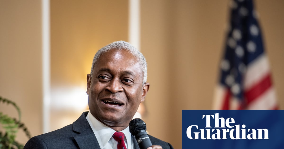
Atlanta Fed chair steps down as Trump attacks central bank’s independence | Federal Reserve
The head of the Federal Reserve Bank of Atlanta announced plans to step down amid an extraordinary campaign by Donald Trump to exert influence over the US central bank.
Raphael W Bostic, president of the Atlanta Fed, will retire from the role in February – creating another vacancy on the Fed’s powerful policy committee.
As the Trump administration continues to demand interest rate cuts, and even target some Fed officials, Bostic is the latest senior figure to depart.
After Adriana Kugler resigned in August from the Fed’s board of governors, Trump replaced her on interim basis with Stephen Miran, one of his top advisers.
The US president will not pick Bostic’s successor, however. The Atlanta Fed will now conduct a nationwide search for its next president.
Bostic, 59, could have remained in post for another six years. He was the first African American and openly gay president of a regional Federal Reserve bank in the Fed’s history.
The Fed’s rate-setting Open Market Committee (FOMC) has 12 members at any one time. These include the central bank’s seven board governors; the president of the New York Fed; and four of the 11 other regional Fed presidents, who rotate through yearly terms.
“I feel incredibly fortunate to have worked with the Atlanta Fed’s outstanding staff to fulfill the Federal Reserve’s mission and serve the Sixth District and the American people,” said Bostic. “I’m proud of what we accomplished during my tenure to turn the lofty goal of an economy that works for everyone into more of a reality, and I look forward to discovering new ways to advance that bold vision in my next chapter.”
Jerome Powell, the Fed chair, paid tribute to Bostic. “His perspective has enriched the Federal Open Market Committee’s understanding of our dynamic economy,” said Powell. “And his steady voice has exemplified the best of public service – grounded in analysis, informed by experience, and guided by purpose.
“His leadership has strengthened our institution and advanced the Federal Reserve’s mission.”
Continue Reading
-

Anthropic announces $50B investment in new US data centers to meet AI demand
SAN FRANCISCO — Artificial intelligence company Anthropic announced a $50 billion investment in computing infrastructure on Wednesday that will include new data centers in Texas and New York.
Anthropic, maker of the chatbot Claude, said it is working with London-based Fluidstack to build the new computing facilities to power its AI systems. It didn’t disclose their exact locations or what source of electricity they will need.
A report last month from TD Cowen said that the leading cloud computing providers leased a “staggering” amount of U.S. data center capacity in the third fiscal quarter of this year, amounting to more than 7.4 gigawatts of energy, more than all of last year combined.
Oracle was securing the most capacity during that time, much of it supporting AI workloads for Anthropic’s chief rival OpenAI, maker of ChatGPT. Google was second and Fluidstack came in third, ahead of Meta, Amazon, CoreWeave and Microsoft.
Anthropic said its projects will create about 800 permanent jobs and 2,400 construction jobs. It said in a statement that the “scale of this investment is necessary to meet the growing demand for Claude from hundreds of thousands of businesses while keeping our research at the frontier.”
The tech industry’s huge amount of spending on computing infrastructure for AI startups that aren’t yet profitable has fueled concerns about an AI investment bubble.
Investors have closely watched a series of intertwined deals over recent months between top AI developers such as OpenAI and Anthropic and the companies building the costly computer chips and data centers needed to power their AI products. Anthropic said it will continue to “prioritize cost-effective, capital-efficient approaches” to scaling up its business.
Continue Reading