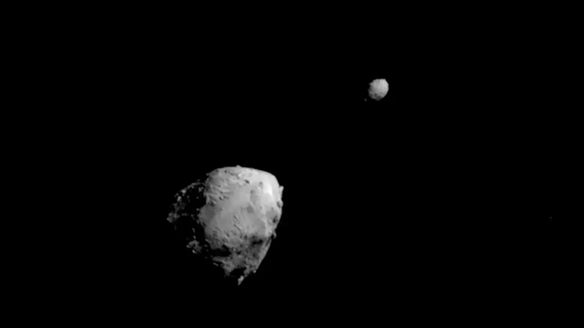
When NASA crashed a spacecraft into the asteroid moonlet Dimorphos in 2022, it altered both Dimorphos’ orbit around its parent asteroid, Didymos, and the two objects’ orbit around the sun, according to new research. NASA’s Jet Propulsion…
Category: 7. Science
-
Scientists Reveal The Oldest Map of The Night Sky Ever Made : ScienceAlert
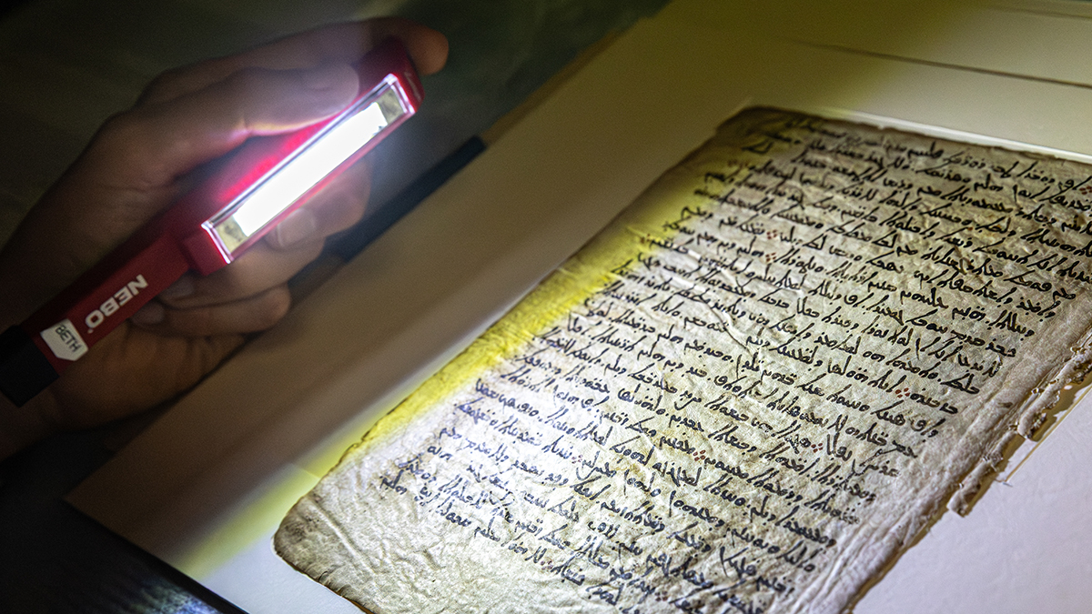
Researchers are painstakingly reconstructing the oldest-known map of the night sky – previously thought lost forever – by X-raying parchment that contains the star catalog hidden beneath other text.
The map of the cosmos is thought to be…
Continue Reading
-
The chromosome-scale genome assembly and annotation of Rosa bracteata (Macartney Rose)
Ku, T. R. K. Rosa (Rosaceae). In Flora of China 9 (eds Wu, Z. Y. & Raven, P. H.) (Missouri Botanical Garden, 2003).
Luo, L. Genus Rosa L. in China (China Forestry Publishing House, 2024).
Cheng, B. et al. Phenotypic and genomic signatures across…
Continue Reading
-
NASA rules out asteroid impact with Moon in 2032
Earlier orbital calculations had indicated a small 4.3 percent chance that the asteroid could strike the Moon. However, improved readings from the James Webb Telescope, collected in February, confirmed that the asteroid will…
Continue Reading
-
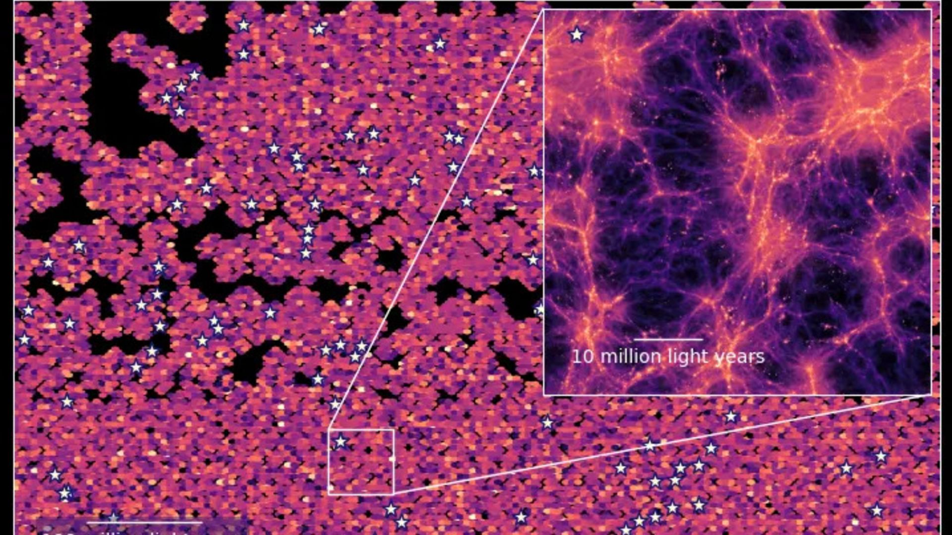
Enormous 3D map of the universe shows brilliant ‘sea of light’ near the cosmic dawn
Astronomers have produced one of the most accurate, comprehensive cosmic maps ever made, revealing a brilliant “sea of light” that permeated the early universe.
Unlike other universal maps, this 3D representation is composed of light emitted by…
Continue Reading
-

Enormous 3D map of the universe shows brilliant ‘sea of light’ near the cosmic dawn
When you buy through links on our articles, Future and its syndication partners may earn a commission.
A section of the line-intensity map created using HETDEX data, showing a swath of the universe as it appeared 10 billion years ago. The inset…
Continue Reading
-
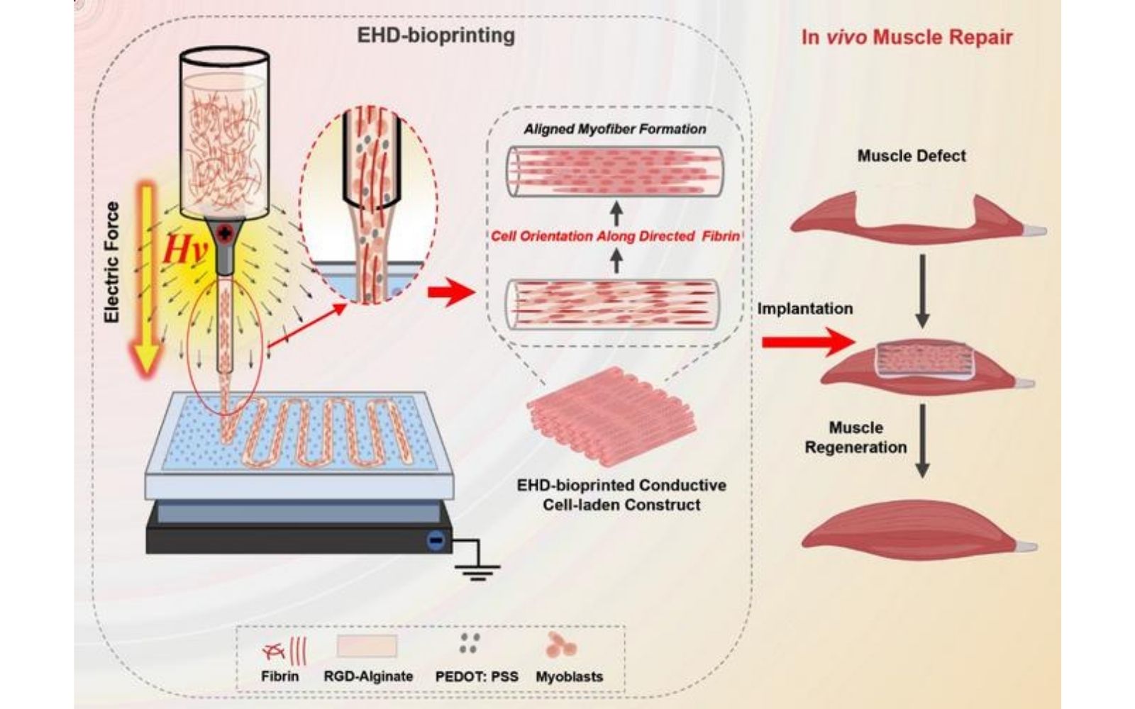
Xi’an Jiaotong University team bioprints aligned skeletal muscle tissue using electric fields | VoxelMatters
Stay up to date with everything that is happening in the wonderful world of AM via our LinkedIn community.A research team from Xi’an Jiaotong University has developed a method for bioprinting skeletal muscle tissue whose cells align in…
Continue Reading
-
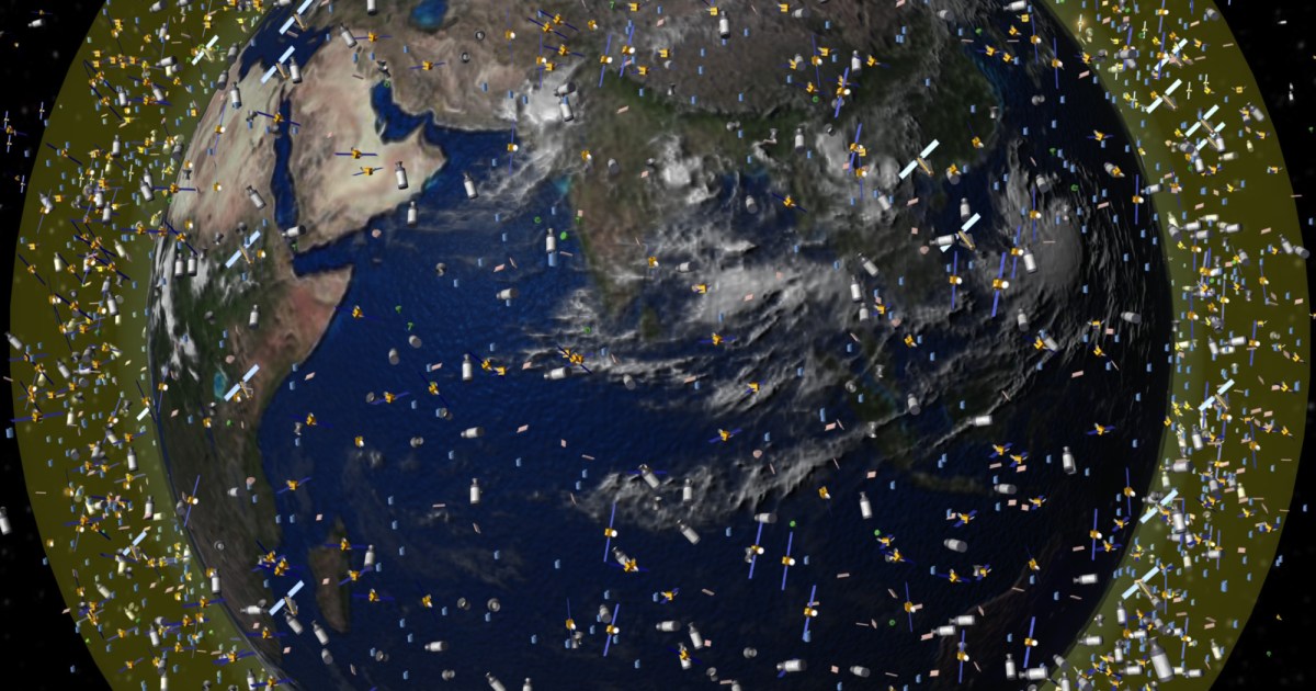
Starts With A Bang podcast #127 – Satellites and space pollution
When most of us were children, and we went to a rural area with clear skies overhead at night, we were all greeted by the same familiar sight: a dark night sky, glittering with many hundreds or even thousands of stars. Depending on…
Continue Reading
-
Digital reconstruction reveals the face of ‘Little Foot,’ a nearly 4 million-year-old human ancestor | science
Scientists have digitally reconstructed the face of Little Foot, a 3.67 million-year-old human ancestor, using high-resolution scanning and 3D modeling techniques. The reconstruction reveals a face that falls between the size of a gorilla and an…
Continue Reading
-
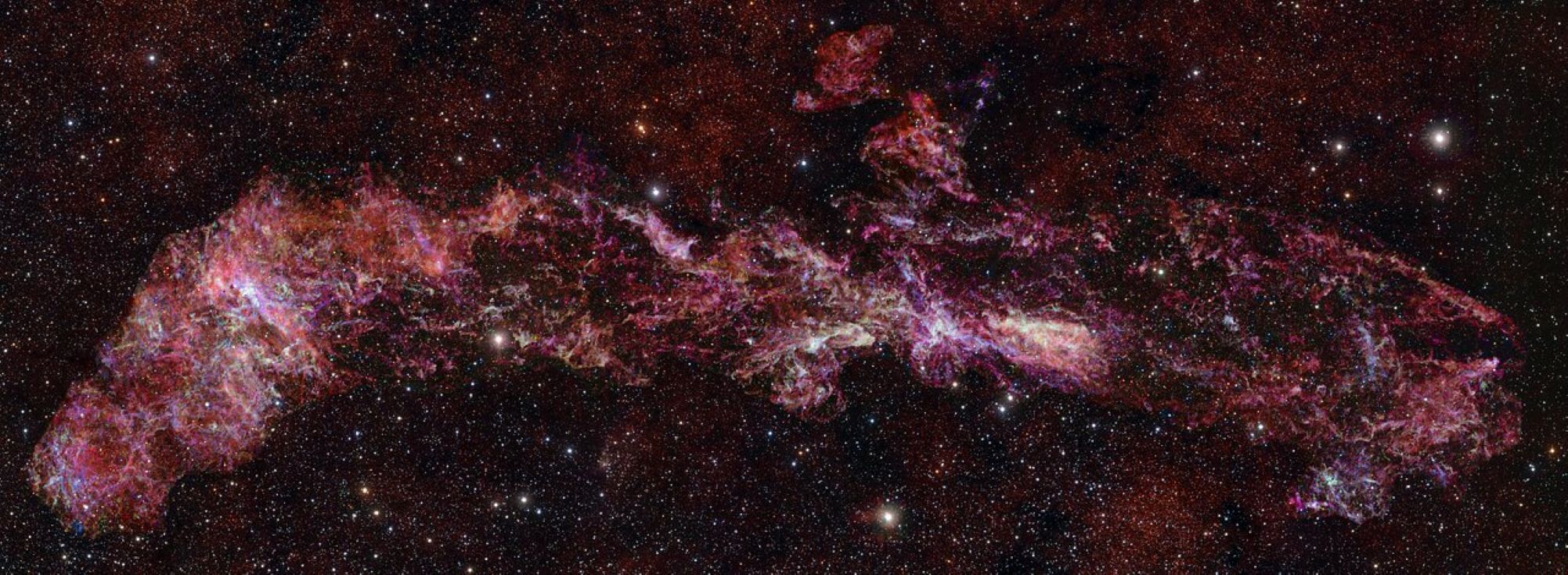
The Milky Way’s center is hiding exotic chemistry, and ALMA just revealed it
Astronomers have just unveiled a breathtaking new view of the very center of our galaxy, the Milky Way. Using the Atacama Large Millimeter/submillimeter Array (ALMA) in Chile’s Atacama Desert, they stitched together the largest ALMA image ever…
Continue Reading