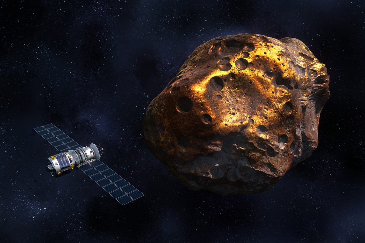
Category: 7. Science

  • A New Method For Producing Oxygen Using Magnets

    A New Method For Producing Oxygen Using Magnets

    In space, the one thing more important than ensuring access to food, water, and waste disposal (combined!) is the need for a steady supply of breathable air. Where the International Space Station (ISS) and other missions in Low Earth Orbit (LEO) can be resupplied regularly, missions operating in deep space will need to produce their own. There are several ways to go about this. One way is to use bioregenerative life support systems (BLSSs), which utilize photosynthetic organisms (like cyanobacteria) that take in carbon dioxide and produce oxygen gas and edible algae.

    Another way is to produce oxygen gas from local resources, a process known as in-situ resource utilization (ISRU). This typically involves electrolysis, a process in which electric current is used to split water molecules into hydrogen and oxygen gas. However, this process is impractical for deep-space missions because of the amount of energy required, not to mention the fact that it does not work well in microgravity. In a recent study, an international team suggests an alternative method that relies on a process known as magnetic phase separation.

    The research was led by Álvaro Romero-Calvo, a recent PhD graduate from the University of Colorado Boulder who is currently an Assistant Professor at the Daniel Guggenheim School of Aerospace Engineering, part of the Georgia Institute of Technology. He was joined by researchers from the Center of Applied Space Technology and Microgravity (ZARM) at the University of Bremen, and the University of Warwick. The paper detailing their findings was published on August 18th in Nature Chemistry.

    Mockup of the Oxygen Generation System Rack. Credit: NASA

    As noted, the current method for producing oxygen in space is through electrolysis, where electrodes immersed in an electrolyte solution (like salt) split water molecules into oxygen and hydrogen. Aboard the ISS, this is done by the Oxygen Generation Assembly (OGA), which spins water in a centrifuge to separate oxygen and hydrogen gas bubbles from the liquid. In microgravity, however, gas bubbles do not float upwards, and those produced in the centrifuge tend to remain suspended and stick to the electrodes. To extract them, a complex fluid management system is used that is heavy, complex, and occupies a lot of space.

    This makes such a system impractical since it would be very difficult to take on long-duration crewed missions. This was the conclusion reached by a team from NASA’s Ames Research Center who evaluated the OGA for crewed missions to Mars. Per the report, the team recommended that the OGA “for Mars should have lower mass, better reliability and maintainability, greater safety, radiation hardening, and capability for quiescent operation. NASA’s methodical, disciplined systems engineering process should be used to develop the appropriate system.”

    To address this problem, the team proposed an alternative solution that uses magnetism, which could make future life support systems lighter, simpler, and more sustainable. As Romero-Calvo said in a Georgia Tech news release:

    One may think that extracting gas bubbles from liquids in space is as simple as opening a can of soda here on Earth. However, the lack of buoyancy makes the extraction process incredibly difficult, undermining the design and operation of oxygen production systems. In this paper, we demonstrate that two largely unexplored magnetic interactions – diamagnetism and magnetohydrodynamics – provide an exciting pathway to solve this problem and develop alternative oxygen production architectures.

    The team created their system using off-the-shelf permanent magnets, which they tested at the ZARM Drop Tower in Bremen – a 146 m (~480 ft) high tower that simulates microgravity. The team also developed a passive phase separation system that collects the bubbles from the electrodes at designated spots. From this, they were able to demonstrate that magnetic fields can enable the separation of gas bubbles from electrodes in a microgravity environment without the need for heavy, complex equipment. The experiments further confirmed that magnetic forces can improve gas bubble detachment and enhance the efficiency of the electrochemical cells by up to 240%.

    41557_2025_1890_Figa_HTML_20250828_224725.png

    A key part of this breakthrough is the two complementary approaches the team developed to collect oxygen bubbles from the electrode. The first takes advantage of how water responds to magnets in microgravity by guiding gas bubbles toward collection points, while the second employs magnetohydrodynamic forces. These forces arise naturally from the interaction between magnetic fields and electric currents generated by electrolysis, creating a spinning motion in the liquid that separates gas bubbles from water through convective effects.

    This process achieves the same phase separation as mechanical centrifuges, like the one used aboard the ISS, but it uses magnetic forces instead of mechanical rotation. “We were able to prove that we do not need centrifuges or any mechanical moving parts for separating the produced hydrogen and oxygen from the liquid electrolyte,” said co-author Katerina Brinkert, an Honorary Professor at the University of Warwick and the Director of ZARM. “We do not even need additional power. Instead, it is a completely passive, low-maintenance system.”

    “Our team was able to prove that magnetic forces can feasibly control electrochemical bubbly flows in microgravity, departing from the state-of-the-art in low-gravity fluid mechanics and enabling future human spaceflight architectures,” Romero-Calvo added. The team’s work is the result of four years of joint research based on Romero-Calvo’s original idea, calculations, and numerical simulations. To validate the theory, Prof. Brinkert’s team at Warwick and ZARM developed the necessary experiments and devices to test the proposed method in microgravity conditions. As Dr. Shaumica Saravanabavan, a Ph.D. researcher at the University of Warwick, said:

    During my trips to ZARM, we confirmed the magnetic buoyancy effect for phase separation in (photo-)electrolysis cells in multiple Drop Tower experiments, using electrode materials we in part fabricated at Warwick. I’m proud to have contributed to advancing sustainable energy technologies beyond Earth applications.

    This research presents opportunities for developing simpler, more cost-effective, and more sustainable life support systems for deep-space missions. In the near future, the team hopes to further validate their method by launching it to orbit, where the system can operate in microgravity for extended periods.

    Further Reading: University of Warwick, Nature Chemistry

    Continue Reading

  • Astronaut puts his mad video game skills to the test

    Astronaut puts his mad video game skills to the test

    When you buy through links on our articles, Future and its syndication partners may earn a commission.

    Expedition 73 flight engineer Kimiya Yui of JAXA (the Japan Aerospace Exploration Agency) captured this view of Earth from on board the International Space Station on Wednesday, Aug. 27, 2025. | Credit: NASA/JAXA Kimiya Yui

    The seven astronauts and cosmonauts that comprise the second half of the International Space Station’s Expedition 73 crew focused this week on a new shipment of supplies and conducting the time-sensitive science that arrived along with the cargo. They also prepared for an upcoming re-boost of the orbital complex’s altitude in orbit.

    Orbital observation

    Before becoming an astronaut, Jonny Kim was a Navy SEAL and a medical doctor. Now aboard the International Space Station, he has revealed he was a gamer, too, and able to put some his boyhood skills to good use.

    “Growing up, I played a lot of video games — and while I still enjoy gaming with my kids, time is scarcer these days,” wrote Kim in a social media post on Wednesday (Aug. 27). “This demo brought me right back, blending elements of real-time strategy, RPGs, and first-person play into something very real.”

    Working with the Surface Avatar team at the European Space Agency (ESA), Kim was able to test how teleoperations could be of use to future missions on the moon and Mars.

    “A joystick and advanced robotic arm controller let me mimic finger and wrist movements with precision. A heads-up display kept me informed with battery levels, location data, and quick access to either an AI assistant or ground teams,” he wrote. “I could enlarge a mini-map to see each robot’s perspective, like a ‘fog of war’ in strategy games, and send parallel commands to different units. With the fine arm controls, I could enter into the perspective of a humanoid robot to manipulate the environment, whether moving science samples or shifting a rock that blocked the way.”

    Kim said his favorite demo was with a rover that had a smaller, deployable unit that could get into tight areas, such as caves — “a feature that felt straight out of a game but with real scientific potential.”

    “It was not just a technology demonstration, but a glimpse into how play, imagination and innovation intersect to shape the future of exploration,” Kim wrote.

    A montage of four photos showing a man with dark hair and light complexion wearing a dark blue short sleeve shirt working with a hand controller and a laptop to teleoperate devices.

    Expedition 73 flight engineer Jonny Kim of NASA works with a laptop and a custom hand controller on board the International Space Station to teleoperate a robotic arm and other devices on Earth. | Credit: NASA/Jonny Kim

    Science status

    Among the research that was conducted by the Expedition 73 crew aboard the space station this week was:

    MVP Cell-07 — Having arrived aboard a SpaceX Dragon spacecraft on Monday (Aug. 25), one of the space station’s newest science experiments quickly got the attention of Expedition 73 flight engineer Zena Cardman with NASA. She set up the Maturation of Vascularized Liver Tissue Construct in Zero Gravity experiment in a portable microgravity glovebag to begin studying how the blood vessels in 3D-printed liver tissue reacts to being weightless.

    Ultrasound 2 — Kimiya Yui of JAXA and Mike Fincke of NASA worked together to test a possible countermeasure to the redistirbution of liquids from the feet to the head in microgravity. Fincke wore a specially designed thigh cuff, while Yui recorded the data from electrodes applied to Fincke’s chest.

    Station keeping

    The Expedition 73 crewmates also took part in activities to maintain the space station’s systems and prepare for future research.

    CRS-33 — NASA astronaut Jonny Kim worked on unloading some of the 5,000 pounds (2,300 kilograms) of supplies delivered on a SpaceX Dragon spacecraft on Monday (Aug. 25). Fellow flight engineers Mike Fincke and Kimiya Yui removed and restowed frozen science samples from Dragon to the freezers on the space station.

    NASA engineers at Mission Control in Houston also took remote control of the space station’s Candarm2 robotic arm to remove and inspect a reboost kit that was launched in the unpressurized trunk of SpaceX’s CRS-33 Dragon. The hardware will later be used to maintain and raise the orbital complex’s altitude.

    Astronaut activity

    a white and black spacecraft is seen approaching a docking to a space station in Earth orbit

    SpaceX’s CRS-33 Dragon cargo spacecraft is seen approaching and docking with the forward-facing port of the Harmony node from a window aboard a Crew Dragon attached nearby at the International Space Station on Monday, Aug. 25, 2025. | Credit: NASA/Zena Cardman

    “Boop! We have a new Dragon at the bow of our International Space Station,” wrote Expedition 73 flight engineer Zena Cardman of NASA in a social media post on Tuesday (Aug. 26).

    The commercial space capsule arrived at the orbiting laboratory on Monday with more than 5,000 pounds (2,300 kilograms) of supplies on board.

    “We’ve been hard at work unpacking ever since. Welcome, SpaceX CRS-33!” wrote Cardman.

    By the numbers

    As of Friday (Aug. 15), there are 7 people aboard the International Space Station: Expedition 73 commander Sergey Ryzhikov of Roscosmos; fellow cosmonauts Alexey Zubritsky and Oleg Platonov; Jonny Kim, Zena Cardman and Mike Fincke of NASA; and Kimiya Yui of JAXA, all flight engineers.

    There are two docked crew spacecraft: SpaceX’s Dragon “Endeavour” attached to the zenith port of the Harmony module and Roscosmos’ Soyuz MS-27 attached to the Earth-facing port of the Prichal node.

    There are three docked cargo spacecraft: Roscosmos’ Progress MS-30 (91P) attached to the aft port of the Zvezda service module, and Progress MS-31 (92P) docked to the space-facing port of the Poisk module. SpaceX’s CRS-33 Dragon spacecraft is docked to the forward port of Harmony Node 2.

    As of Friday, the space station has been continuously crewed for 24 years and 9 months and 27 days.

    Continue Reading

  • Liming Returns as Climate and Yield Strategy

    Liming Returns as Climate and Yield Strategy

    Kaleigh Harrison

    A traditional agricultural method—liming—may be key to addressing both food security and climate goals, according to new research out of Georgia Tech. The process, which involves applying crushed limestone to correct soil acidity, has long been used to improve crop performance. But its overlooked potential to remove carbon dioxide from the atmosphere is now gaining traction with researchers, agribusinesses, and supply chain leaders.

    Far from being just a soil amendment, liming is proving to be a dual-purpose tool: enhancing crop yields while contributing to verified emissions reductions. That’s a rare combination in climate-smart agriculture, particularly for regions like sub-Saharan Africa where degraded soils cost billions annually in lost productivity.

    This positions liming as a high-impact, lower-cost intervention for agricultural producers, food processors, and corporate sustainability teams. As pressure increases for measurable Scope 3 emissions reductions and nature-based solutions, liming presents a practical, data-backed opportunity.

    Data-Driven Models Unlock Commercial Potential

    What sets this new phase of liming apart is the role of advanced analytics. Research partnerships across the U.S.—including trials in southern Georgia, North Carolina, and the Corn Belt—are using precision data collection to map variables like soil pH, nutrient levels, trace elements, and greenhouse gas fluxes.

    These inputs are feeding machine learning models that can forecast how liming will impact both productivity and emissions under specific soil, crop, and climate scenarios. One of the key breakthroughs: not all liming leads to carbon removal, and the net climate impact depends on timing, soil chemistry, and other conditions.

    For agtech firms and consultants, this opens the door to tailored liming recommendations and precision tools that drive both agronomic and environmental outcomes. Because many farms already collect relevant soil and crop data, implementing enhanced liming strategies doesn’t require expensive new infrastructure—lowering barriers to entry and accelerating adoption.

    There’s also a clear path to monetization. With growing interest from voluntary carbon markets and regulatory bodies, verified liming practices could qualify for credits tied to CO₂, methane, and nitrous oxide reductions. For businesses managing agricultural supply chains, that means potential for cost savings, revenue generation, and stronger ESG reporting—all from an existing farm input.


    Continue Reading

  • Fat Microscopy: Visualizing Cell Lipids

    Fat Microscopy: Visualizing Cell Lipids

    New technique to image single lipids: Lipids are notoriously difficult to detect with light microscopy. Using a new chemical labeling strategy, the Dresden team has overcome this limitation, enabling novel insights into where specific lipids are located and how they are transported in cells.

    Map of lipid flow: The researchers used the new lipid imaging method to answer the long-standing question how cells transport specific lipids to their target organelle membranes. The study revealed that non-vesicular lipid transport by proteins is the primary mechanism that maintains the membrane composition of specific organelles.

    Understanding the role of lipids in diseases: Lipid imbalances play a role in several metabolic or neurodegenerative diseases. The new lipid-imaging technique will help understand the role of lipid transport in health and disease. The identification of the proteins involved in selective lipid transport can accelerate further discoveries of new drug targets for lipid-associated diseases.


    Lipid molecules, or fats, are crucial to all forms of life. Cells need lipids to build membranes, separate and organize biochemical reactions, store energy, and transmit information. Every cell can create thousands of different lipids, and when they are out of balance, metabolic and neurodegenerative diseases can arise. It is still not well understood how cells sort different types of lipids between cell organelles to maintain the composition of each membrane. A major reason is that lipids are difficult to study, since microscopy techniques to precisely trace their location inside cells have so far been missing.

    In a long-standing collaboration André Nadler , a chemical biologist at the Max Planck Institute of Molecular Cell Biology and Genetics (MPI-CBG) in Dresden, Germany teamed up with Alf Honigmann , a bioimaging specialist at Biotechnology Center (BIOTEC) at the TUD Dresden University of Technology , to develop a method that enables visualizing lipids in cells using standard fluorescence microscopy. After the first successful proof of concept, the duo brought mass-spectrometry expert Andrej Shevchenko (MPI-CBG), Björn Drobot at the Helmholtz-Zentrum Dresden-Rossendorf (HZDR) , and the group of Martin Hof from the J. Heyrovsky Institute of Physical Chemistry in Prague on board to study how lipids are transported between cellular organelles.

    Artificial lipids under the sunbed

    “We started our project with synthesizing a set of minimally modified lipids that represent the main lipids present in organelle membranes. These modified lipids are essentially the same as their native counterparts, with just a few different atoms that allowed us to track them under the microscope,” explains Kristin Böhlig, a PhD student in the Nadler group and chemist who was in charge of creating the modified lipids.

    The modified lipids mimic natural lipids and are “bifunctional,” which means they can be activated by UV light, causing the lipid to bind or crosslink with nearby proteins. The modified lipids were loaded in the membrane of living cells and, over time, transported into the membranes of organelles. The researchers worked with human cells in cell culture, such as bone or intestinal cells, as they are ideal for imaging.

    “After the treatment with UV light, we were able to monitor the lipids with fluorescence microscopy and capture their location over time. This gave us a comprehensive picture of lipid exchange between cell membrane and organelle membranes,” concludes Kristin.

    In order to understand the microscopy data, the team needed a custom image analysis pipeline. “To address our specific needs, I developed an image analysis pipeline with automated image segmentation assisted by artificial intelligence to quantify the lipid flow through the cellular organelle system,” says Juan Iglesias-Artola, who did the image analysis.

    Speedy lipid transport by proteins

    By combining the image analysis with mathematical modeling, done by Björn Drobot at the HZDR, the research team discovered that between 85% and 95% of the lipid transport between the membranes of cell organelles is organized by carrier proteins that move the lipids, rather than by vesicles. This non-vesicular transport is much more specific with regard to individual lipid species and their sorting to the different organelles in the cell. The researchers also found that the lipid transport by proteins is ten times faster than by vesicles. These results imply that the lipid compositions of organelle membranes are primarily maintained through fast, species-specific, non-vesicular lipid transport.

    In a parallel set of experiments, the group of Andrej Shevchenko at the MPI-CBG used ultra-high-resolution mass spectrometry to see how the different lipids change their structure during the transport from the cell membrane to the organelle membrane.

    A boost for lipids in cell biology and disease

    This new approach provides the first-ever quantitative map of how lipids move through the cell to different organelles. The results suggest that non-vesicular lipid transport has a key role in the maintenance of each organelle membrane composition.

    Alf Honigmann, research group leader at the BIOTEC says, “Our lipid-imaging technique enables the mechanistic analysis of lipid transport and function directly in cells, which has been impossible before. We think that our work opens the door to a new era of studying the role of lipids within the cell.”

    Imaging of lipids will allow further discoveries and help to reveal the underlying mechanisms in diseases caused by lipid imbalances. The new technique could potentially help to develop new druggable targets and therapeutic approaches for lipid-associated diseases, such as nonalcoholic fatty liver disease.

    “We knew that we were onto something big”

    André Nadler, research group leader at MPI-CBG, looks back at the start of the study, “Imaging lipids in cells has always been one of the most challenging aspects of microscopy. Our project was no different. Alf Honigmann and I started discussing about solving the lipid imaging problem as soon as we got hired in close succession at MPI-CBG in 2014/15 and we quickly decided to go for it. It still took us almost five years from the start of the project to the point in autumn 2019 when the two of us finally produced a sample with a beautiful plasma membrane stain. That’s when we knew that we were onto something big. As a reward, certain well known global events meant we were required to shut down our laboratories a few months later. In the end, the delay was for the best. Before the revolution in the use of artificial intelligence in image segmentation, we would not have been able to properly quantify the imaging data, so our conclusions would have been much more limited.”

    Researchers still need to determine which lipid-transfer proteins drive the selective transport of different lipid species. They also need to identify the energy sources that power lipid transport and ensure that each organelle keeps its own unique membrane composition.

    /Public Release. This material from the originating organization/author(s) might be of the point-in-time nature, and edited for clarity, style and length. Mirage.News does not take institutional positions or sides, and all views, positions, and conclusions expressed herein are solely those of the author(s).View in full here.

    Continue Reading

  • NASA Astronaut Megan McArthur Retires

    NASA Astronaut Megan McArthur Retires

    NASA astronaut Megan McArthur has retired, concluding a career spanning more than two decades. A veteran of two spaceflights, McArthur logged 213 days in space, including being the first woman to pilot a SpaceX Dragon spacecraft and the last person to “touch” the Hubble Space Telescope with the space shuttle’s robotic arm.

    McArthur launched as pilot of NASA’s SpaceX Crew-2 mission in April 2021, marking her second spaceflight and her first long-duration stay aboard the International Space Station. During the 200-day mission, she served as a flight engineer for Expeditions 65/66, conducting a wide array of scientific experiments in human health, materials sciences, and robotics to advance exploration of the Moon under Artemis and prepare to send American astronauts to Mars.

    Her first spaceflight was STS-125 in 2009, aboard the space shuttle Atlantis, the fifth and final servicing mission to Hubble. As a mission specialist, she was responsible for capturing the telescope with the robotic arm, as well as supporting five spacewalks to update and repair Hubble after its first 19 years in space. She also played a key role in supporting shuttle operations during launch, rendezvous with the telescope, and landing.

    “Megan’s thoughtful leadership, operational excellence, and deep commitment to science and exploration have made a lasting impact,” said Steve Koerner, acting director of NASA’s Johnson Space Center in Houston. “Her contributions have helped shape the future of human space exploration, and we are incredibly grateful for her service.”

    In addition to her flight experience, McArthur has served in various technical and leadership roles within NASA. In 2019, she became the deputy division chief of the Astronaut Office, supporting astronaut training, development, and ongoing spaceflight operations. She also served as the assistant director of flight operations for the International Space Station Program starting in 2017.

    Since 2022, McArthur has served as the chief science officer at Space Center Houston, NASA Johnson’s official visitor center. Continuing in this role, she actively promotes public engagement with space exploration themes, aiming to increase understanding of the benefits to humanity and enhance science literacy.

    “Megan brought a unique combination of technical skill and compassion to everything she did,” said Joe Acaba, chief of the Astronaut Office at NASA Johnson. “Whether in space or on the ground, she embodied the best of what it means to be an astronaut and a teammate. Her contributions will be felt by the next generation of explorers she helped train.”

    McArthur was born in Honolulu and raised as a “Navy kid” in many different locations worldwide. She earned a Bachelor of Science in aerospace engineering from the University of California, Los Angeles, and a doctorate in oceanography from the Scripps Institution of Oceanography at the University of California, San Diego. Before being selected as an astronaut in 2000, she conducted oceanographic research focusing on underwater acoustics, which involved shipboard work and extensive scuba diving.

    McArthur is married to former NASA astronaut Robert Behnken, who also flew aboard the Dragon Endeavour spacecraft during the agency’s SpaceX Demo-2 mission in 2020.

    “It was an incredible privilege to serve as a NASA astronaut, working with scientists from around the world on cutting-edge research that continues to have a lasting impact here on Earth and prepares humanity for future exploration at the Moon and Mars,” said McArthur. “From NASA’s Hubble Space Telescope to the International Space Station, our research lab in low Earth orbit, humanity has developed incredible tools that help us answer important scientific questions, solve complex engineering challenges, and gain a deeper understanding of our place in the universe. Seeing our beautiful planet from space makes it so clear how fragile and precious our home is, and how vital it is that we protect it. I am grateful I had the opportunity to contribute to this work, and I’m excited to watch our brilliant engineers and scientists at NASA conquer new challenges and pursue further scientific discoveries for the benefit of all.”

    To learn more about NASA’s astronauts and their contributions to space exploration, visit:

    https://www.nasa.gov/astronauts

    -end-

    Shaneequa Vereen
    Johnson Space Center, Houston
    281-483-5111
    shaneequa.y.vereen@nasa.gov

    Continue Reading

  • Double-charge-transfer molecule may help improve solar fuels

    Double-charge-transfer molecule may help improve solar fuels


    For decades, scientists have been trying to use the sun’s energy to turn water and carbon dioxide into fuel. While nature makes photosynthesis seem effortless, it requires choreographing multiple complex charge-transfer steps.

    To study these charge-transfer processes and better understand how to harness them for productive chemistry, researchers often turn to model systems such as donor-photosensitizer-acceptor complexes. These structures are made up of a metal complex flanked by an electron-accepting molecular piece and an electron-donating piece such that when the metal center absorbs light energy, the acceptor and donor can store it in an electron-hole pair.


    This new complex is designed so that electrons flow from the strongest donor (left) to the strongest acceptor (right).

    University of Basel chemist Oliver Wenger and his graduate student Mathis Brändlin have now created a molecule that can capture and store up to two electron-hole pairs at once (Nat. Chem. 2025, DOI: 10.1038/s41557-025-01912-x). Previous researchers had succeeded in jamming one electron and two holes into a single molecule, or two electrons and one hole, but two electrons and two holes remained elusive.

    Wenger and Brändlin’s approach was deceptively simple: just add a second donor group and a second acceptor group to either end of a donor-photosensitizer-acceptor complex. The trick was to choose the right molecular pieces and arrange them so that they’d efficiently shuttle charges away from the central ruthenium photosensitizer.

    The final molecule is large and complicated, and Wenger says making it was “a heroic effort” on Brändlin’s part.

    The researchers characterized the complex extensively using a suite of methods including cyclic voltammetry and ultraviolet–visible spectroscopy. The “final experiment that nailed it” involved firing two lasers—one continuous, the other pulsed—at the molecule and monitoring its spectroscopic signal to confirm that it could indeed hold two electrons and two holes at once, Wenger says.

    From the spectroscopic data, the researchers determined that the molecule can hold one electron-hole pair for about 120 µs and two pairs for a little less than 1 µs—which may sound vanishingly short, but it would be plenty long enough to do chemistry, Wenger says.

    Daniel G. Nocera of Harvard University, an expert in artificial photosynthesis who was not involved in the work, calls it “an interesting fundamental study” in molecular energy storage. Practical charge transfer for solar fuels is better accomplished using semiconductors, he says, but Brändlin and Wenger’s cleverly designed complex nevertheless provides useful insights into charge-transfer mechanisms.

    The researchers have not yet used the complex to drive any chemical reactions. But that would be the natural next step, Wenger says—that is, if he can find someone to take over the project after Brändlin defends his dissertation later this year.

    Continue Reading

  • Proteomic Study Uncovers New Gastric Cancer Targets

    Proteomic Study Uncovers New Gastric Cancer Targets

    Gastric signet ring cell carcinoma (GSRCC) is a distinct subtype of gastric cancer (GC) with unique epidemiological and pathogenic characteristics. Despite its clinical significance, large-scale proteomic studies on GSRCC remain scarce, limiting our molecular understanding of the disease. Advanced mass spectrometry (MS)-based proteomics is crucial for identifying key biomarkers and drug targets, thereby enabling more effective therapeutic strategies.

    In a recent study published in Genes & Diseases, researchers from several institutions, including Tianjin University, Chinese Academy of Sciences, Zhejiang Cancer Hospital, Fudan University, Diagnosis and Therapy of Upper Gastrointestinal Cancer of Zhejiang Province, Chinese Academy of Medical Sciences and Peking Union Medical College, and University of Houston, characterizes the proteomic features and molecular mechanisms of GSRCC to date.

    Initially, the research team analyzed clinical data from over 10,000 patients with GC between January 2010 and December 2019. An in-depth proteomic analysis was conducted on tumor tissues from 112 GSRCC patients, each with over 70% signet ring cell content. Using advanced MS, the team identified 7322 proteins, establishing the largest tissue-specific peptide spectral library for GSRCC. Additionally, through unsupervised clustering, the team identified four novel proteomic subtypes of GSRCC: Metabolism (S-Mb), Microenvironment Dysregulation (S-Me), Migration (S-M) and Proliferation (S-PF).

    Two key prognostic biomarkers were identified and validated in an independent cohort of 75 patients: PRDX2, a protein associated with favorable prognosis; and DDX27, linked to poor survival outcomes. Furthermore, proteomic profiling of 79 biomarker-negative GSRCC cases revealed marked tumor heterogeneity. Notably, unsupervised clustering identified three distinct proteomic clusters, with cluster 2 linked to the poorest prognosis.

    Focusing on HER2-negative, EBV-negative, and pMMR GSRCC cases (LMT [Lack of Medical Treatment]-GSRCC), the study identified four potential drug targets: eukaryotic translation initiation factor 2 subunit gamma (EIF2S3), eukaryotic translation initiation factor 6 (EIF6), and nuclear factor kappa B subunit 2 (NFKB2). Remarkably, high expression of these proteins was associated with poor prognosis, underscoring their relevance as promising therapeutic candidates.

    Interestingly, molecular docking and cytotoxicity testing singled out neratinib—a drug approved for breast cancer treatment—as the most promising candidate. Furthermore, in vitro and in vivo studies demonstrated neratinib’s potent ability to inhibit tumor growth, cell migration, and invasion, while promoting cancer cell apoptosis, all with minimal side effects.

    In conclusion, this is the first study to focus specifically on the LMT-GSRCC population, uncovering potential biomarkers and drug targets through proteomic analysis. The findings from this study not only provide a foundation for developing novel targeted therapies but also personalized treatment strategies for GSRCC.

    Reference

    Title of Original Paper: A comprehensive proteomic analysis uncovers novel molecular subtypes of gastric signet ring cell carcinoma: Identification of potential prognostic biomarkers and therapeutic targets

    Journal: Genes & Diseases

    Genes & Diseases is a journal for molecular and translational medicine. The journal primarily focuses on publishing investigations on the molecular bases and experimental therapeutics of human diseases. Publication formats include full length research article, review article, short communication, correspondence, perspectives, commentary, views on news, and research watch.

    DOI: https://doi.org/10.1016/j.gendis.2025.101717

    /Public Release. This material from the originating organization/author(s) might be of the point-in-time nature, and edited for clarity, style and length. Mirage.News does not take institutional positions or sides, and all views, positions, and conclusions expressed herein are solely those of the author(s).View in full here.

    Continue Reading

  • Microalgae Key to Carbon Absorption in Southern Ocean

    Microalgae Key to Carbon Absorption in Southern Ocean

    Some Fourteen thousand years ago, algal blooms in the Southern Ocean helped to massively reduce the global carbon dioxide content of the atmosphere – as has now been revealed by new analyses of ancient DNA published by a team from the Alfred Wegener Institute in the journal Nature Geoscience. In the ocean around the Antarctic continent, these algal blooms had a significant impact on global carbon dynamics. The current and expected future decline in sea ice in this region now poses a serious threat to these algae, which could incur global consequences.

    At the end of the last ice age, the warming in the southern hemisphere slowed temporarily in a phase known as the Antarctic Cold Reversal (ACR). A new study by the Alfred Wegener Institute, Helmholtz Centre for Polar and Marine Research (AWI) reveals that the special climatic conditions of this period – in particular involving a vast sea ice cover in winter, followed by strong seasonal melting in the springtime – favoured massive algal blooms of the genus Phaeocystis in the Southern Ocean. These blooms absorbed large amounts of carbon dioxide, markedly slowing the increase of this climate-damaging gas in the atmosphere. The AWI research team was able to prove this connection for the first time by examining so-called sedimentary ancient DNA (sedaDNA) – genetic material that has been preserved in the seabed for thousands of years. This is due to the fact that Phaeocystis does not leave behind classic microfossils and therefore remained invisible in previous climate archives. To date, it has not been possible to detect its presence by way of classic geochemical methods.

    In conducting their study, the AWI team examined a sediment core from a depth of almost 2,000 metres in the Bransfield Strait north of the Antarctic Peninsula. The core contains sedaDNA from the last 14,000 years. The researchers extracted this from the sediment cores to study changes in biological communities over time. “Our study shows that these algal blooms contributed to a significant reduction in global atmospheric CO₂ levels during a climatically important transition phase characterized by high sea ice extent,” explains Josefine Friederike Weiß from AWI, lead author of the study. This is because the sediment core exhibits a high ratio of barium (Ba) to iron (Fe) for this phase – a ratio considered as an indicator of organic carbon input and deposition, due to the fact that it is linked to biological productivity in surface water.

    “The further the sea ice expands in winter, the larger the area in spring where nutrient-rich meltwater enters the surface sea – and therefore the zone where Phaeocystis algae find ideal growth conditions. As a result, greater sea ice extent leads directly to a larger region with high algal productivity.” Such biological processes in the ocean are closely linked to global climate events – even if they remain invisible to the human eye. In addition, the large-scale Phaeocystis blooms impacted on food webs and nutrient distribution in the Southern Ocean, triggering a complex chain reaction: From changes in plankton composition and shifted biogeochemical cycles through to increased carbon transport into the depths, they influenced the ecological balance and the carbon cycle over long periods of time.

    Today, Phaeocystis is particularly endangered in Antarctica, given that the long-term trend towards sea ice loss and, in particular, the recent dramatic decline in the Southern Ocean is altering its living conditions significantly. The loss of these important algal blooms could destabilise local ecosystems. Although other algae species such as diatoms could benefit from ice-free conditions, the structure of the food web would change fundamentally. What is more, Phaeocystis is particularly efficient in transporting carbon to the deep sea. Therefore, a decline in its blooms could mean that less carbon is stored in the ocean overall, which could exacerbate climate change in the long term.

    Furthermore, Phaeocystis produces dimethyl sulphide (DMS), a gas that promotes cloud formation, thereby increasing the reflection of sunlight. Consequently, the loss of algal blooms could also incur a negative impact on cloud formation and therefore on climate regulation, which in turn would lead to an additional, amplifying impact on climate.

    On the one hand, the study by the AWI scientists provides new insights into the role of the Southern Ocean and its microorganisms in the global climate events of the past, which could not have previously been detected using traditional methods in sediment archives. On the other hand, it shows for the first time that previous geological investigation methods, in combination with sedimentary ancient DNA, give rise to a more realistic reconstruction of past ecosystems and our understanding of earlier carbon dioxide fluctuations. This will pave the way for more differentiated assessments of future developments in the climate system. The analysis of these geological processes underscores the crucial role that biological processes play in climate regulation. This finding highlights the significance of giving greater consideration to marine ecosystems in current climate research and in future forecasts.

    With regard to further research, this means that the combination of DNA analyses and geological methods should be further improved in order to obtain and outline an even more accurate picture of past climate changes. In addition, individual significant organisms, such as Phaeocystis, should be studied in greater detail to be able to better understand their influence on the carbon cycle and climate. This will not only result in better climate predictions, but also allow potential profound ecological changes in the ocean to be identified at an early juncture and their impacts assessed accordingly.

    /Public Release. This material from the originating organization/author(s) might be of the point-in-time nature, and edited for clarity, style and length. Mirage.News does not take institutional positions or sides, and all views, positions, and conclusions expressed herein are solely those of the author(s).View in full here.

    Continue Reading

  • Seismic Waves Slow Right After Earthquake

    Seismic Waves Slow Right After Earthquake

    Solid as they are, rocks are not static materials with constant properties. Even small loads are enough to alter their mechanical properties; their reaction to being deformed is a loss of stiffness. Rocks which have been damaged in such a way are then less able to withstand loads, such as gravity or tectonic stresses. This phenomenon is therefore of relevance for understanding the occurrence of material failure, as in landslides or earthquakes. Such changes in elastic properties are commonly observed in heterogeneous and granular materials such as rocks, concrete or sediments. As a result, they play a role in geotechnics as well as in the stability of man-made structures.

    These effects have been observed in laboratory experiments for years using acoustic methods. The development of seismic interferometry made such observations possible in the field by exploiting the so-called “seismic noise”. A key observation using these methods is the sudden decrease in the velocity of seismic waves in the subsurface in the wake of an earthquake (damage). This decrease is followed by a slow re-increase, which can extend over several years (recovery).

    Despite these studies and many years of research, the physical origins of these processes have still not been clearly determined. It is however commonly agreed that the contrast between the very stiff grains and vastly softer grain contact planes, as well as the stress concentrations at these contact points, is responsible for these effects. Manuel Asnar and a team of collaborators from the GFZ, the University of Edinburgh in the UK, and the Université de Lorraine in France, have managed to make a breakthrough in laboratory experiments carried out at the GFZ’s High-Pressure Labs. Their experimental setup allowed them to measure wave velocities in a 10-centimetre sandstone cylinder along various directions of propagation to an extremely high degree of precision.

    The sample was subjected to varying levels of stress along the cylinder’s axis. Doing so showed that, as expected, the static effects of the loading strongly impacted waves along the main axis, while leaving the waves along the diameter relatively unaffected. The time-dependent effects however, that is, the sudden damage and long healing, were consistently observed along every direction of propagation.

    These results show that the time-dependent effects are not caused by grain contacts that are being more or less compressed against each other. Rather, these effects can be traced back to contact planes sliding against each other, irrespective of whether the load is being applied or released.

    The effects of friction along contacts within the material have long been suggested as being responsible for these time-dependent changes in wave velocities; but this study of the anisotropy of velocity changes – meaning, their directional dependence – provides meaningful evidence in favour of this interpretation. Based on those findings, models can be developed to better describe and predict the time-dependent changes of mechanical properties in rocks and geotechnical materials.

    Original study: Asnar, M., Sens-Schönfelder, C., Bonnelye, A. et al. Anisotropy reveals contact sliding and aging as a cause of post-seismic velocity changes. Nat Commun 16, 7587 (2025). https://doi.org/10.1038/s41467-025-62667-0

    /Public Release. This material from the originating organization/author(s) might be of the point-in-time nature, and edited for clarity, style and length. Mirage.News does not take institutional positions or sides, and all views, positions, and conclusions expressed herein are solely those of the author(s).View in full here.

    Continue Reading

  • Blazing Rainbow in Space Hints at Earth’s Origins : ScienceAlert

    Blazing Rainbow in Space Hints at Earth’s Origins : ScienceAlert

    The glorious guts of a dying star are the means by which astronomers are hoping to discover the very earliest origins of how our planet was born.

    In the midst of the Butterfly Nebula NGC 6302, located some 3,400 light-years from Earth in the southern constellation of Scorpius, astronomers have found compelling evidence of dust crystallizing as it cools from hot gas.

    “For years, scientists have debated how cosmic dust forms in space. But now, with the help of the powerful James Webb Space Telescope, we may finally have a clearer picture,” says astrophysicist Mikako Matsuura of Cardiff University in the UK.

    “We were able to see both cool gemstones formed in calm, long-lasting zones and fiery grime created in violent, fast-moving parts of space, all within a single object. This discovery is a big step forward in understanding how the basic materials of planets come together.”

    Related: New Images of Interstellar Dust Look Like Something Out of a Dream

    Hubble Space Telescope image of the Butterfly Nebula in optical wavelengths. You can see the thick band of dark dust in the center. (ESA/Webb, NASA & CSA, K. Noll, J. Kastner, M. Zamani/CC BY 4.0)

    Cosmic dust is what it sounds like: dust that drifts around the space between the stars. It’s thought to form primarily in the outer layers of dying stars, seeding the nebular material that is taken up into newly forming stars and the worlds that orbit them.

    The Butterfly Nebula is the gorgeous swansong of just such a dying star. It’s what we call a planetary nebula (because the first known examples of its kind were round, like planets). This is the expanding cloud of material that forms around a star as it shucks its outer layers into space as it dies.

    At the center of this nebula is a white dwarf – the remnant of a giant star that has already completed its death throes. You’ll also notice that the nebula is not nice and neat and round, but a pair of violently expelled outflows, like the wings of a butterfly.

    From right to left, observations of the nebula in optical, near-infrared, and combined infrared and radio wavelengths. (ESA/Webb, NASA & CSA, M. Matsuura, ALMA (ESO/NAOJ/NRAO), N. Hirano, M. Zamani/CC BY 4.0)

    Wrapped around the central white dwarf – which burns exceedingly hot with the residual heat of its death and reformation – is a thick torus of dust. Matsuura and her colleagues used the infrared power of JWST to peer into this dust to literally see what it is made of.

    Most wavelengths of light are blocked and scattered by the dust, but long, infrared wavelengths can pierce through, making JWST the perfect tool to probe this enigmatic environment.

    The researchers combined JWST infrared observations with radio observations from the Atacama Large Millimeter/submillimeter Array (ALMA). These observations revealed new details about the processes taking place in the heart of the Butterfly Nebula.

    The dusty donut, the researchers found, has infrared signatures of both amorphous dust grains, like soot, and lovely neat crystalline structures. The glinting light suggests also that these grains are quite large for dust, on the scale of microns – suggesting that it has been hanging out there and growing for some time.

    The composition of the dust is also fascinating, containing crystals of the silicate minerals forsterite, enstatite, and quartz.

    An annotated version of the combined JWST/ALMA observations detailing the features at the nebula’s center. (ESA/Webb, NASA & CSA, M. Matsuura, ALMA (ESO/NAOJ/NRAO), N. Hirano, M. Zamani/CC BY 4.0)

    Around the outside of the torus, there’s a clear gradation in the atoms and molecules. Those ions that require the most energy to form are closer to the center of the nebula, while the ions that don’t require much energy to form concentrate farther from the center.

    Other features identified in the JWST data include large jets of iron and nickel streaming away from the star in opposite directions; and quite a significant concentration of polycyclic aromatic hydrocarbons, or PAHs. This is particularly interesting.

    PAHs are sooty molecules based on rings of carbon atoms that drift through space in high abundance. They feature heavily, therefore, in theories about the origin of carbon-based life. Finding them in the heart of the oxygen-rich Butterfly Nebula gives us new clues about how the building blocks of life might be formed: when powerful winds from the star slam into the material around it.

    We can’t rewind the Solar System to find out how it all came together from a cloud in space. Instruments like JWST, and objects like the Butterfly Nebula, give scientists the crucial insight to figure out how we all got here, from dust from a dying star.

    The research has been published in The Monthly Notices of Royal Astronomical Society.

    Continue Reading