IPCC (Intergovernmental Panel on Climate Change). Climate Change 2014: Impacts, Adaptation, and Vulnerability. Working Group II Contribution to the Fifth Assessment Report of the Intergovernmental Panel on Climate Change (Cambridge University…
Category: 7. Science
-
Factors influencing herders’ willingness to engage in grassland ecological restoration in Ruoergai County
China Government Network. Ecological Environment. Available online: (2005). https://www.gov.cn/test/2005-07/28/content_17792.htm (Accessed 9 Aug 2025).
Ministry of Agriculture and Rural Affairs of the People’s Republic of China. Reply to…
Continue Reading
-
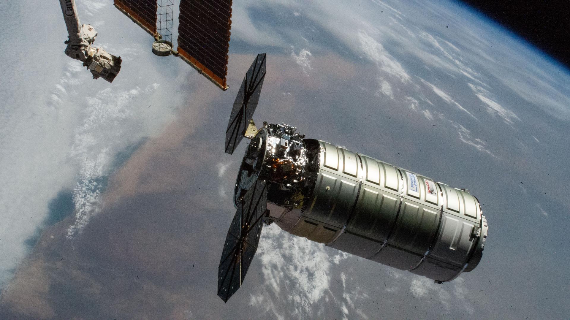
NASA Invites Media to Northrop Grumman CRS-24 Station Resupply Launch
Media accreditation is open for the next launch to deliver NASA science investigations, supplies, and equipment to the International Space Station. A Northrop Grumman Cygnus XL spacecraft will launch in April to the orbital laboratory on a…
Continue Reading
-
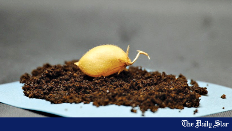
Scientists grow chickpeas in ‘moon dirt’
Scientists working to cultivate the field of extraterrestrial agriculture have grown chickpeas in dirt made mostly of simulated lunar soil, a step toward enabling astronauts on long-term moon missions to produce their own food.
Researchers said…
Continue Reading
-
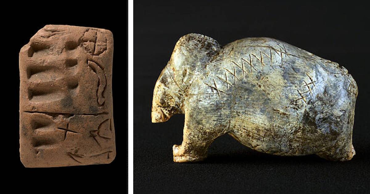
40,000-Year-Old Ice Age Symbols May Be First Step in Writing

The mammoth figurine from Vogelherd Cave. (Photo: University of Tübingen / Hildegard Jensen CC BY 4.0)
Long before cities, agriculture, or record keeping, Ice Age humans carved small lines and dots into ivory and bone….
Continue Reading
-
NASA Defense Test Kicked Asteroid Off Course — And Changed Its Orbit Around The Sun – Barron's
- NASA Defense Test Kicked Asteroid Off Course — And Changed Its Orbit Around The Sun Barron’s
- It’s Official: Humanity Has Changed The Path Of A Celestial Body Around The Sun For The First Time IFLScience
- You Can Probably Breathe Easier About…
Continue Reading
-
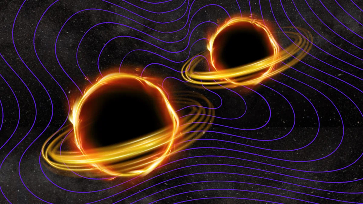
Scientists just doubled our catalog of black hole and neutron star collisions
When you buy through links on our articles, Future and its syndication partners may earn a commission.
Scientists have discovered over 100 more gravitational wave events. | Credit: Robert Lea (created with Canva)
Our catalog of spacetime ripples…
Continue Reading
-
Salt may have pushed us further into Snowball Earth 700 million years ago – Phys.org
- Salt may have pushed us further into Snowball Earth 700 million years ago Phys.org
- Snowball Earth’s liquid seas dipped way below freezing Space
- What Lies Beneath the Ocean Floor? 66 Million Years of Climate History Explained The Economic Times
Continue Reading
-
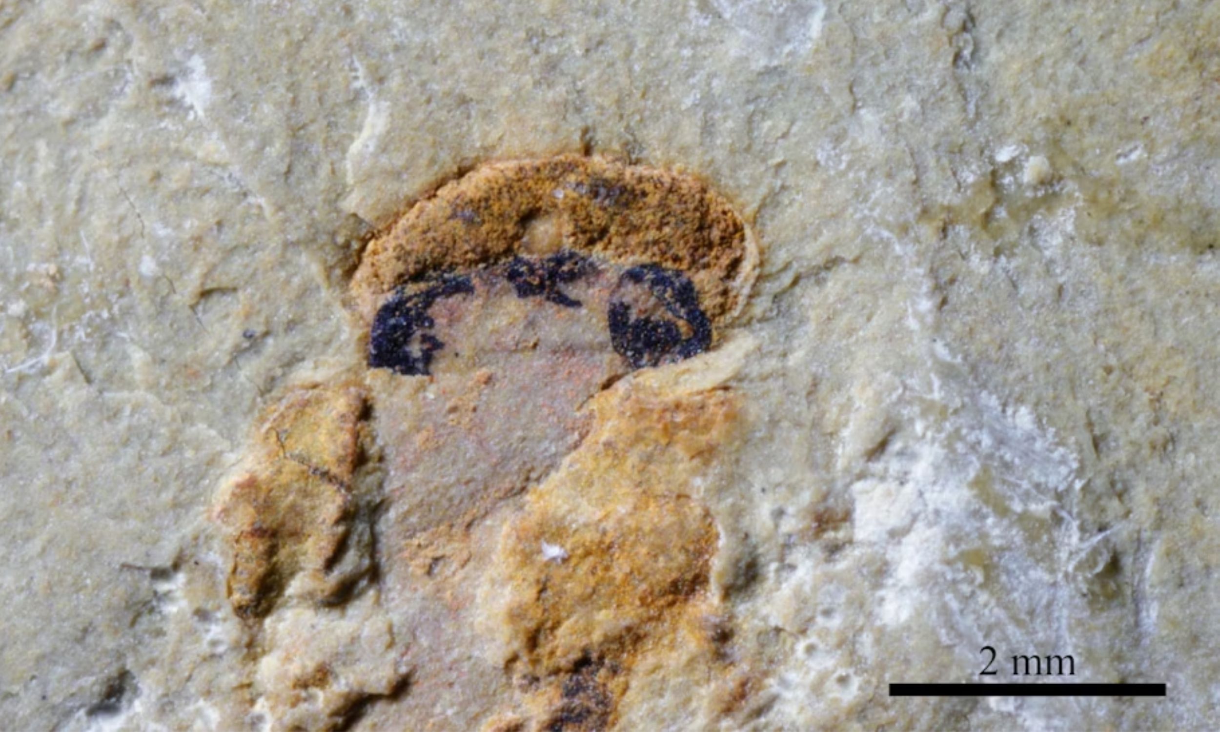
Fossil shows early vertebrates had four eyes
A 518-million-year-old fossil has revealed that some of the earliest vertebrates possessed four image-forming eyes instead of two.
That configuration recasts a small brain structure humans still carry as the remnant of a once fully visual organ.
Continue Reading