
Here on Earth, signal degradation is a real problem whenever we transmit information to one another. Signals like sound, light, and gravity spread out through space in three dimensions, becoming weaker and weaker as you travel…
Category: 7. Science
-
Astronomers discover giant cosmic sheet around the Milky Way
Nearly a century ago, astronomer Edwin Hubble discovered that almost all galaxies are receding from the Milky Way. This observation became a cornerstone of modern cosmology because it provided key evidence that the universe is expanding and that…
Continue Reading
-

Electrons catapult across solar materials in just 18 femtoseconds
Scientists have discovered that electrons can be propelled across solar materials at speeds close to the fastest nature allows, a result that challenges long accepted ideas about how solar energy systems operate.
The finding could open new paths…
Continue Reading
-

Let’s teach neuroscientists how to be thoughtful and fair reviewers
I used to joke, “Someone, somewhere, is trashing my paper right now at a journal club.” All joking aside, I often felt that it was uncomfortably plausible. Years of journal clubs had taught me that when you put on the “reviewer…
Continue Reading
-
Strange 90-Million-Year-Old Dinosaur Fossil Rewrites History – SciTechDaily
- Strange 90-Million-Year-Old Dinosaur Fossil Rewrites History SciTechDaily
- Argentine fossil rewrites evolutionary history of a baffling dinosaur clade Nature
- ONLINE EMBARGO 16.00 GMT, 25/02/26 themercury.com
- Pint sized amid giants: Newly…
Continue Reading
-
Cold microwave plasma jets for wound healing: antimicrobial efficacy, mechanisms and changes in microbial cells
Boeckmann, L. et al. Cold Atmospheric Pressure Plasma in Wound Healing and Cancer Treatment. Appl. Sci. https://doi.org/10.3390/app10196898 (2020).
Slater, A. M., Barclay, S. J., Granfar, R. M. S. & Pratt, R. L. Fascia as a regulatory system in…
Continue Reading
-
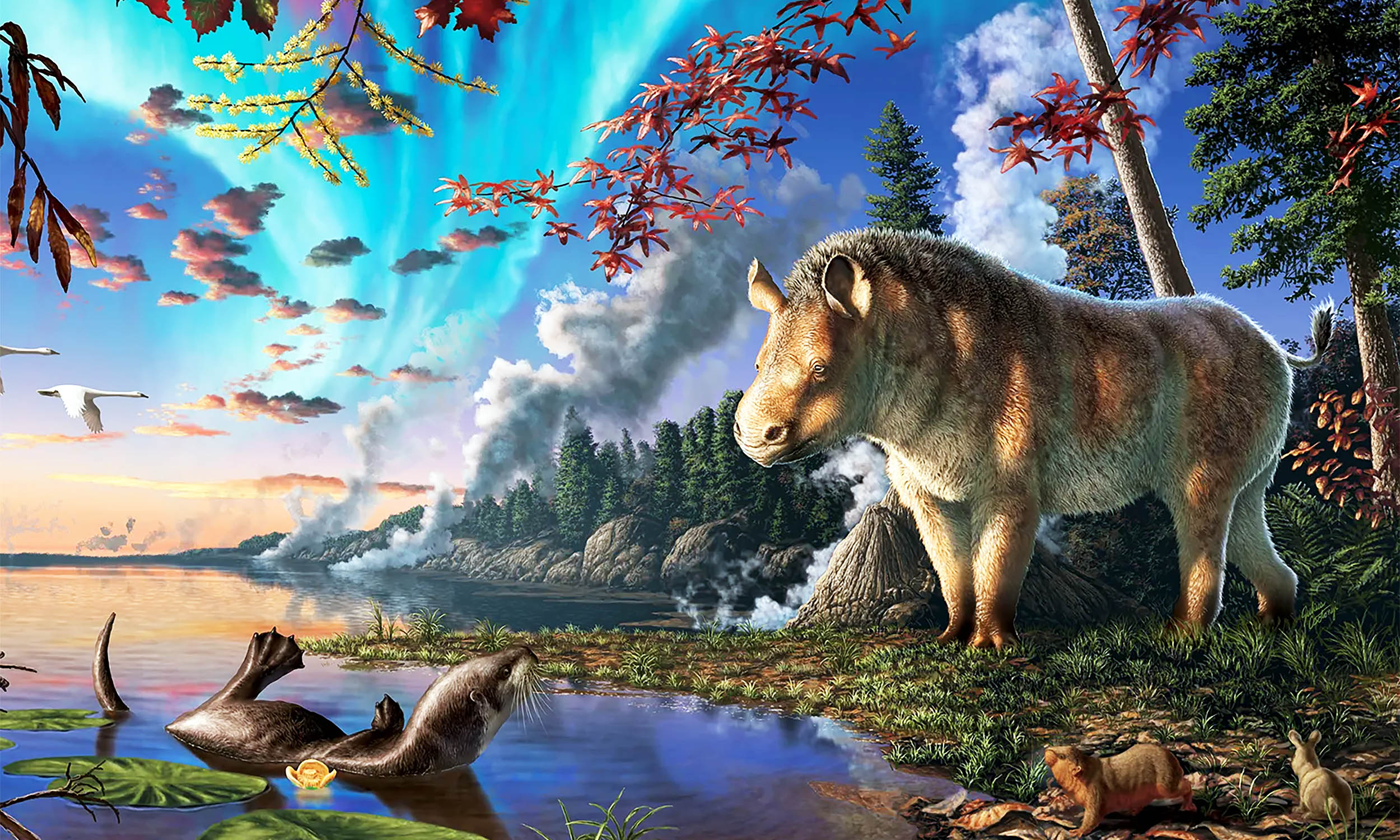
23-million-year-old ‘frosty rhino’ discovered in the High Arctic
Deep within the frozen ground of Devon Island in Canada’s High Arctic, researchers found the nearly complete skeleton of a rhinoceros, Epiaceratherium itjilik, that lived there around 23 million years ago.
The discovery, made by a team from the
Continue Reading
-
Mysteries Of March Cosmos – The Rising Nepal
- Mysteries Of March Cosmos The Rising Nepal
- What’s Up: March 2026 Skywatching Tips from NASA NASA Science (.gov)
- Can you spot the planets shining in our March night skies? The Weather Network
- March 2026 calendar: St. Patrick’s Day, daylight…
Continue Reading
-
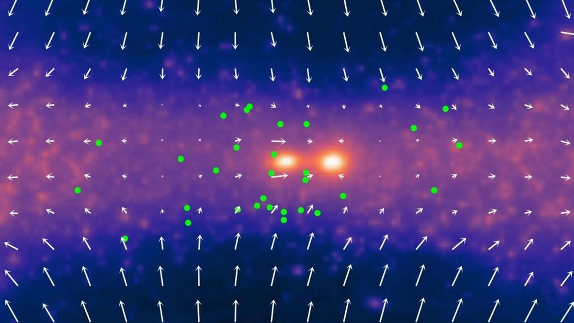
ALMA captures the most detailed image ever of the Milky Way’s turbulent core
Astronomers have unveiled a striking new view of the center of the Milky Way, exposing an intricate network of cosmic gas filaments in unprecedented detail. The image was produced using the Atacama Large Millimeter/submillimeter Array (ALMA) and…
Continue Reading
-
ALMA captures the most detailed image ever of the Milky Way’s turbulent core
Astronomers have unveiled a striking new view of the center of the Milky Way, exposing an intricate network of cosmic gas filaments in unprecedented detail. The image was produced using the Atacama Large Millimeter/submillimeter Array (ALMA) and…
Continue Reading