Photos of Earth taken from outer space usually show the beautiful blue and green planet against a field of black — perhaps its most idyllic setting.
In contrast, scientists at NASA’s Jet Propulsion Laboratory in Pasadena are zeroing in on a…

Photos of Earth taken from outer space usually show the beautiful blue and green planet against a field of black — perhaps its most idyllic setting.
In contrast, scientists at NASA’s Jet Propulsion Laboratory in Pasadena are zeroing in on a…
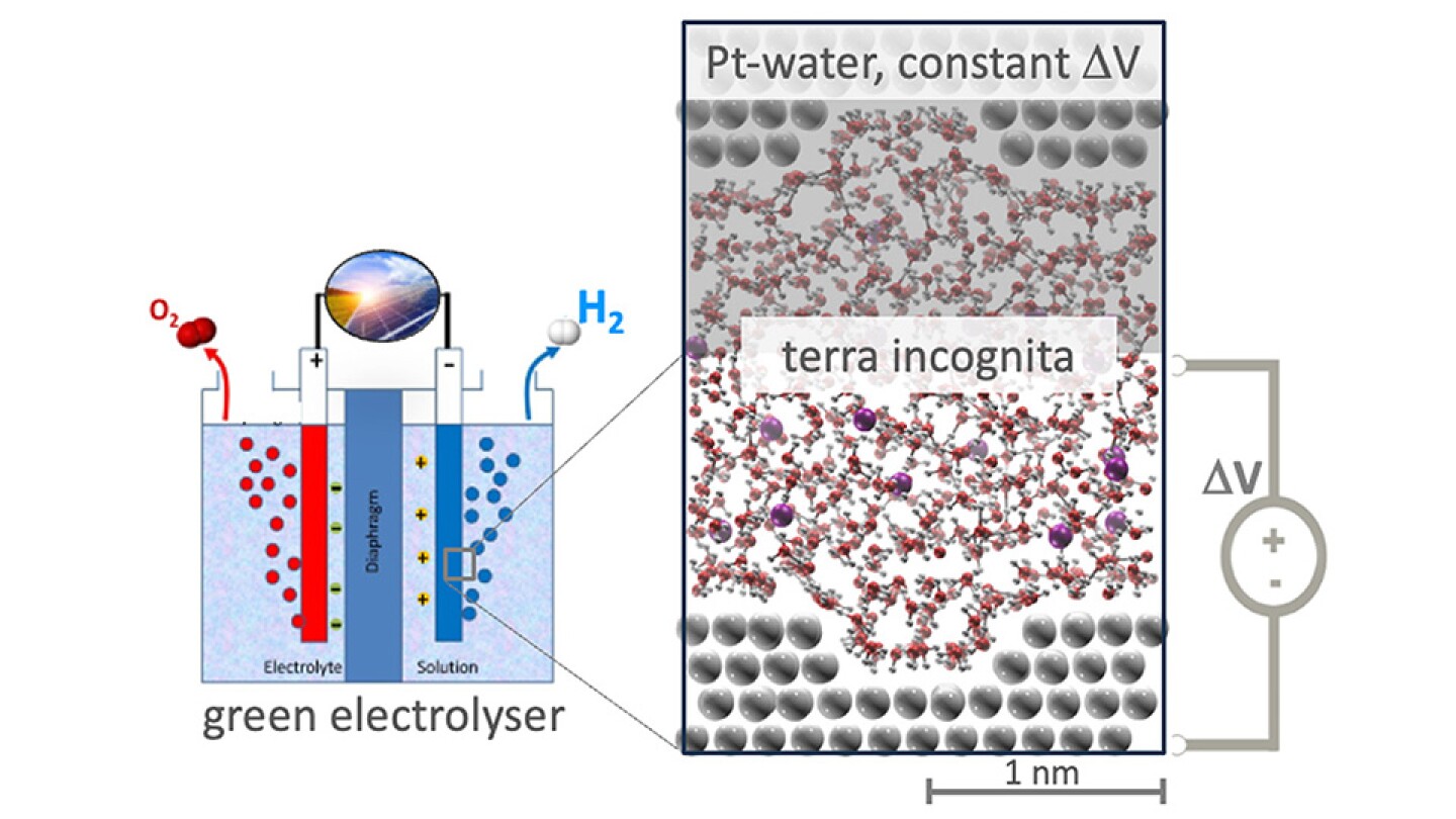
Nanoscale surface structure at the platinum-water interface has an important effect on electrical properties.
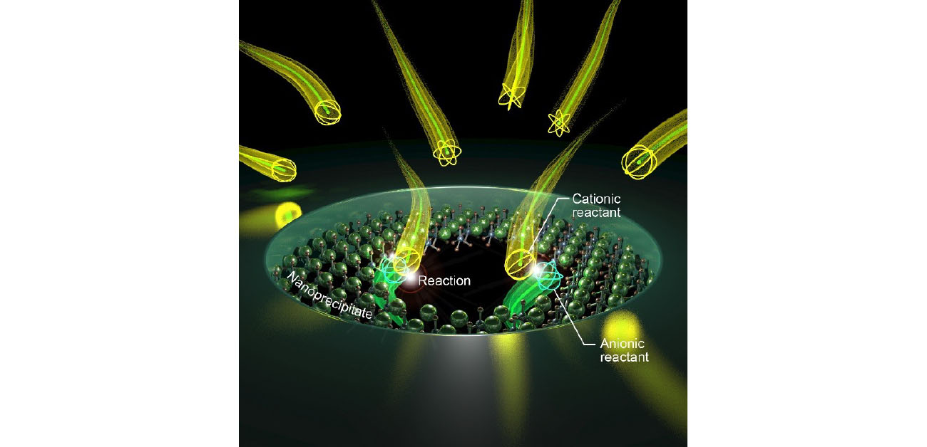
Many chemical reactions,…
Watch On
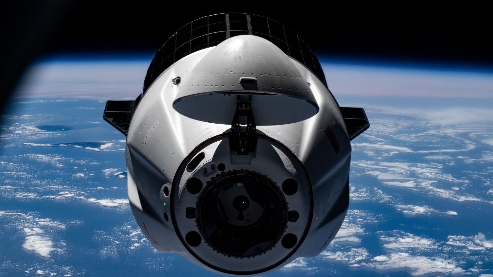
A SpaceX Dragon cargo capsule will undock from the International Space Station today (Feb. 26), and you can watch its departure live.

3D printing is no longer a novel concept – whether it is tech enthusiasts…
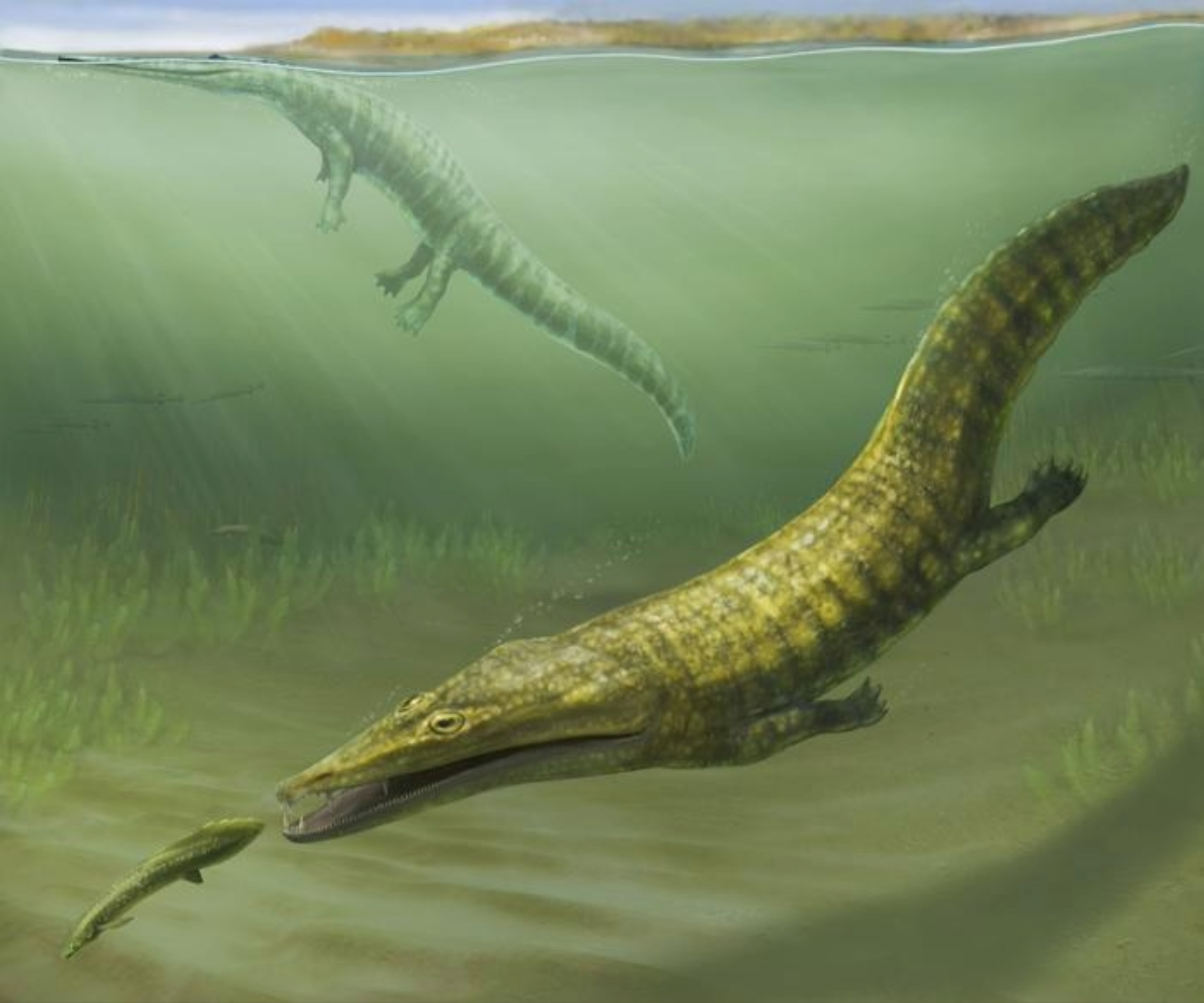
Fossils found in northwestern Australia show that 250 million years ago, the red, dusty Kimberley region was nothing like it is today. Instead of a desert, it was a shallow coastal bay, with tropical waters lapping over the area and…
Ion channels are narrow passageways that play a pivotal role in many biological processes. To model how ions move through these tight spaces, pores need to be fabricated at very small length scales. The narrowest regions of ion channels can be…


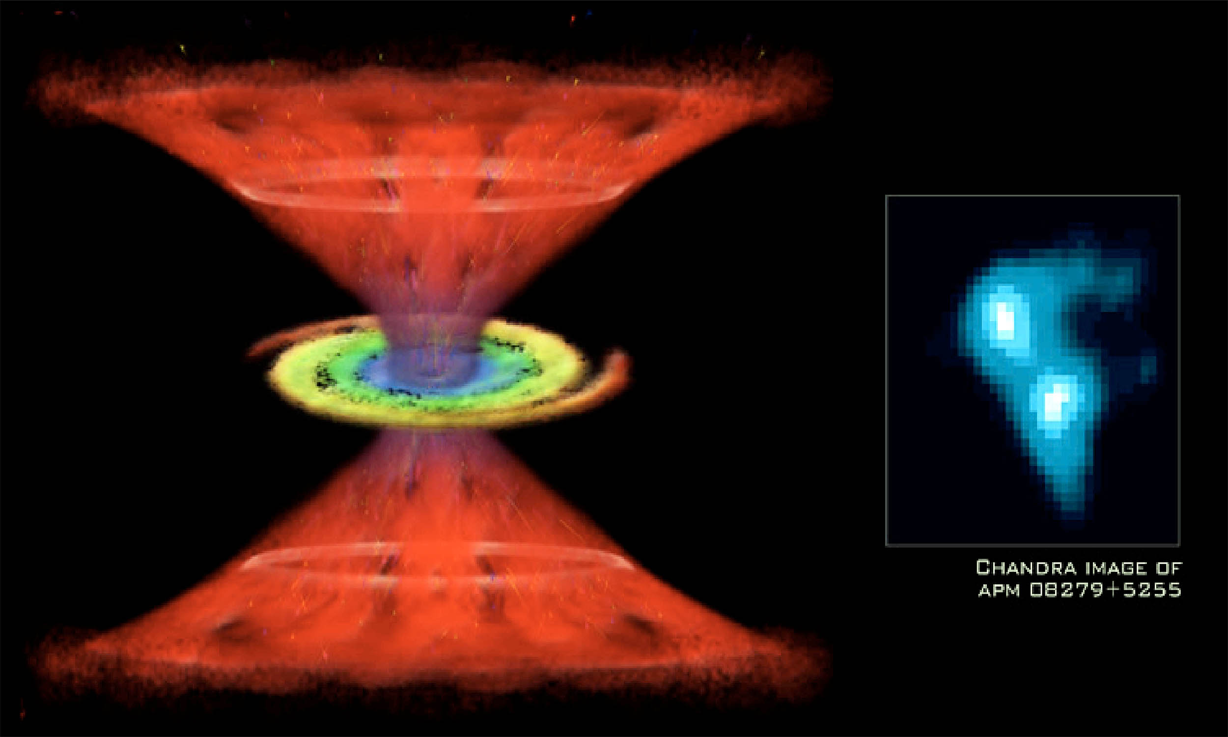
Astronomers enjoy it when the universe throws a curveball, and this object does exactly that. Working in two teams, they have found the largest, most distant stash of water ever seen in the cosmos. APM 08279+5255 is a quasar – an active galaxy…