When you look up at the night sky, it appears unchanging. But if you look deep enough you will find that the sky is in fact constantly shifting. Satellites, asteroids and interstellar objects pass by. Stars not only shine brightly, they can…
Category: 7. Science
-
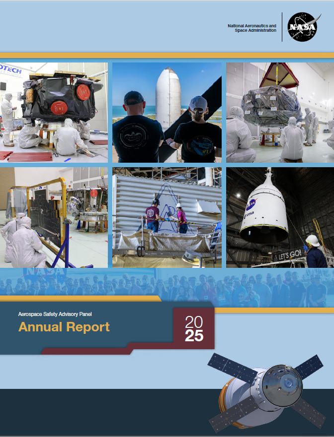
NASA’s Aerospace Safety Advisory Panel Releases 2025 Annual Report
The Aerospace Safety Advisory Panel (ASAP), which advises NASA and Congress on safety, has released its 2025 annual report on NASA’s performance and challenges.
While the panel acknowledged NASA’s safety achievements, it warned that…
Continue Reading
-
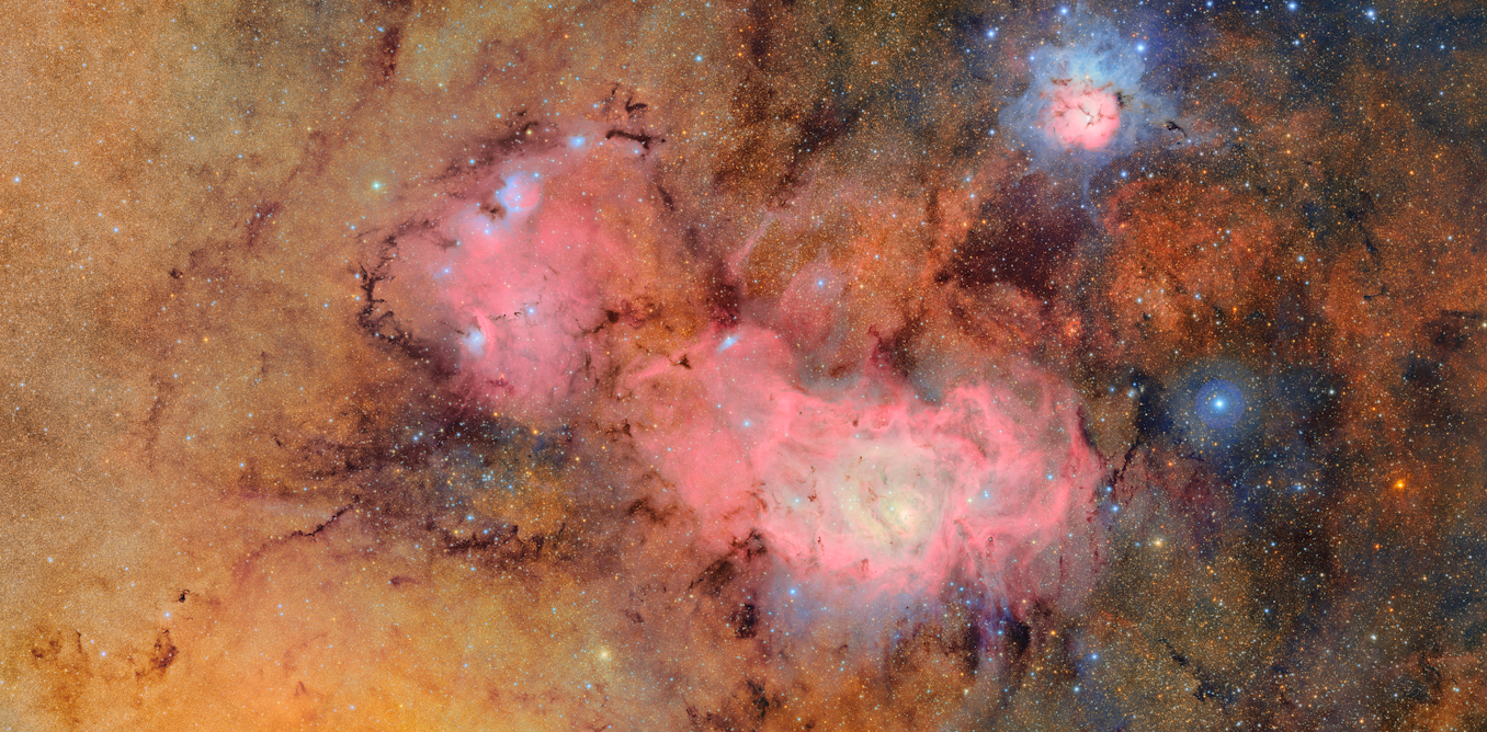
Largest ALMA image ever shows cold gas in our galactic center – Astronomy Magazine
- Largest ALMA image ever shows cold gas in our galactic center Astronomy Magazine
- AP Trending SummaryBrief at 12:02 p.m. EST The News-Item
- World’s largest radio telescope array pierces heart of our Milky Way: ‘This is just the beginning’ Yahoo
Continue Reading
-
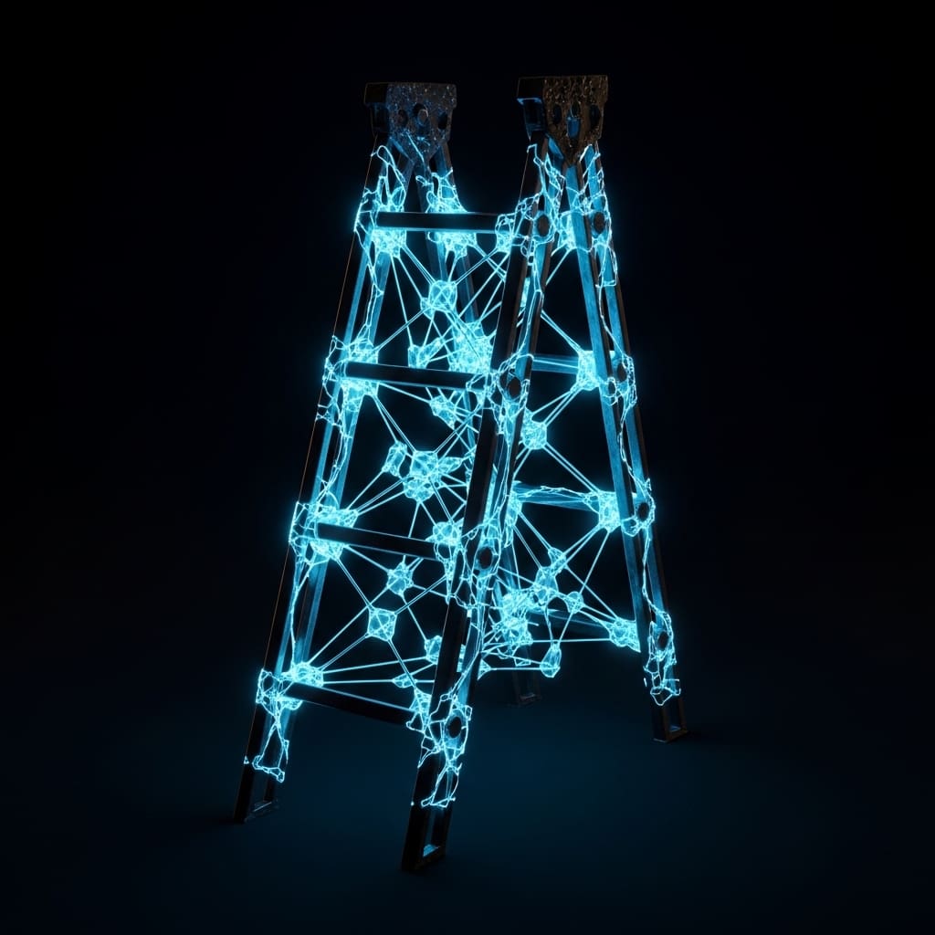
Ladder Lattices Reveal Robust Insulating Phases Of Matter
Researchers are increasingly focused on understanding strongly correlated quantum systems, and a new study details the Mott-insulating phases within the Bose-Hubbard model on quasi-one-dimensional ladder lattices. Lorenzo Carfora, from the…
Continue Reading
-

Chinese astronauts finally reveal why spacecraft left them ‘stranded’ for 437 days in space
Crew members of China’s Shenzhou-20 mission…
Continue Reading
-
A 300-Million-Year-Old Find: It Lived on Land and Was the First to Eat Plants – Explorersweb »
- A 300-Million-Year-Old Find: It Lived on Land and Was the First to Eat Plants Explorersweb »
- 307-million-year-old plant-eater: Scientists found “Tyrannoroter heberti” could be the first… Moneycontrol.com
- This creature may have been the first…
Continue Reading
-
Rubin Observatory launches real-time alerts for night sky monitoring – Stanford University
- Rubin Observatory launches real-time alerts for night sky monitoring Stanford University
- NSF-DOE Vera C. Rubin Observatory launches real-time discovery machine for monitoring the night sky National Science Foundation (.gov)
- NSF–DOE Vera C….
Continue Reading
-
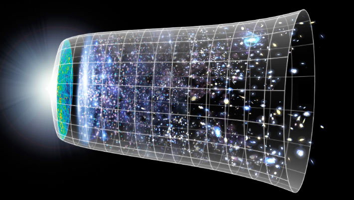
Astrophysicists Propose New Method to Measure Hubble Constant
Astrophysicists from the University of Illinois and the University of Chicago have developed an innovative method to measure the Hubble constant — the rate at which the Universe is expanding — using the subtle background hum of…
Continue Reading
-
Mouse study finds some brain regions recover better after myelin damage – Multiple Sclerosis News Today
- Mouse study finds some brain regions recover better after myelin damage Multiple Sclerosis News Today
- Stunning New Maps of Myelin-Making Mouse Brain Cells Advance Understanding of Nervous System Disorders Johns Hopkins Medicine
- Mapping the…
Continue Reading
-

Centenarians have blood profiles closer to 30-year-olds
Only a tiny slice of Switzerland’s population makes it past 100 years old – about 0.02 percent. That rarity raises a simple question: Do people who reach 100 have measurable biological traits that set them apart from everyone else?
A team…
Continue Reading