- Rubin Observatory launches real-time alerts for night sky monitoring Stanford University
- NSF-DOE Vera C. Rubin Observatory launches real-time discovery machine for monitoring the night sky National Science Foundation (.gov)
- NSF–DOE Vera C….
Category: 7. Science
-
Rubin Observatory launches real-time alerts for night sky monitoring – Stanford University
-

Astrophysicists Propose New Method to Measure Hubble Constant
Astrophysicists from the University of Illinois and the University of Chicago have developed an innovative method to measure the Hubble constant — the rate at which the Universe is expanding — using the subtle background hum of…
Continue Reading
-
Mouse study finds some brain regions recover better after myelin damage – Multiple Sclerosis News Today
- Mouse study finds some brain regions recover better after myelin damage Multiple Sclerosis News Today
- Stunning New Maps of Myelin-Making Mouse Brain Cells Advance Understanding of Nervous System Disorders Johns Hopkins Medicine
- Mapping the…
Continue Reading
-

Centenarians have blood profiles closer to 30-year-olds
Only a tiny slice of Switzerland’s population makes it past 100 years old – about 0.02 percent. That rarity raises a simple question: Do people who reach 100 have measurable biological traits that set them apart from everyone else?
A team…
Continue Reading
-

Chinese astronauts describe moment a crack was discovered on Shenzhou-20 spacecraft
Chinese astronauts have described what happened when they were nearly stranded in space last year after a suspected piece of space junk struck their return capsule.
Chen Dong, Chen Zhongrui and Wang Jie, the crew of the ill-fated Shenzhou-20…
Continue Reading
-

New Discoveries on Mars and What They Say About Habitability and Life
Mars dominates the planetary news with exciting reports. Two NASA rovers, working nearly 3,700 kilometers apart, made discoveries that addressed the question: how far did Mars come toward conditions that, on Earth, supported…
Continue Reading
-

Rubin Observatory launches real-time monitoring of the sky with thousands of alerts – UW News
The Vera C. Rubin Observatory sits on its mountain peak in Chile during observation activities in April 2025. The observatory will soon begin real-time nightly monitoring of the entire Southern Hemisphere sky. Photo:… Continue Reading
-
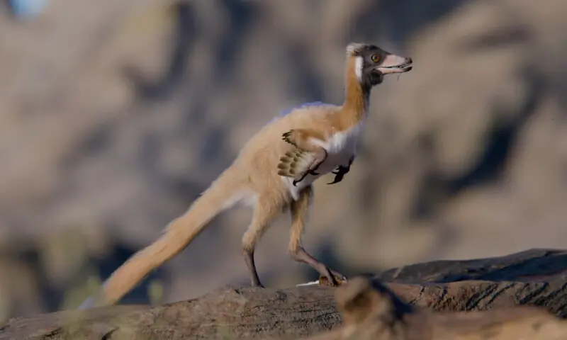
‘Exquisite’ fossil of one of the smallest dinosaurs found in Argentina – Life & Style
In Argentina’s Patagonia region 95 million years ago, some huge dinosaurs roamed the landscape, including fearsome meat-eater Giganotosaurus, at about eight tons, and immense long-necked plant-eater Argentinosaurus, perhaps 70 tons. But this…
Continue Reading
-

Sky to display rare 6-planet alignment forming ‘planetary parade’ on February 28
Sky to display rare 6-planet alignment forming ‘planetary parade’ on February 28 Six planets, including Mercury, Venus, Jupiter, Saturn, Uranus, and Neptune, will align along the ecliptic this Saturday, February…
Continue Reading
-
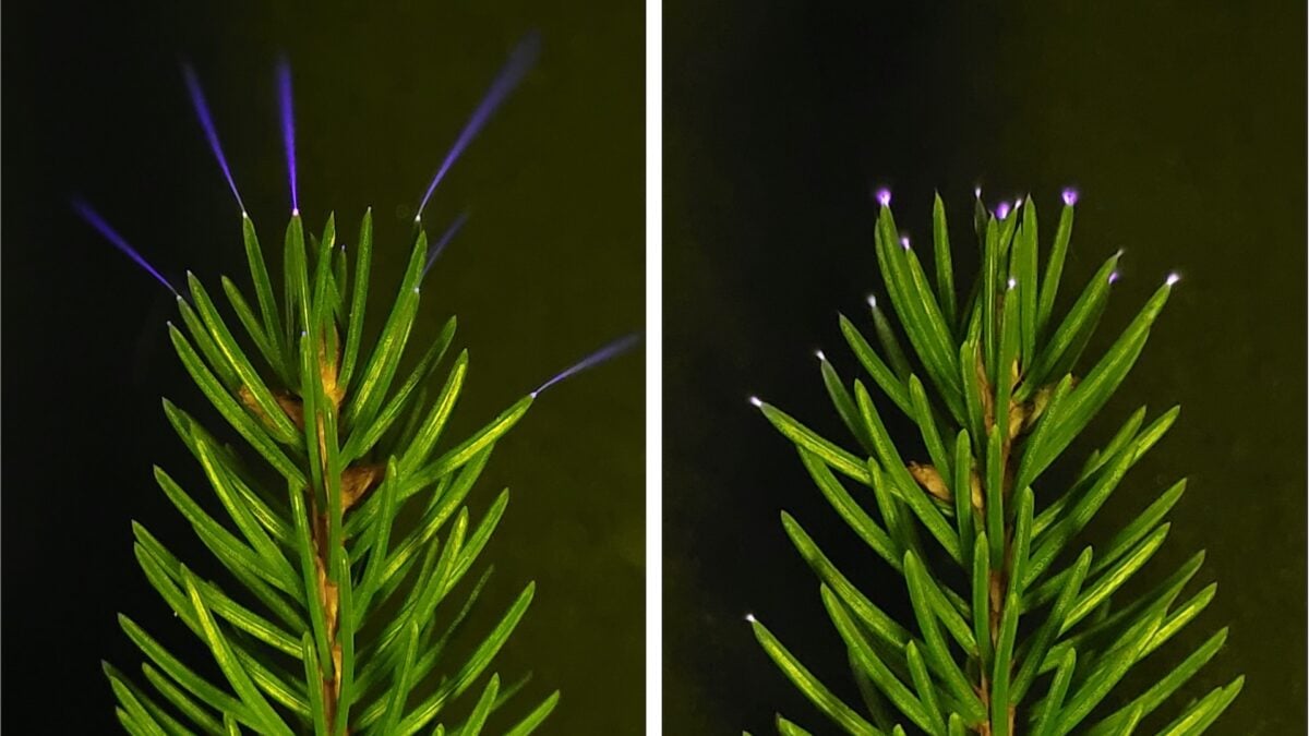
Scientists Just Caught Trees Firing Off Ultraviolet Sparkles During Thunderstorms
In the summer of 2024, a team of researchers went storm-chasing in a Toyota Sienna minivan in search of tiny, faint sparks lighting up the tips of leaves. As a thunderstorm raged overhead, the researchers pointed their camera at…
Continue Reading