Key takeaways
- Precious transition metals like platinum and palladium are used as catalysts to speed up chemical reactions that produce carbon-nitrogen bonds.
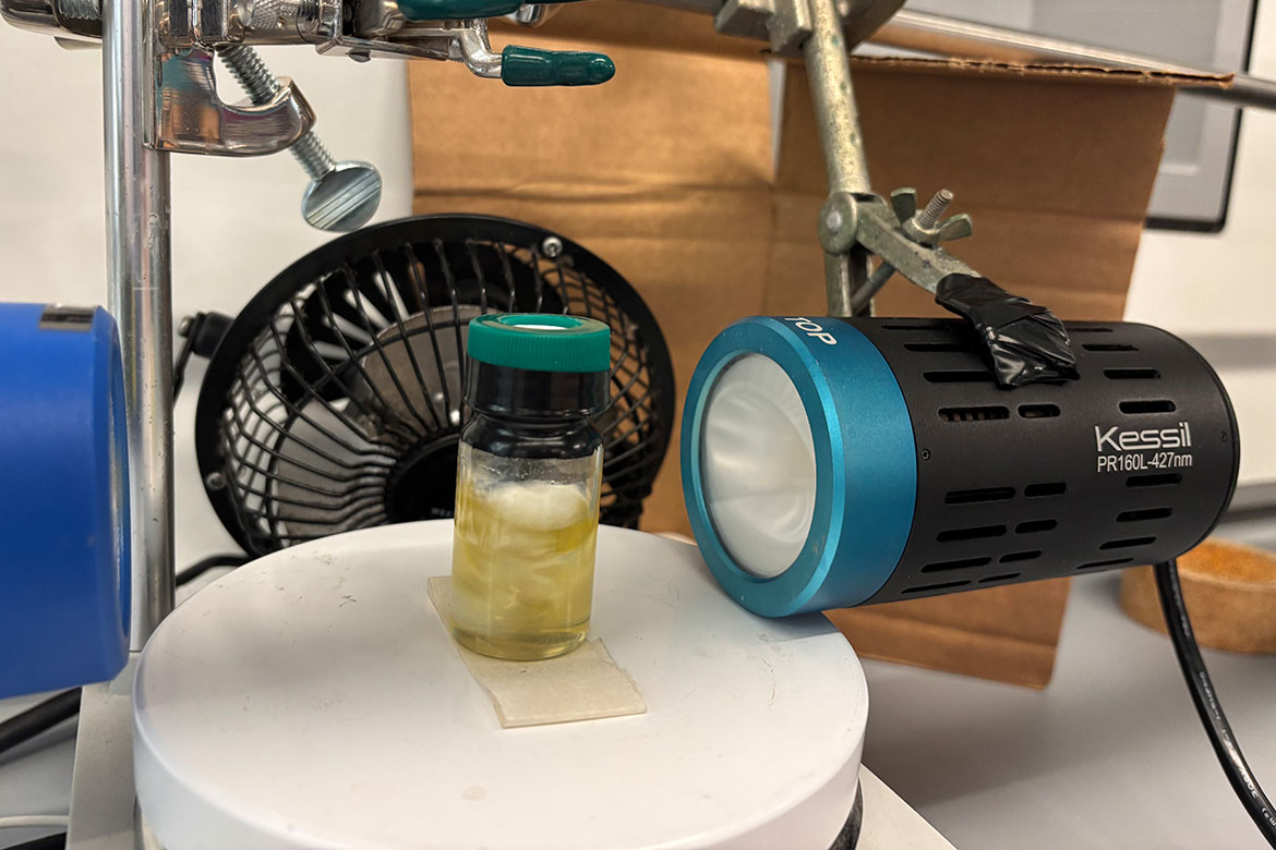
- UCLA organic chemists have figured out how to make inexpensive phosphine act…
Key takeaways
ChatGPT Health launched in January 2026 as OpenAI’s consumer health tool, reaching millions of users. Here, we conducted a structured stress test of triage recommendations using 60 clinician-authored vignettes across 21 clinical domains under…
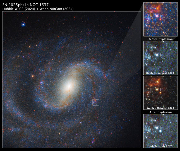
Using the NASA/ESA/CSA James Webb Space Telescope, astronomers have for the first time identified the progenitor of a nearby supernova — a red supergiant star cloaked in thick, dust-rich shrouds that made it invisible to previous…
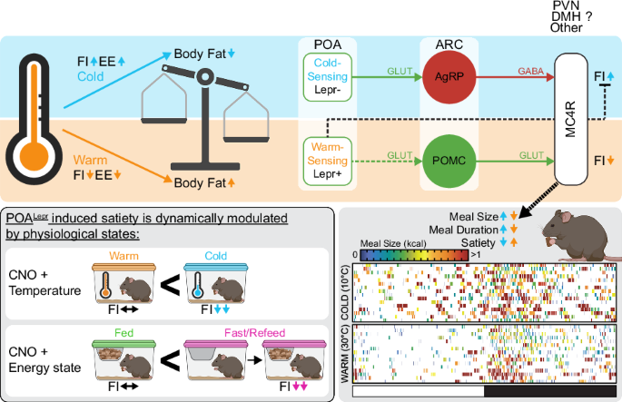
Nakamura, K. Central circuitries for body temperature regulation and fever. Am. J. Physiol. Regul. Integr. Comp. Physiol. 301, R1207–R1228 (2011).
Yu, S., Francois, M., Huesing, C. &…
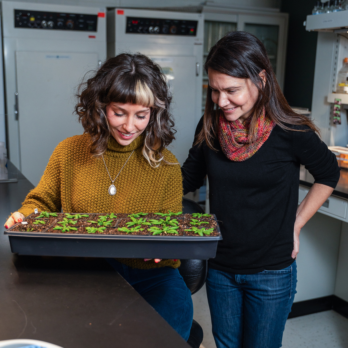
The study was funded by the National Science Foundation and led by Grace Johnston, who conducted the research as a student. Johnston was recruited into Argueso’s lab as an undergraduate biology student and wrote the paper as her…
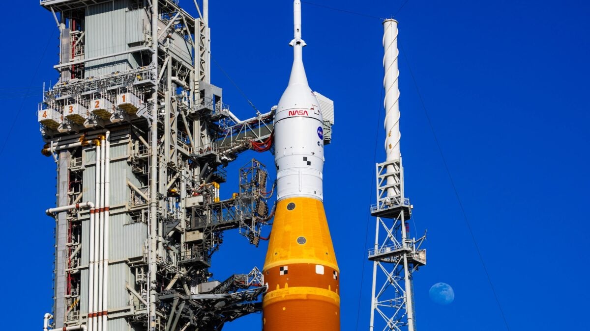
Well, I guess we jinxed it.
One day after NASA completed a wet dress rehearsal for the Artemis 2 mission with no major issues, the Space Launch System (SLS) rocket started acting up again. This time, it wasn’t a hydrogen leak….
Earthquakes are one of many natural phenomena that, despite technological advances, we’ve yet to predict in advance. Researchers in Japan—a country frequently hit by devastating earthquakes—propose we look for an…

Summary: Neurons are high-energy cells that must find ways to survive when nutrients are scarce. New research has discovered a fascinating survival mechanism: neurons pair up their protein factories (ribosomes) into inactive “disomes” to save…
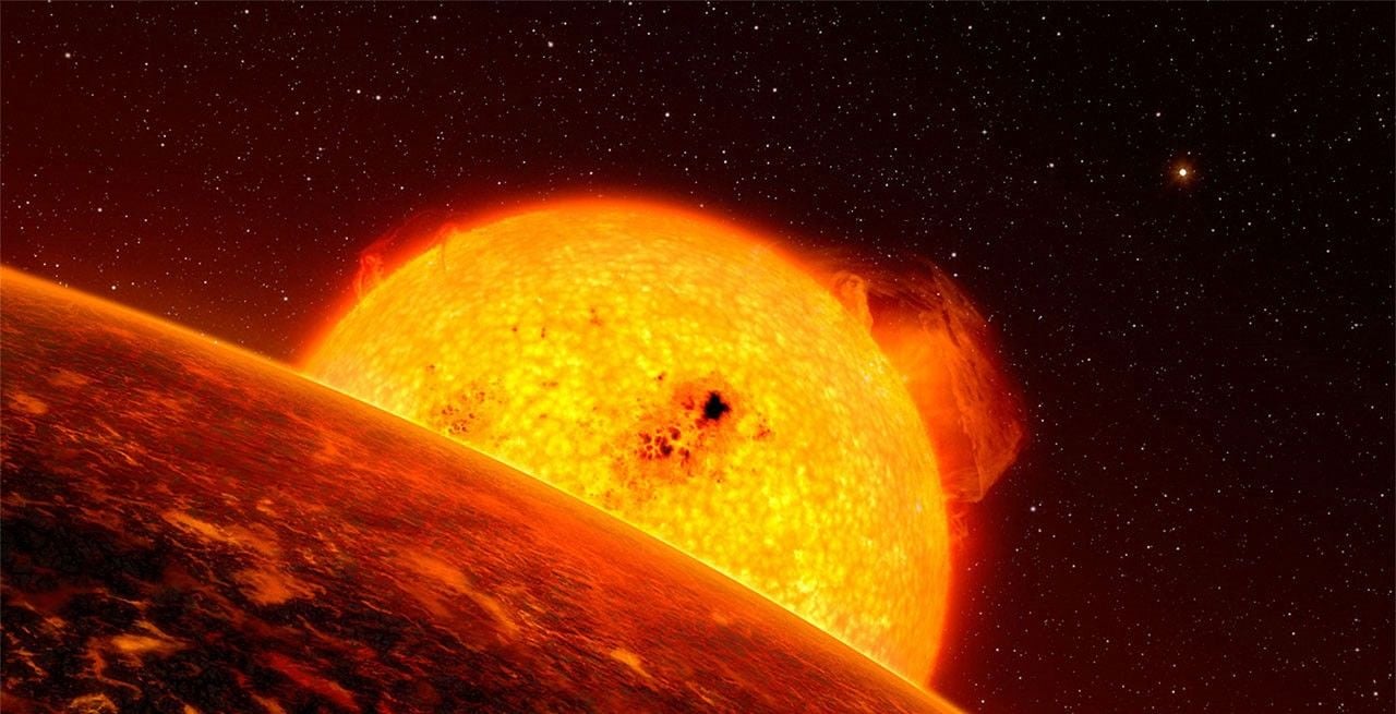
When stars like our Sun reach the end of their main sequence, they enter their Red Giant Branch phase and expand to become several times their original size. During this time, the star will undergo chemical changes in its interior,…