Key points
- Genome sequencing of several wheat stem rust strains shows some outbreaks arose independently, overturning assumptions and changing how scientists track disease threats.
- A new gene atlas reveals why resistance failed in the…
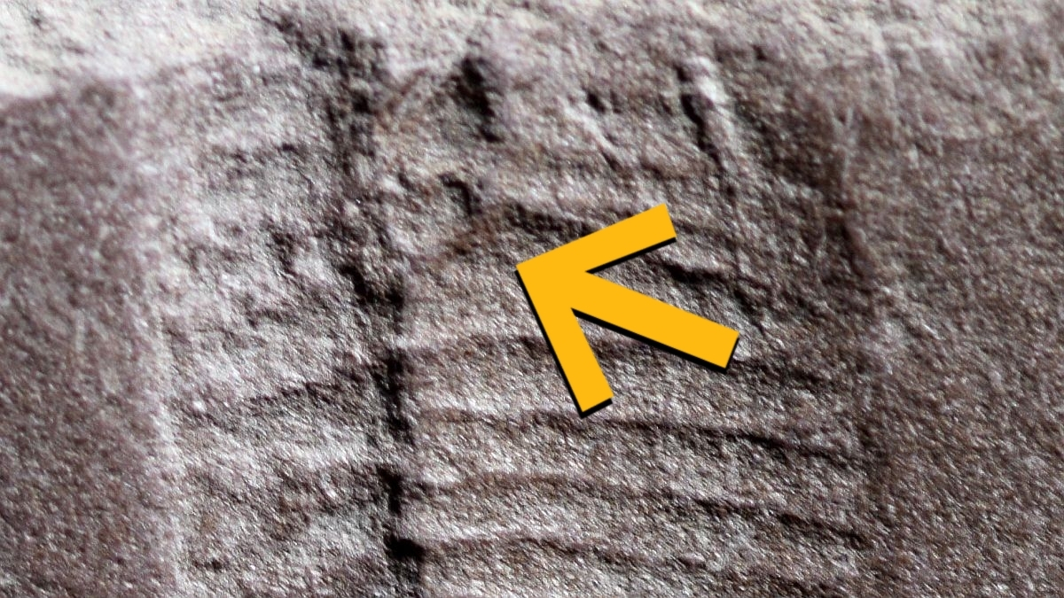
Once, long ago, a little reptile going about its business plopped itself down in the mud before getting up and carrying on with its day.
Nearly 300 million years later, that brief rest has yielded the world’s earliest known fossilized imprint…
Jappe, U. Pathological Mechanisms of Acne with Special Emphasis on Propionibacterium acnes and Related Therapy. Acta Derm. Venereol. 83, 241 (2003).
Morshed, S. M. Understanding the impact of…
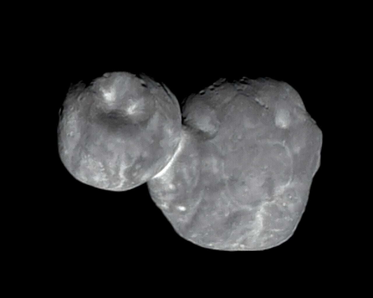
On a frigid orbit beyond Neptune, some of the solar system’s smallest worlds project a strange silhouette. Two rounded lobes, pressed together with a narrow “neck,” like a snowman that never melted.
Those shapes are common enough to demand…

When you step outside on a winter morning or pop a mint into your mouth, a tiny molecular sensor in your body springs into action, alerting your brain to the sensation of cold. Scientists have now captured the first detailed images…

It looks like a March launch is no longer in the cards for Artemis II, NASA’s first crewed trip to the moon’s vicinity since the final Apollo mission over 50 years ago. While preparations were underway at the Kennedy Space Center for a launch as…
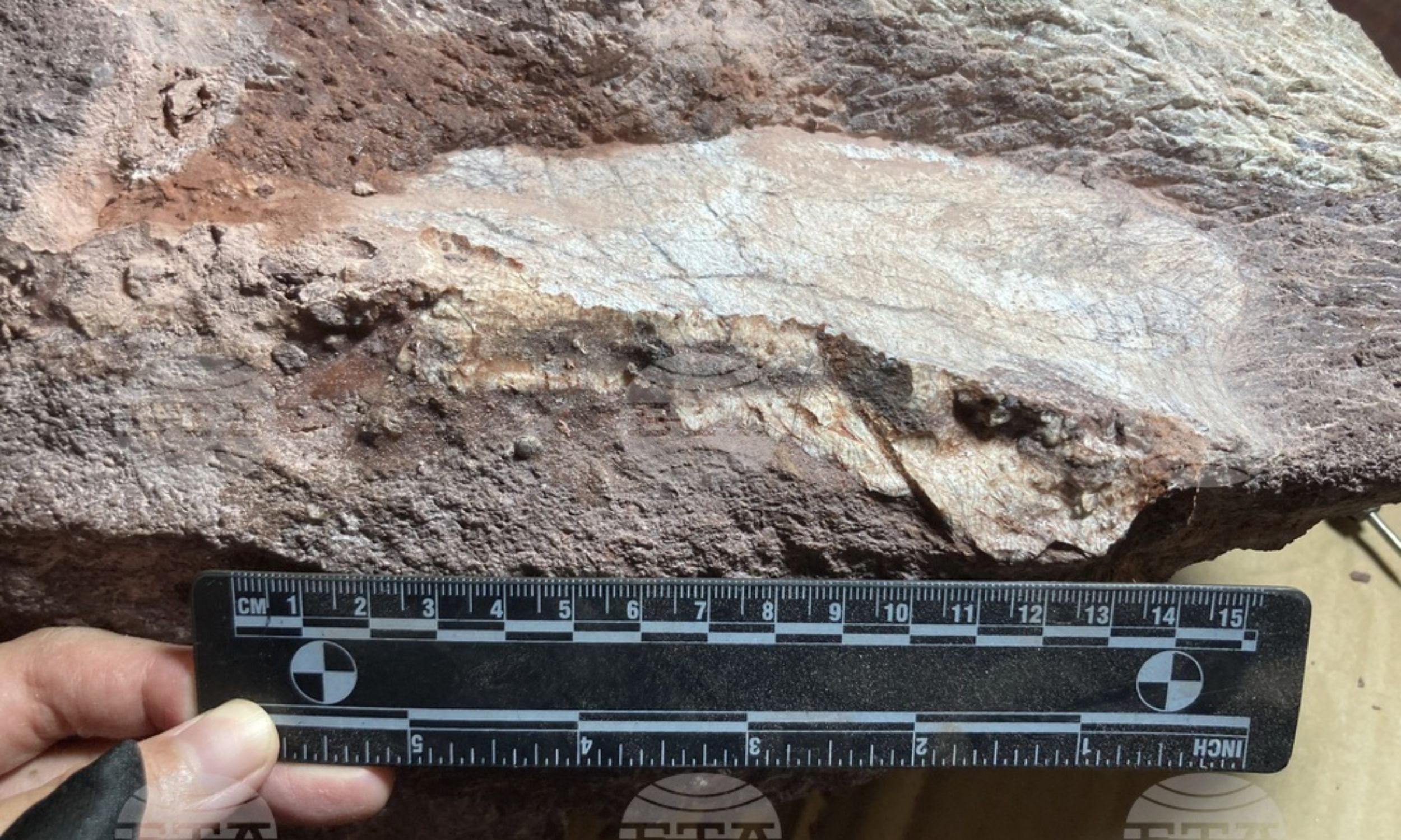
Scientists have found traces of a tough material that once formed the outer shells of ancient sea creatures called trilobites, preserved inside fossils that are more than 500 million years old.
The discovery shows that parts of living organisms…