- Deep-sea fish break the mold with novel visual system Arab News
- Deep-sea fish larvae rewrite the rules of how eyes can be built Phys.org
- Scientists Discover Fish with Revolutionary Eye Cells That Rewrite Biology Textbooks Fine Day 102.3
- How do…
Category: 7. Science
-
Deep-sea fish break the mold with novel visual system – Arab News
-
UAE extends hope probe’s Mars mission to 2028 – middle-east-online.com
- UAE extends hope probe’s Mars mission to 2028 middle-east-online.com
- Gulf states build on UAE’s Mars breakthrough Semafor
- UAE extends Mars probe mission until 2028 Phys.org
- UAE Space Agency Extends Hope Probe Mission to 2028 Yemen Online
- UAE…
Continue Reading
-
Annular solar eclipse seen over Antarctica – Dawn
- Annular solar eclipse seen over Antarctica Dawn
- What time is the annular solar eclipse on Feb. 17? Space
- ‘Ring of fire’ solar eclipse: February 2026 path, visibility over Antarctica and partial eclipse for South Africa CNN
- First solar eclipse…
Continue Reading
-
Annular solar eclipse seen over Antarctica – Dawn
- Annular solar eclipse seen over Antarctica Dawn
- What time is the annular solar eclipse on Feb. 17? Space
- First solar eclipse of 2026 to miss Pakistan skies Pakistan Today
- First solar eclipse of 2026 blazes a ‘ring of fire’ above Antarctica
Continue Reading
-
Rare ‘Ring Of Fire’ Solar Eclipse Sweeps Across Antarctica And Africa – Forbes
- Rare ‘Ring Of Fire’ Solar Eclipse Sweeps Across Antarctica And Africa Forbes
- Annular ‘ring of fire’ solar eclipse February 2026 – Live updates Space
- ‘Ring of fire’ solar eclipse: February 2026 path, visibility over Antarctica and partial…
Continue Reading
-
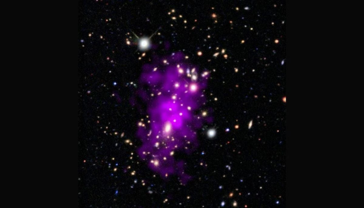
New study suggests universe can end in ‘Big Crunch’ in 20bn years
A new astrophysics study predicts the universe could eventually collapse in a…
Continue Reading
-
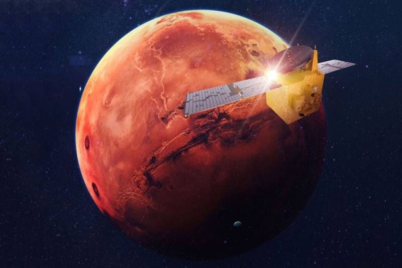
UAE extends Mars probe mission until 2028
The United Arab Emirates announced on Tuesday that it would extend its Mars probe mission, now in its fifth year, for an additional three years, underlining the oil-rich state’s space ambitions.
The UAE’s Hope probe arrived in orbit around Mars…
Continue Reading
-
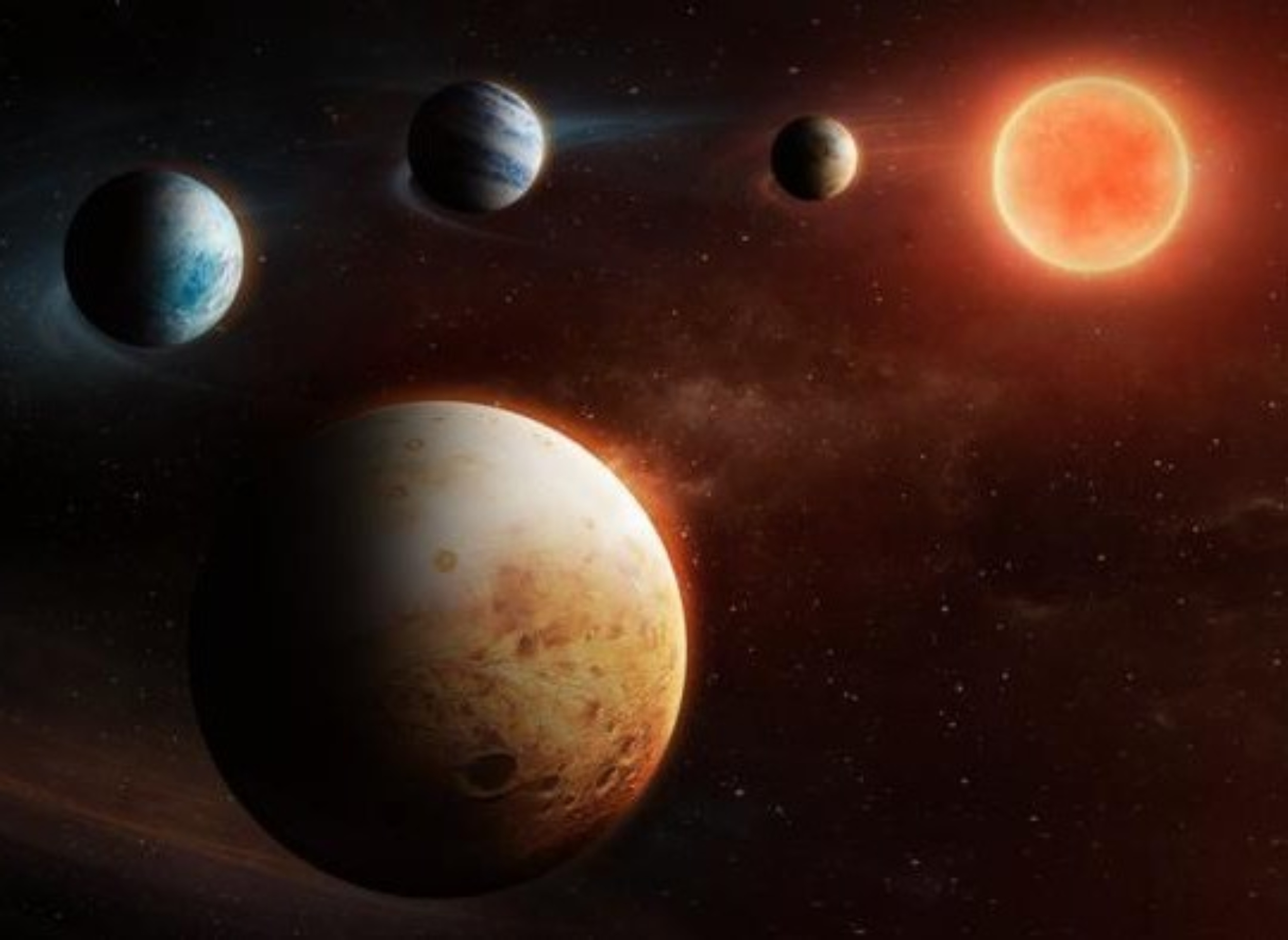
‘Inside-out’ planetary system breaks the rules of planet formation
Most of us memorized the order of the planets in our Solar System using playful phrases like, “My Very Educated Mother Just Served Us Nachos.”
It may sound like nonsense, but the sentence neatly encodes a familiar sequence: Mercury, Venus,…
Continue Reading
-
UAE extends Emirates Mars Mission until 2028
DUBAI, Feb. 17 (Xinhua) — The United Arab Emirates (UAE) Space Agency on Tuesday announced the extension of the Emirates Mars Mission (EMM) until 2028, according to local daily Al Etihad.
The mission, carried out by the Hope…
Continue Reading
-
UAE extends Emirates Mars Mission until 2028-Xinhua
DUBAI, Feb. 17 (Xinhua) — The United Arab Emirates (UAE) Space Agency on Tuesday announced the extension of the Emirates Mars Mission (EMM) until 2028, according to local daily Al Etihad.
The mission, carried out by the Hope Probe, was…
Continue Reading