A series of viral social media posts drew attention to an old…
Category: 7. Science
-
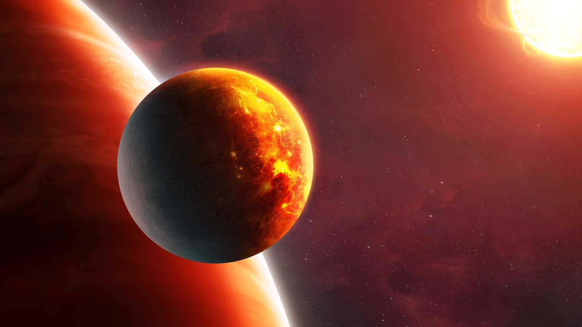
Hydrogen atmospheres keep rogue moons warm for billions of years
A new study led by researchers at the Max Planck Institute for Extraterrestrial Physics and Giulia Roccetti of the European Space Agency suggests rogue planets may host habitable moons.
The team found that thick hydrogen atmospheres can trap…
Continue Reading
-
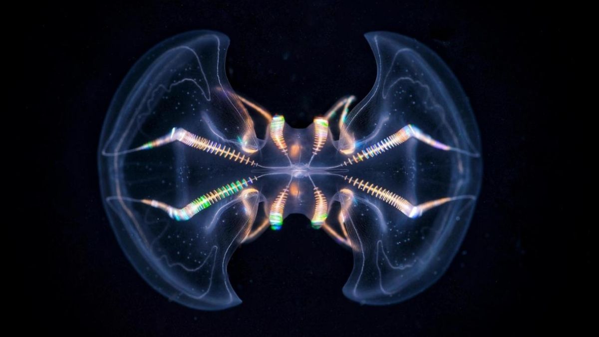
The Comb Jelly ‘Brain’ Is Far More Complex Than We Ever Realized : ScienceAlert
Comb jellies – very simple, gelatinous creatures best-known for their hypnotic underwater light shows – first appeared in Earth’s oceans around 550 million years ago.
For a long time, biologists have kind of considered them the living…
Continue Reading
-
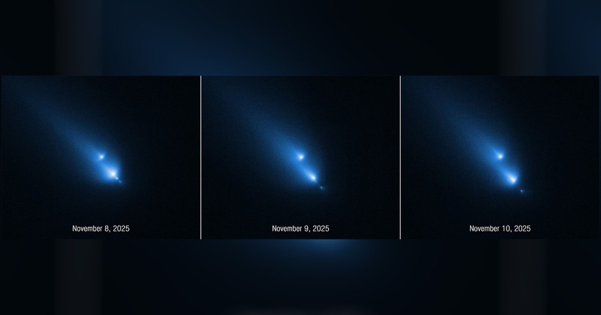
Scientists Startled by What Happens When They Point Hubble at Comet
Lady Fortune was on astronomers’ side when they pointed the Hubble Space Telescope at a comet…
Continue Reading
-

What if the next great astronomer isn’t human? How AI is revolutionizing our study of the cosmos
How do you unravel the universe’s deepest secrets when the data piles up faster than we can make sense of it? It’s a bit like being handed a zillion puzzle pieces from a cosmic explosion and being told to recreate the original star.
Modern…
Continue Reading
-
Neanderthals May Have Used Birch Tar As Ancient Antibiotic, Study Finds – SciTechDaily
- Neanderthals May Have Used Birch Tar As Ancient Antibiotic, Study Finds SciTechDaily
- Neanderthals may have used a prehistoric glue as a topical antibiotic Chemical & Engineering News
- Neanderthals May Have Used the World’s First Antibiotic…
Continue Reading
-
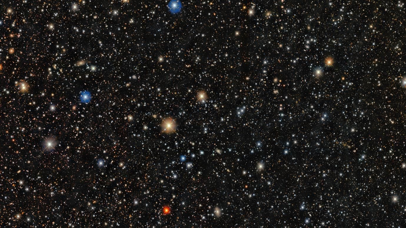
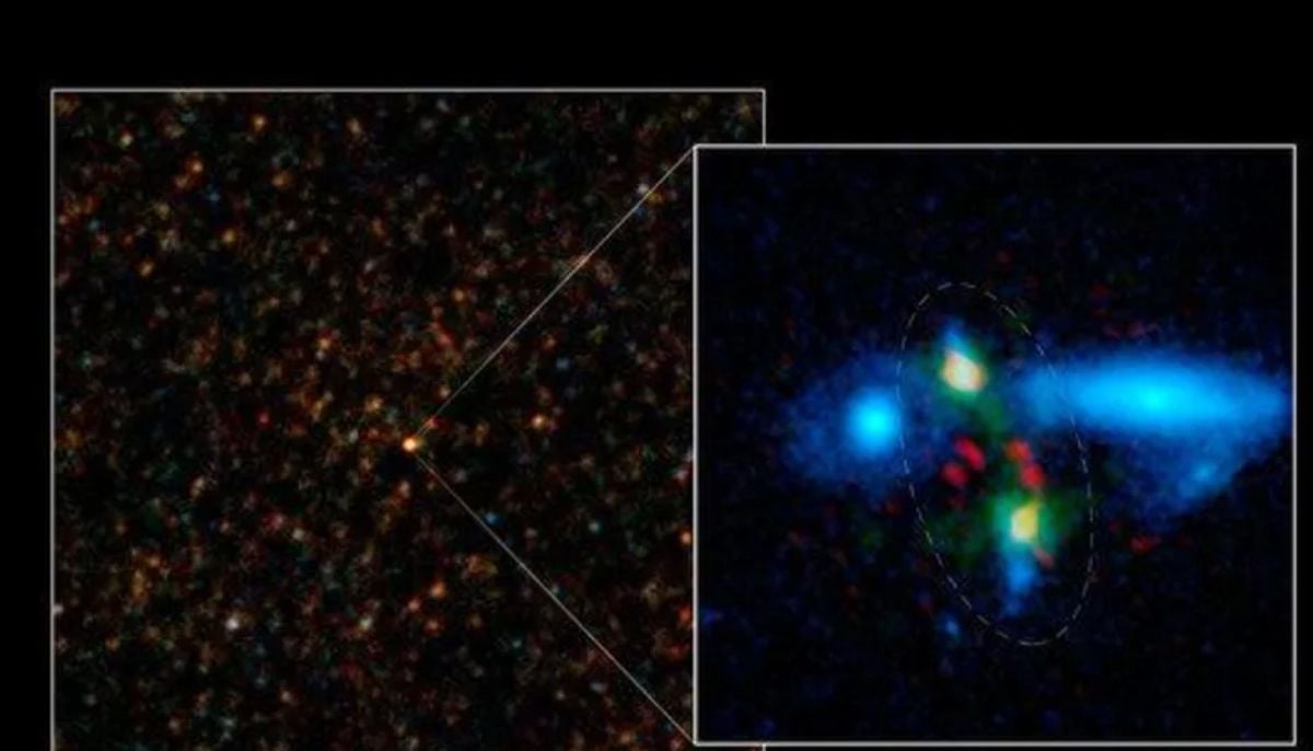
Rare star spotted in its original galaxy could answer a key question about the ingredients of life: Space photo of the week
Quick Facts
What it is: Star PicII-503 inside the Pictor II dwarf galaxy
Where it is: 150,000 light-years from Earth in the Pictor constellation
When it was shared: March 16, 2026
This gorgeous snap, taken by the Dark Energy Camera (DECam) mounted…
Continue Reading
-
‘This is really intolerable’: Astronomers protest giant orbiting mirror project and SpaceX’s million AI satellites
Astronomers are up in arms, protesting against a proposed constellation of tens of thousands of orbiting mirrors intended to reflect light onto ground-based solar power plants and SpaceX’s envisioned one million orbiting data centers.
The…
Continue Reading
-

Scientists discover hidden freshwater beneath the Great Salt Lake
The Great Salt Lake in Utah is the largest inland saltwater body in the Western Hemisphere. However, beneath its shimmering, salty surface, scientists have discovered something surprising. There is a large layer of freshwater that reaches depths…
Continue Reading