- Ancient Rocks Reveal Hidden Climate Motion During Snowball Earth SciTechDaily
- Earth had seasons during Ice Age, according to new study BBC
- Earth’s coldest ocean conditions quantified China Daily
- Snowball Earth wasn’t fully frozen: ice-free…
Category: 7. Science
-
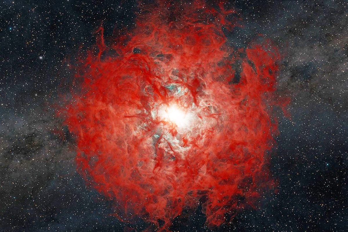
Ancient Rocks Reveal Hidden Climate Motion During Snowball Earth – SciTechDaily
-
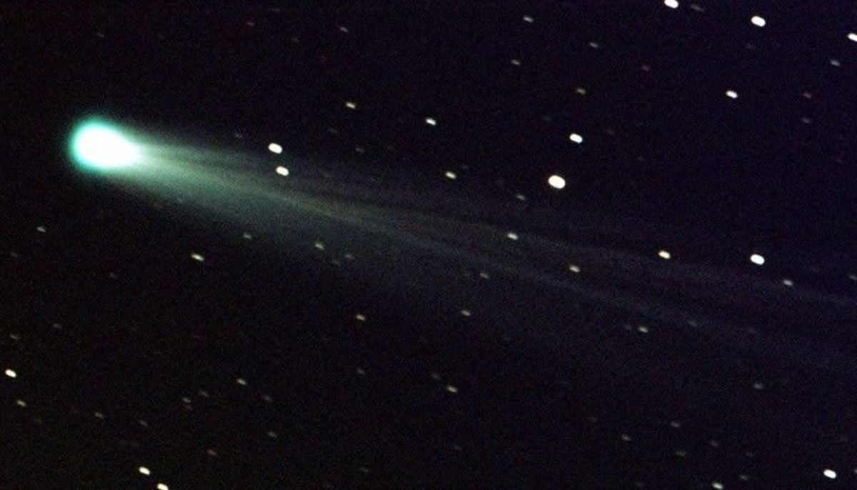
Bright green comet C/2024 E1 nears closest approach before leaving solar system
The bright green comet C/2024 E1 (Wierzchoś) displays increasing…
Continue Reading
-
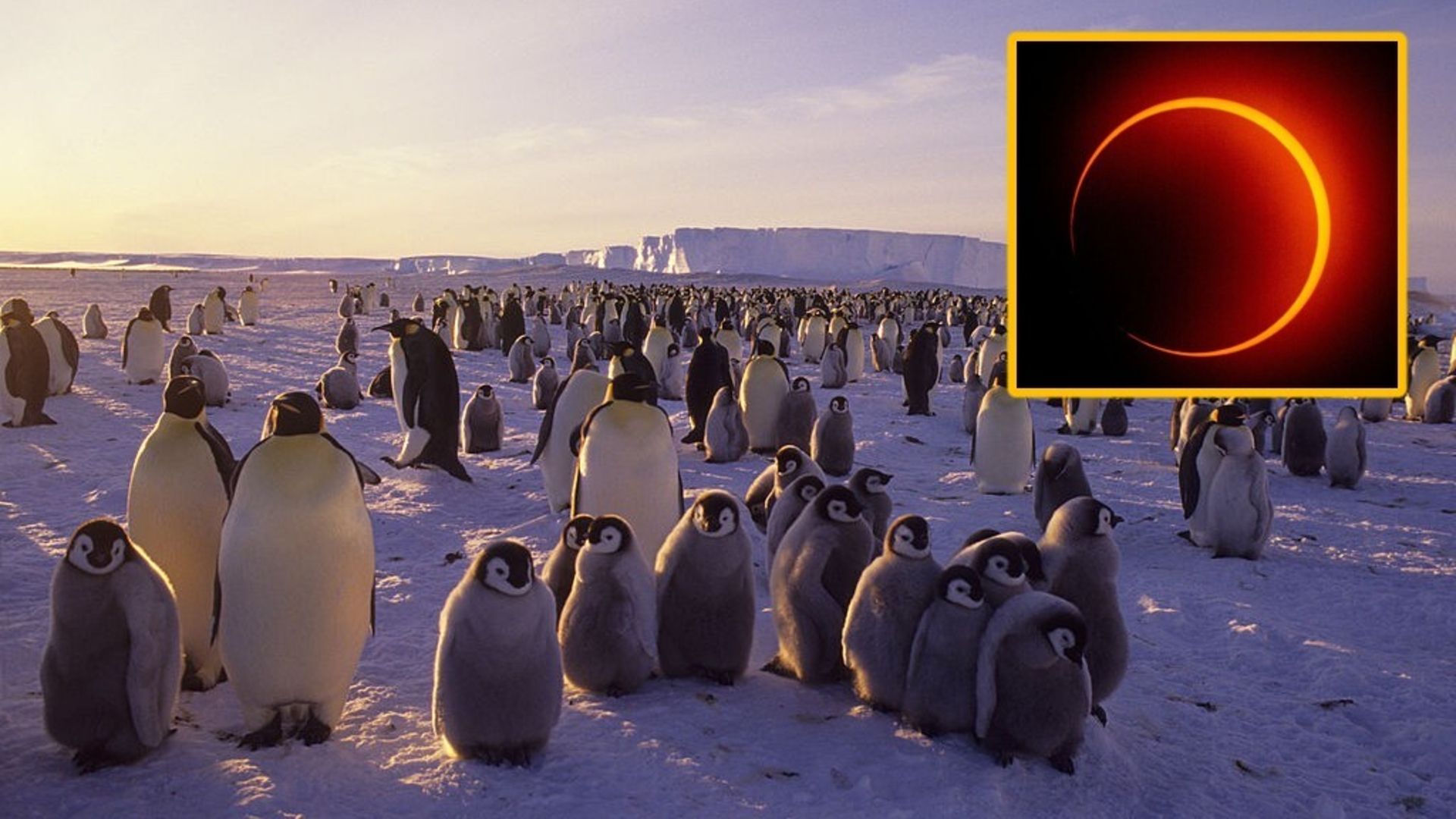
Lucky few to see ‘ring of fire’ solar eclipse over Antarctica on Feb. 17
While the new moon this Tuesday (Feb. 17) will pass without much fanfare in most of the world, something more exciting will be taking place over a sliver of Antarctica: a “ring of fire” solar eclipse, also known as an annular solar…
Continue Reading
-
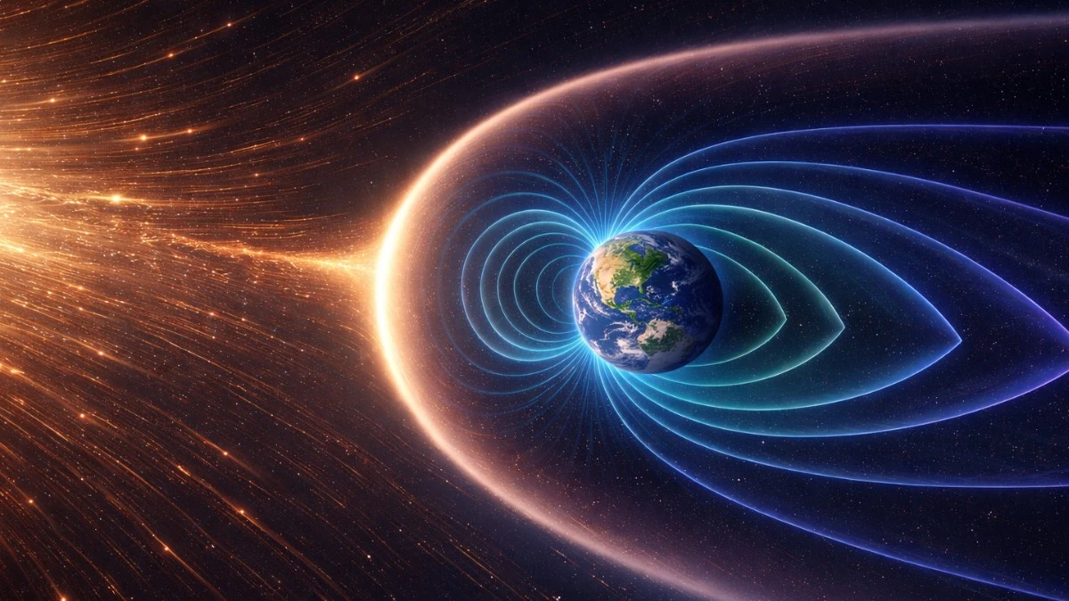
Long-term correlation found between Earth’s magnetic field strength and atmospheric oxygen
An analysis of geological proxy records shows that Earth’s atmospheric oxygen levels and geomagnetic field strength evolved in parallel over the past 540 million years. The study compares reconstructions of atmospheric oxygen concentration…
Continue Reading
-
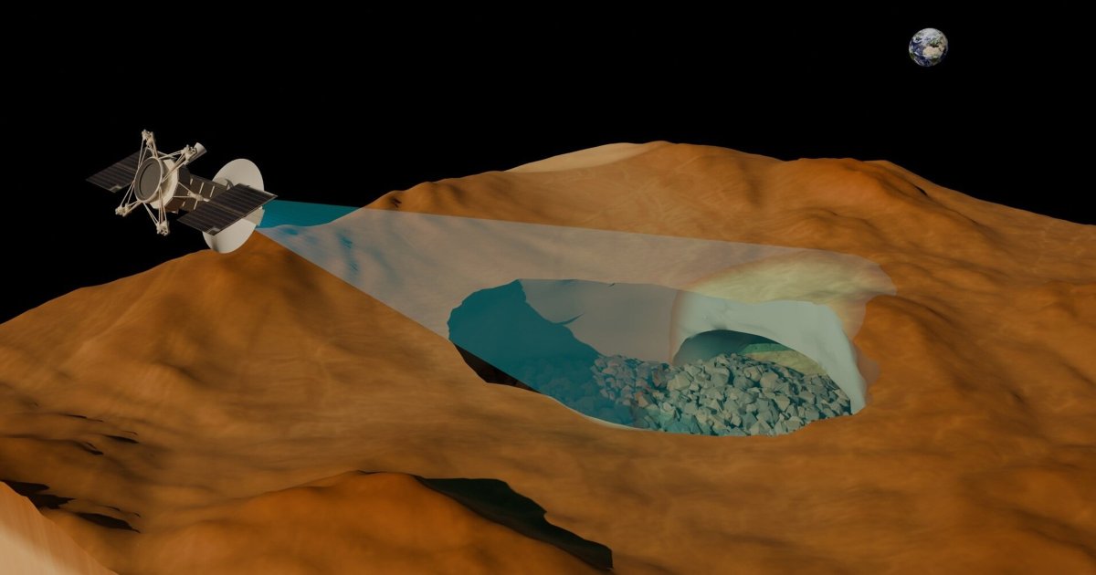
Scientists Spot Huge Cave on Venus
RSLab / University of Trento Astronomers say they’ve spotted a vast cave lurking beneath the surface of Venus — providing the strongest evidence yet that the planet is tunneled with lava tubes.
The…
Continue Reading
-
Scientists get clearest view ever of star collapsing into a black hole. And they very nearly missed it
Astronomers have captured their clearest view yet of a star collapsing and forming a black hole.
The event was discovered in archive data from 2014, captured by a now-defunct NASA asteroid-hunting space telescope.
Using the data, astronomers…
Continue Reading
-
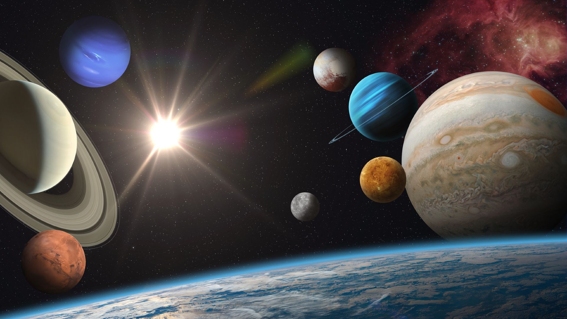
How long do most planets last?
Planets go through different life stages: They form, evolve and eventually meet an end. But the timelines for these processes differ widely between Earth-like planets and worlds that orbit less-powerful stars.
So, how long do most planets last?
Continue Reading
-
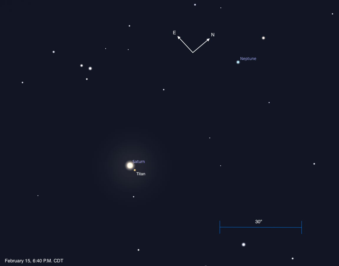
Saturn and Neptune stand close
Saturn and Neptune are less than a degree apart in a perfect north-south line this evening, visible with binoculars or any telescope.
This chart shows…
Continue Reading
-

NASA replaces evacuated astronauts with fast-track SpaceX launch
A new crew rocketed toward the International Space Station on Friday to replace the astronauts who returned to Earth early in NASA’s first medical evacuation.
SpaceX launched the replacements as soon as possible at NASA’s…
Continue Reading
-

Exotic white dwarf binary systems may produce mysterious radio pulses | Science News
Astronomers have been struggling to explain mysterious repeating bright radio pulses with unusually long periods. New research suggests that such flashes…
Continue Reading