Researchers have addressed a critical limitation in fault-tolerant quantum computing arising from hook errors within the rotated surface code. Gilad Kishony and Austin Fowler, both of Classiq Technologies, alongside their colleagues,…
Category: 7. Science
-
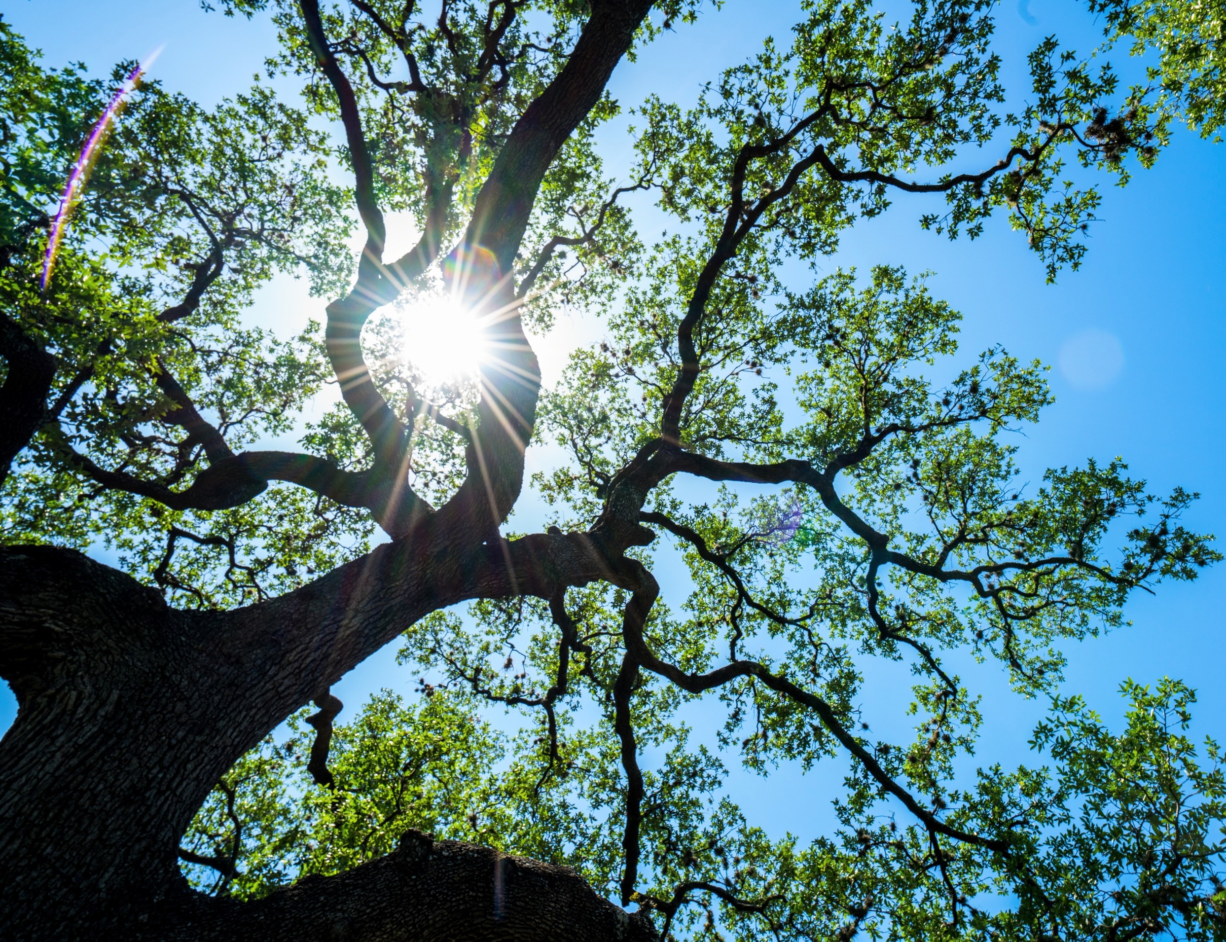
Oak trees rely on microbes to survive drought and disease
Oak trees can live for hundreds of years, yet climate change brings drought, poor soil, and disease at a much faster pace. Scientists now believe that tiny microbes living on and inside oak trees may help protect them from such stress.
A recent…
Continue Reading
-
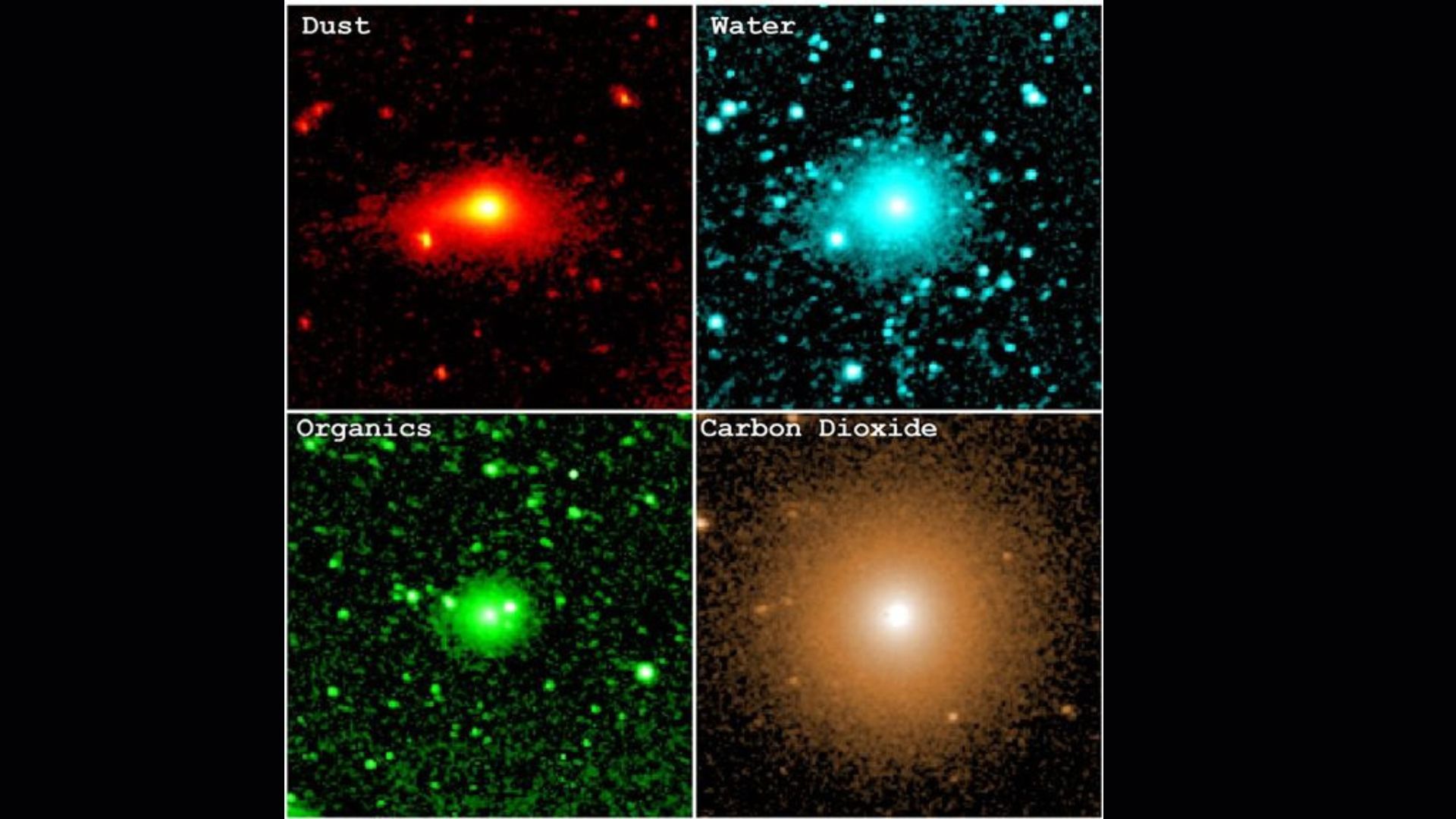
NASA telescope spots the building blocks for life spewing out of comet 3I/ATLAS
The interstellar comet 3I/ATLAS shed the building blocks of life as it flew past Earth last year, according to new data from NASA’s SPHEREx space telescope.
From its position in orbit, SPHEREx watched the rare interstellar visitor swing around…
Continue Reading
-
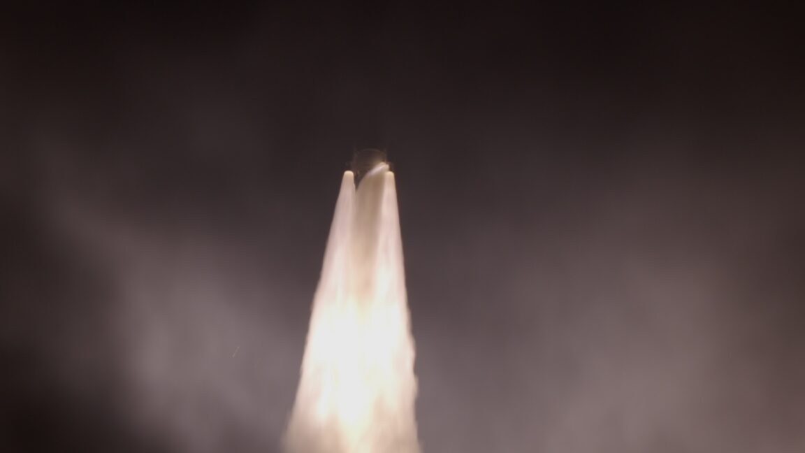
ULA’s Vulcan rocket suffers another booster problem on the way to orbit
Moments after liftoff from Florida’s Space Coast early Thursday morning, a shower of sparks emerged in the exhaust plume of United Launch Alliance’s Vulcan rocket. Seconds later, the…
Continue Reading
-
Gravitational lensing technique unveils supermassive black hole pairs – Phys.org
- Gravitational lensing technique unveils supermassive black hole pairs Phys.org
- Astronomers move closer to pinpointing where giant black holes merge Yahoo
- New method could reveal hidden supermassive black hole binaries Nanowerk
- Detection system…
Continue Reading
-
Strange 'inside-out' planetary system baffles astronomers – France 24
- Strange ‘inside-out’ planetary system baffles astronomers France 24
- Weird inside-out planet system may have formed one world at a time New Scientist
- Inside out planetary system upends notions of formation EarthSky
- Faraway solar system challenges…
Continue Reading
-
Mini-Tanking Test On Tap Following Artemis II SLS Leak Repair
Mini-Tanking Test On Tap Following Artemis II SLS Leak Repair | Aviation Week Network