Researchers at the University of Southampton have found new evidence that Earth’s climate did not completely grind to a halt during its most extreme ice age, a time often called Snowball Earth.
This dramatic chapter unfolded during the Cryogenian…

Researchers at the University of Southampton have found new evidence that Earth’s climate did not completely grind to a halt during its most extreme ice age, a time often called Snowball Earth.
This dramatic chapter unfolded during the Cryogenian…

Micelle-forming polymers such as poloxamer 407 (P407) are promising drug nanocarriers, yet their sol–gel transition under physiological conditions remains poorly understood. In a recent study, researchers from Japan experimentally analyzed…
Tao, S. Exploration and development of unconventional oil and gas resources: latest advances and prospects. Energies 18, 3933. https://doi.org/10.3390/en18153933 (2025).
Zhang, N. et al….
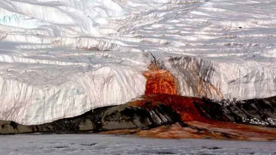
(Web Desk) – One of the most mysterious natural phenomena on Earth, known as “Blood Falls,” continues to intrigue scientists in Antarctica’s McMurdo Dry Valleys, one of the coldest and driest places on the…

How’s your golf swing? Before you tee off, check your form – because poor mechanics can hurt you more than that hook shot into the trees.
“The golf swing is a complex total-body movement,” says Martin Boehm, a physical therapist at the…
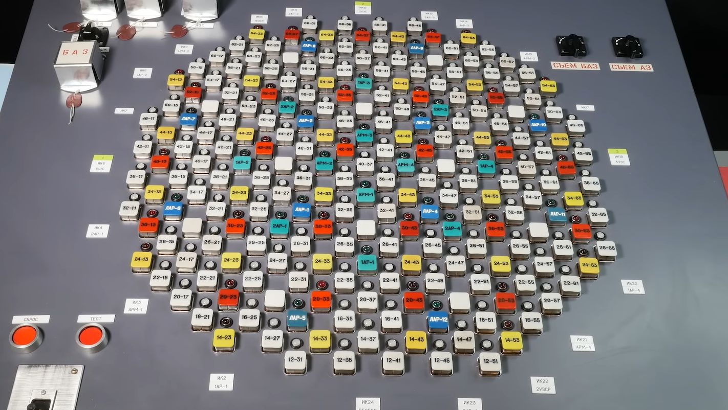
Control panels of a pre-digitalization nuclear plant look quite daunting, with countless dials, buttons and switches that all make perfect sense to a trained operator, but seem as random…
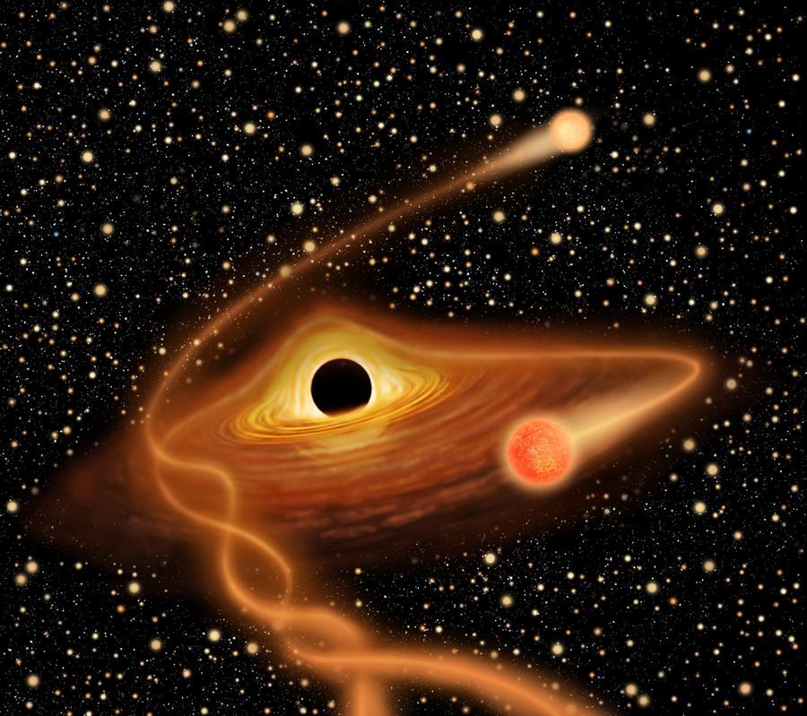
BEIJING, Feb. 12 (Xinhua) — China’s Tianguan satellite — also named the Einstein Probe — has likely captured an intermediate-mass black hole tearing apart and devouring a white dwarf star, marking the first time such an extreme event has…
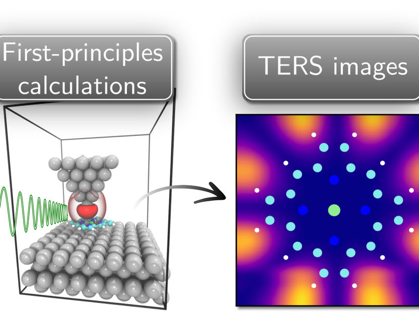
Probing the vibration of atoms provides detailed information on local structure and bonding that define material properties. Tip-enhanced Raman spectroscopy (TERS) offers extremely high resolution to probe such vibrations….

Abstract
Near-infrared spectroscopy has become a critical enabler of modern biopharmaceutical analysis through its non-destructive, rapid, and information-rich measurement capabilities. In 2025, a series of influential publications has…