<(From Left) Dr. Sukkyung Kang, Professor Sanha Kim from Department of Mechanical Engineering>
The performance and stability of smartphones and artificial intelligence (AI) services depend on how uniformly and precisely semiconductor…

<(From Left) Dr. Sukkyung Kang, Professor Sanha Kim from Department of Mechanical Engineering>
The performance and stability of smartphones and artificial intelligence (AI) services depend on how uniformly and precisely semiconductor…
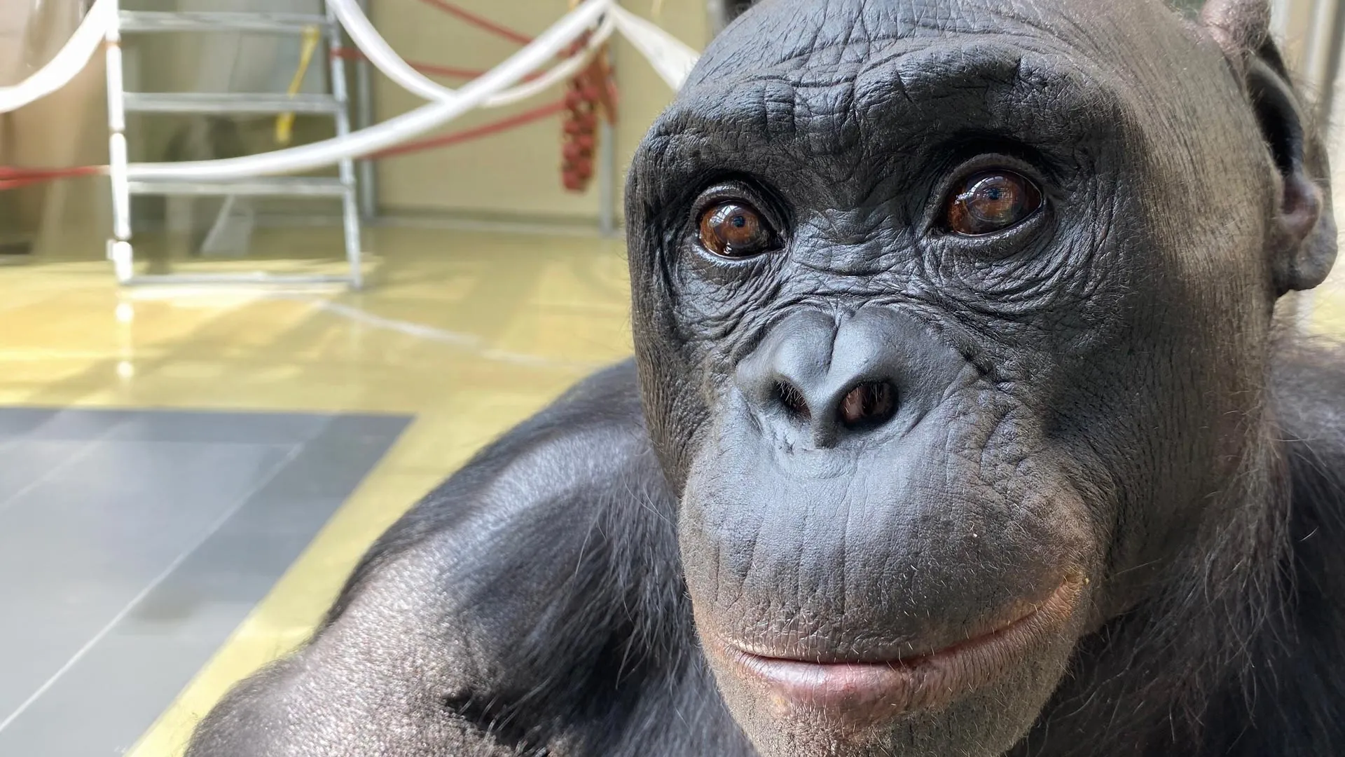
In a set of playful experiments modeled after children’s tea parties, researchers at Johns Hopkins University have shown for the first time that apes can use imagination and take part in pretend play. This ability was long believed to belong only…

Known as the “Queen of Climbers,” the genus Clematis boasts over 300 species widely distributed across the globe. From tropical rainforests to sub-arctic regions, these plants are celebrated by gardeners for their vibrant flowers and…
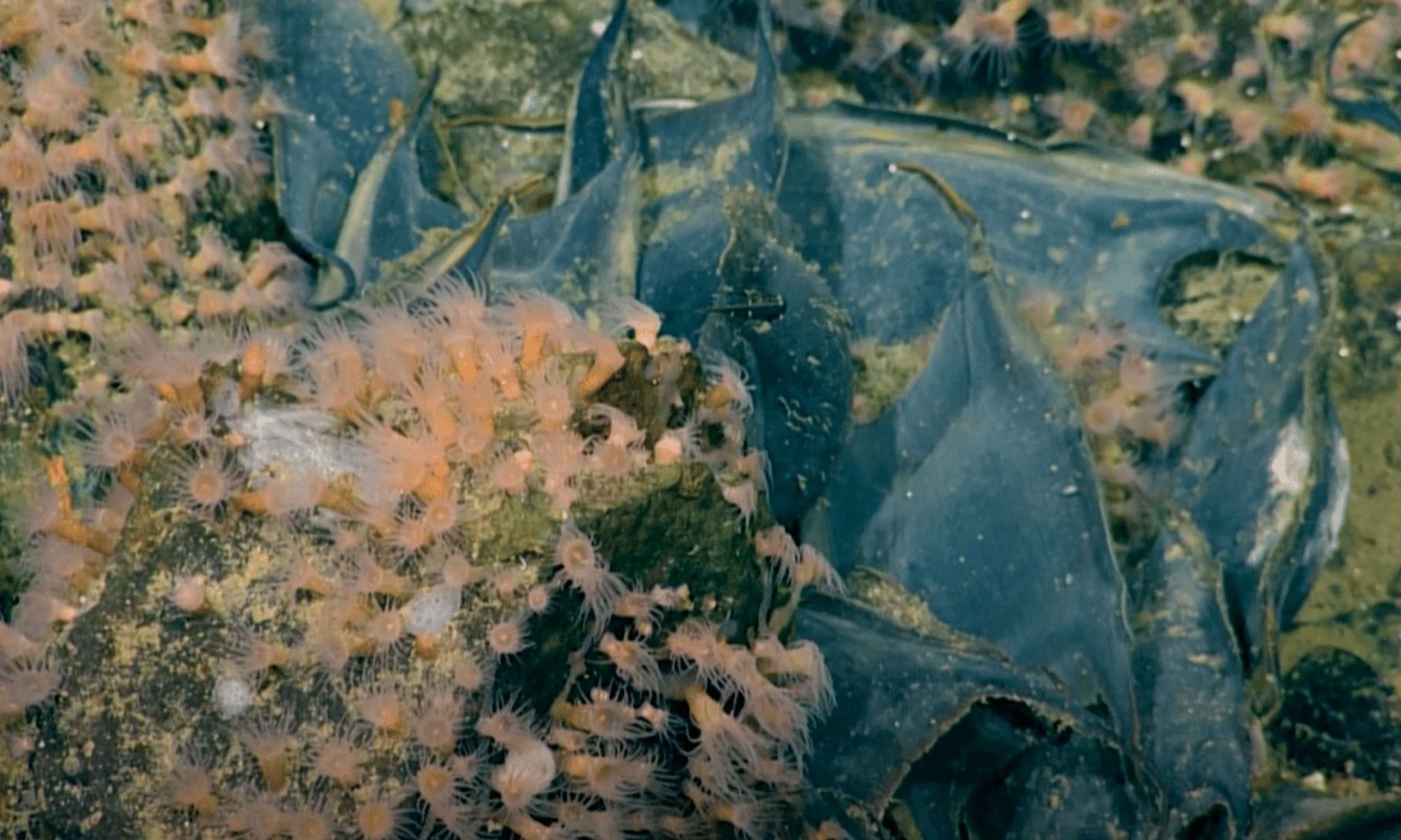
More than one million giant, leathery skate egg capsules from a deep-sea ray-like fish have been documented incubating at a single underwater volcanic site off Canada’s Pacific coast.
The concentration links volcanic heat to reproduction at a…
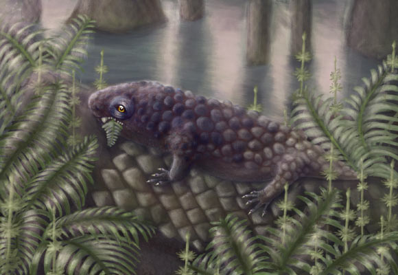
Tyrannoroter heberti, a new species of pantylid ‘microsaur’ from the Carboniferous period, shows that some of Earth’s earliest land vertebrates had already evolved complex teeth for grinding plants, suggesting terrestrial herbivory…

The deadline of the ADEX Voice of the Ocean Photo+Video+Art Competition 2026 is just a month away! You’ve only got till March 10th (11:59PM PST) to show us your amazing art—images, short films and artworks (analog or digital)!
This is…