…
Category: 7. Science
-
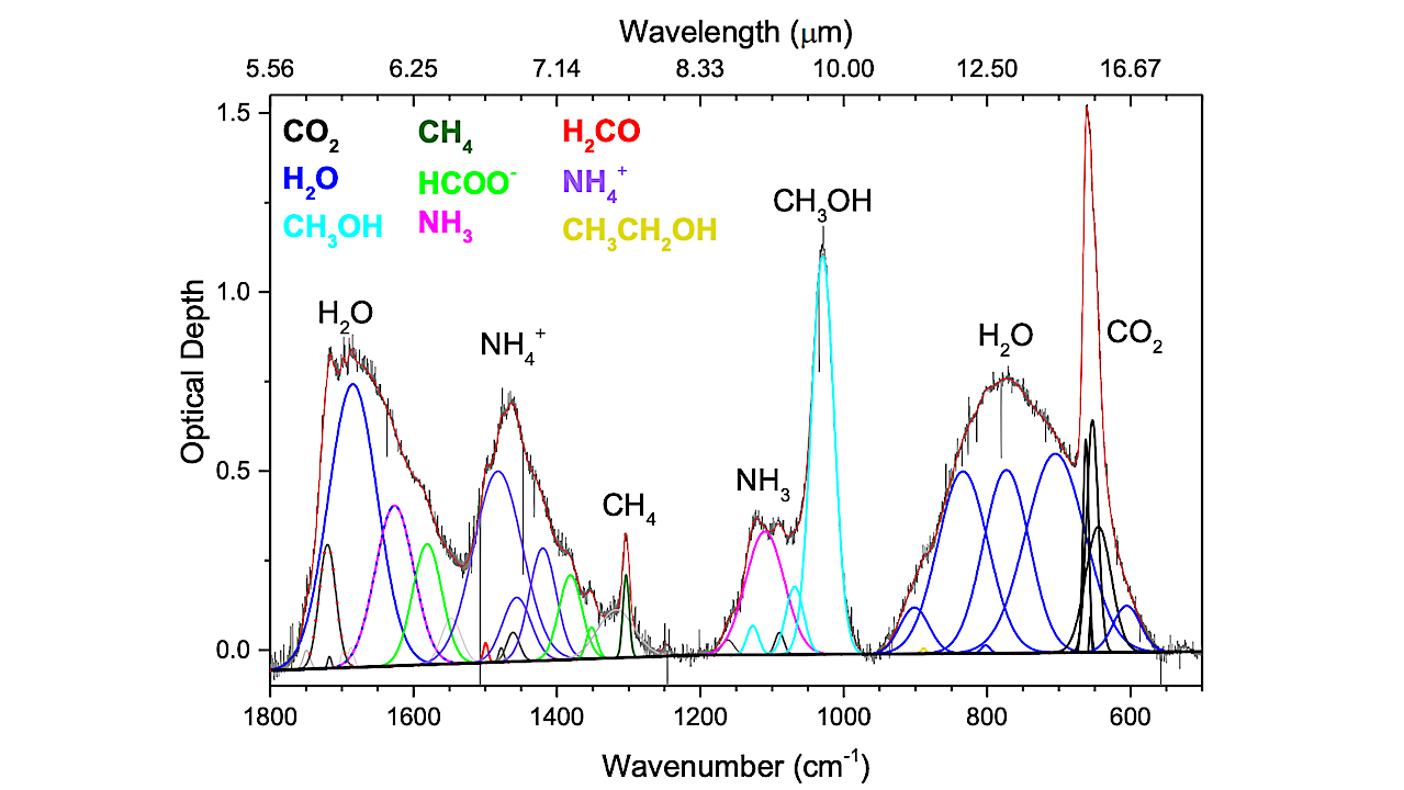
Mars Curiosity Seeks Evidence Of Organics – astrobiology.com
- Mars Curiosity Seeks Evidence Of Organics astrobiology.com
- Curiosity Blog, Sols 4788-4797: Welcome Back from Conjunction NASA Science (.gov)
- Mars rover conducts test for life-related material on the Red Planet Mashable
- Curiosity Rover Reconnects…
Continue Reading
-
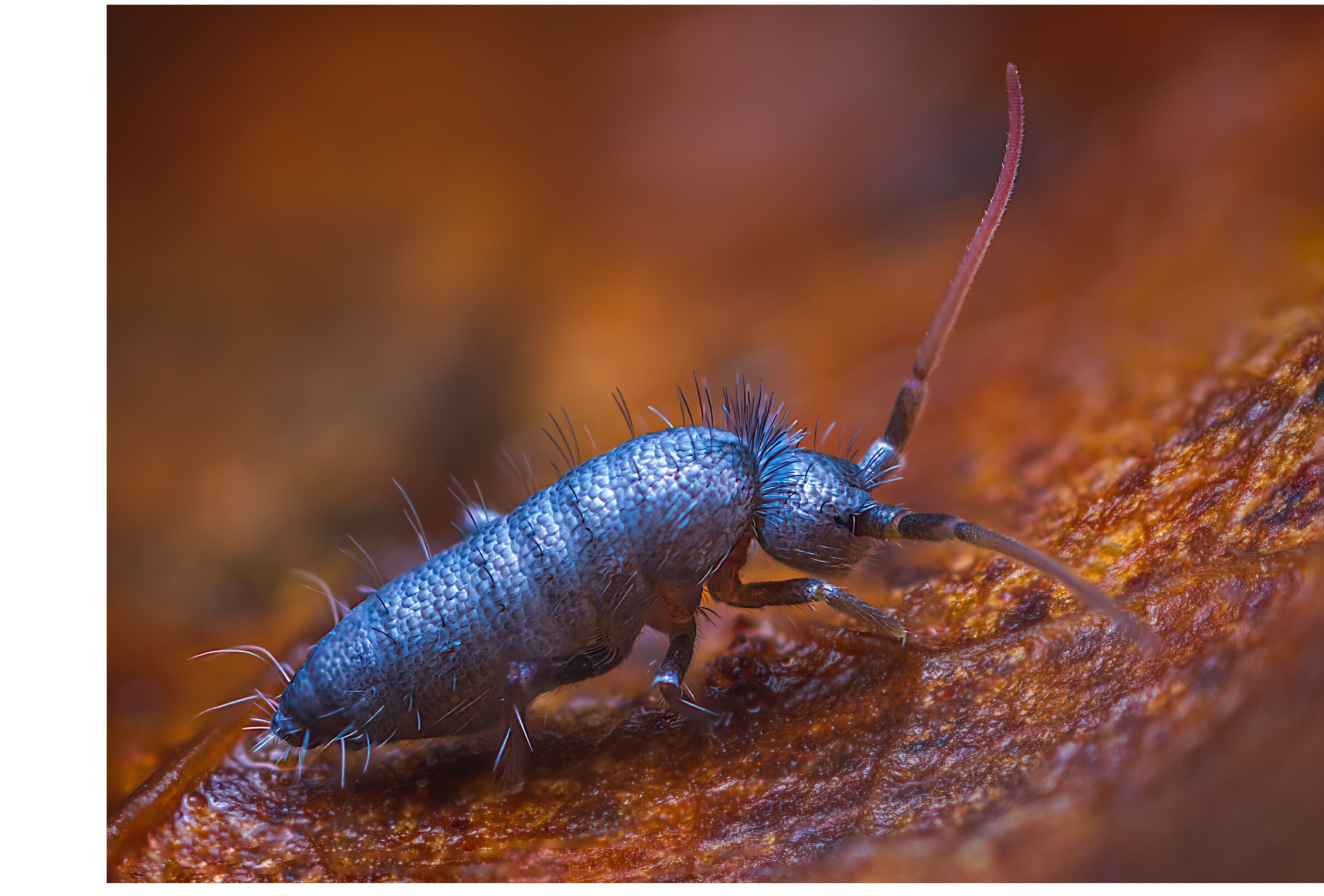
New genus of insect-like springtails discovered in China- Earth.com
Scientists have documented four previously unknown species of tiny, insect-like springtails in China. Each one is smaller than a grain of rice and lives almost entirely out of sight.
Known as springtails, these animals inhabit the thin layer of…
Continue Reading
-

Scientists finally solve a 100-year-old mystery in the air we breathe
Researchers at the University of Warwick have developed a new method that makes it possible to predict how irregularly shaped nanoparticles move through the air. These particles are a major category of air pollution and have long been difficult…
Continue Reading
-
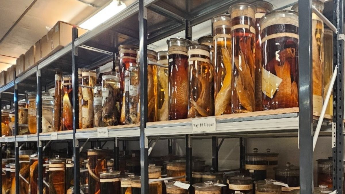
Scientists Fired Lasers at Charles Darwin’s Priceless Specimens. Here’s Why. : ScienceAlert
Rows of preserved specimen jars from Charles Darwin’s iconic Galapagos voyage have sat, unopened, in the archives of London’s Natural History Museum (NHM) for 200 years. Now, lasers have given us an unprecedented look inside.
Darwin himself is…
Continue Reading
-
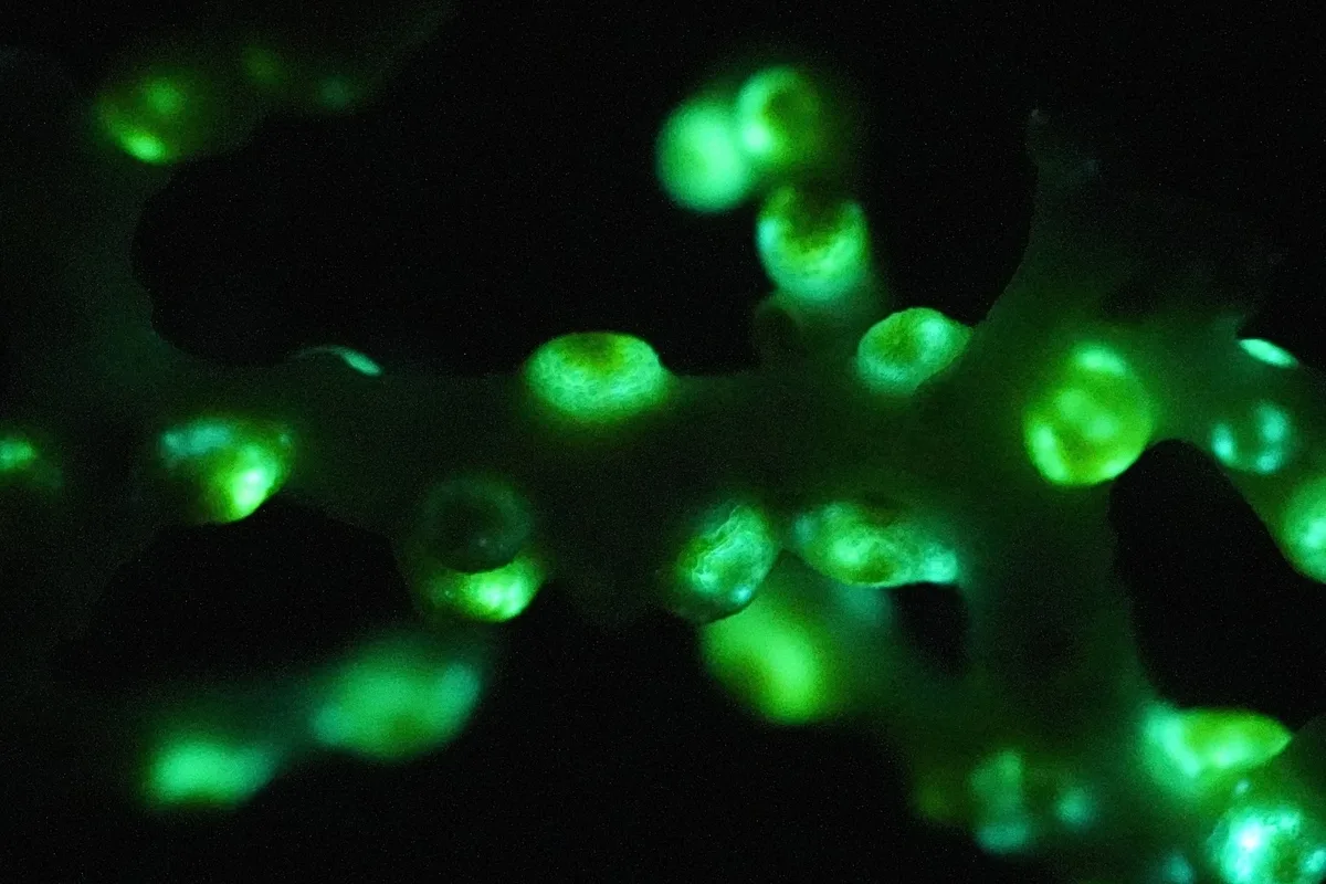
Scientists discover a new deep sea creature that glows in the dark
Scientists have confirmed a bright yellow deep-sea animal, Corallizoanthus aureus, as a new species of marine coral. It emits green light when disturbed, marking the first known case of bioluminescence documented inside a deep-sea cave.
The…
Continue Reading
-
AI Neanderthal Images Still Lag Behind Archaeology
Getting your audio player ready…
It is impressive how Generative AI can conjure a “day in the life” image of a Neanderthal in seconds. But a new study suggests those scenes often come with a dash of built-in time travel. Researchers…
Continue Reading