
Researchers are increasingly investigating alternatives to black holes to resolve the singularity problem at the heart of gravitational collapse. Shounak Ghosh from the Directorate of Legal Metrology, Rikpratik Sengupta from the Indian…
Category: 7. Science
-
First solar eclipse of 2026 set for February 17
The first solar eclipse of 2026 will take place on February 17, when the Moon will partially cover the Sun and create a Ring of Fire, a striking celestial event also known as an annular solar eclipse.
Astronomers explain that this phenomenon…
Continue Reading
-
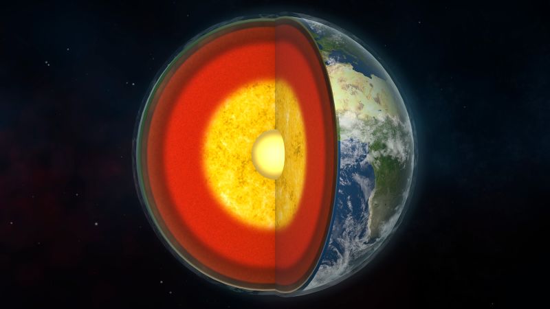
Earth’s core may be the planet’s biggest source of hydrogen, scientists estimate
Picture all of Earth’s oceans, which cover about 70% of the planet and are mostly made of hydrogen. Now multiply that by nine. That may be the amount of hydrogen in Earth’s core, possibly making it the…
Continue Reading
-

NASA, SpaceX delay Crew-12 launch to Friday amid weather concerns
Feb. 10 (UPI) — NASA and SpaceX on Tuesday moved their launch from Florida’s Cape Canaveral Space Station to the International Space Station from Thursday to no earlier than Friday due to weather conditions.
NASA announced Tuesday that the
Continue Reading
-
Chang'e-6 samples constrain lunar impact flux and illuminate early impact history – Phys.org
- Chang’e-6 samples constrain lunar impact flux and illuminate early impact history Phys.org
- Samples from Chang’e-6 on the far side of the Moon reveal traces of a giant impact Universe Space Tech
- Samples from the far side of the Moon shed light…
Continue Reading
-
Canadian fossil reveals one of the first plant-eating animals – Reuters
- Canadian fossil reveals one of the first plant-eating animals Reuters
- Football-sized fossil creature may have been one of the first land animals to eat its veggies Field Museum
- Newly Discovered Fossil Among The Earliest Land Creatures to Enjoy a…
Continue Reading
-
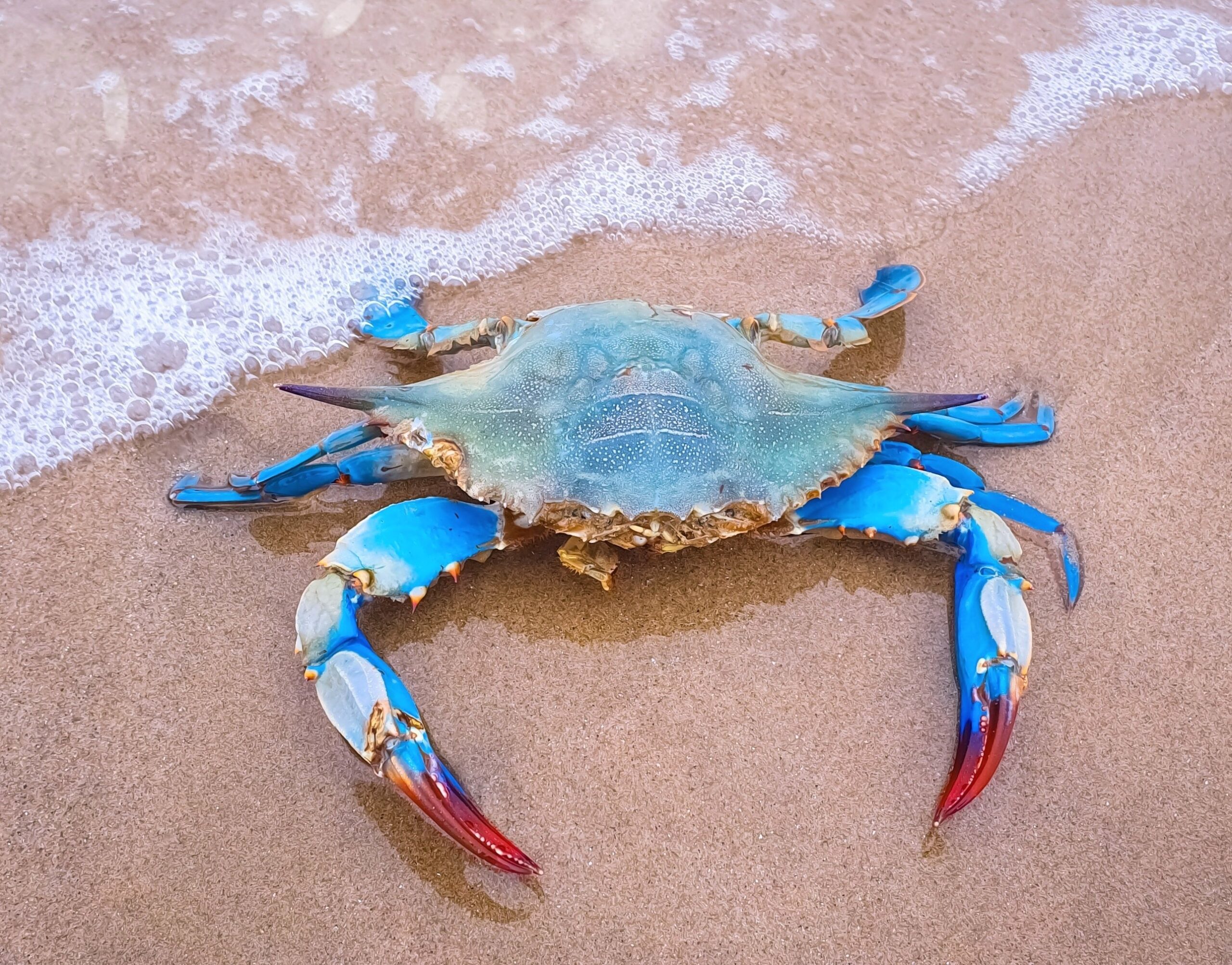
Oysters help protect blue crabs from a deadly parasite
Oysters have a solid reputation. They sit quietly on the bottom, pulling algae and excess nutrients out of the water, helping keep coastal bays from turning into green soup.
For years, that filtering job has been the main reason people care about…
Continue Reading
-
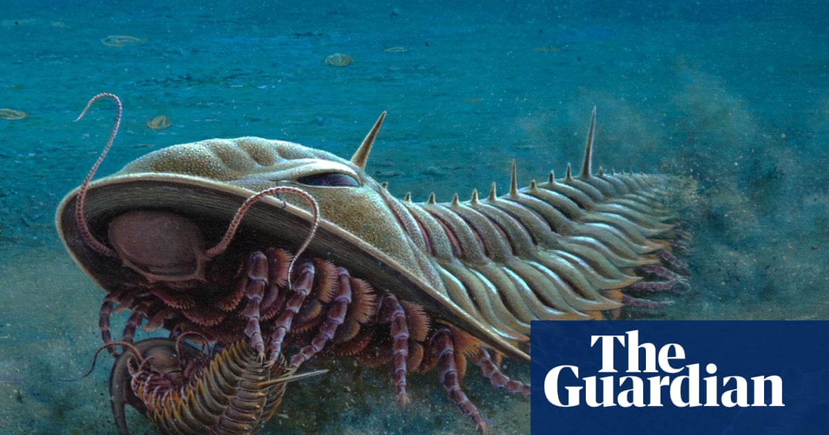
Drawings of Kangaroo Island fossils provide inspiration for stamp series | South Australia
Prehistoric fossils from Kangaroo Island have been transformed into vibrant creatures of the deep for a series of artworks, which in turn have been made into a series of stamps.
Dr Peter Trusler has used the latest research on the 512m-year-old…
Continue Reading
-

Methane spiked after 2020 and the cause was unexpected
Methane concentrations in the atmosphere climbed at an unprecedented pace in the early 2020s due to a combination of weaker natural removal and rising emissions from warming wetlands, rivers, lakes, and agricultural areas. An international group…
Continue Reading
-
MIT Scientists Shrink Terahertz Light To Reveal Hidden Quantum “Jiggles” – SciTechDaily
- MIT Scientists Shrink Terahertz Light To Reveal Hidden Quantum “Jiggles” SciTechDaily
- Terahertz microscope reveals the motion of superconducting electrons MIT Physics
- Imaging a terahertz superfluid plasmon in a two-dimensional…
Continue Reading