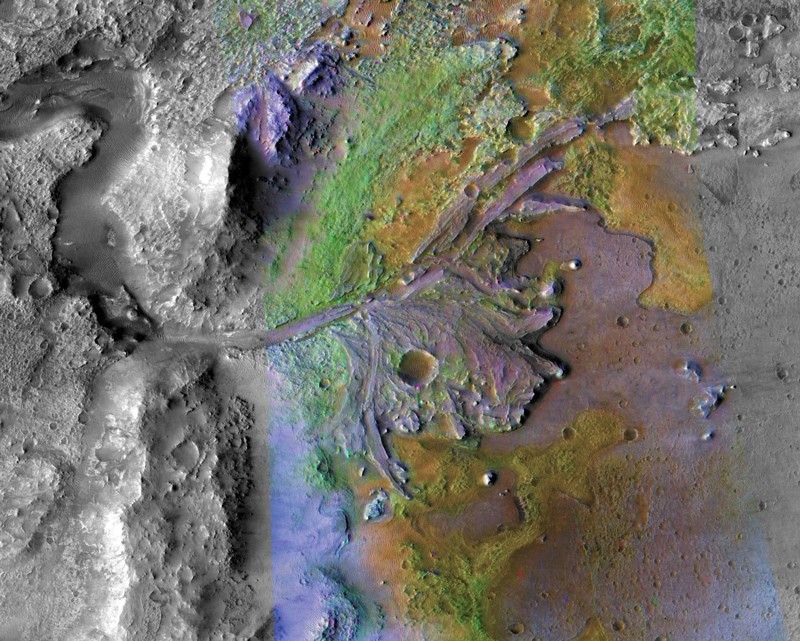
When NASA’s Perseverance rover landed in Jezero Crater in 2021, its primary mission was to scour the remnants of a dried-up Martian lakebed for signs of ancient life. Scientists have…
Category: 7. Science
-

NASA Brings Jared Isaacman, Super Guppy, and T-38 Talon to SUN ’n FUN 2026
In January, NASA Administrator Jared Isaacman issued a new workforce directive underscoring the enduring importance of aviation heritage and public engagement within the agency, outlining expanded recognition programs, aviation-focused…
Continue Reading
-
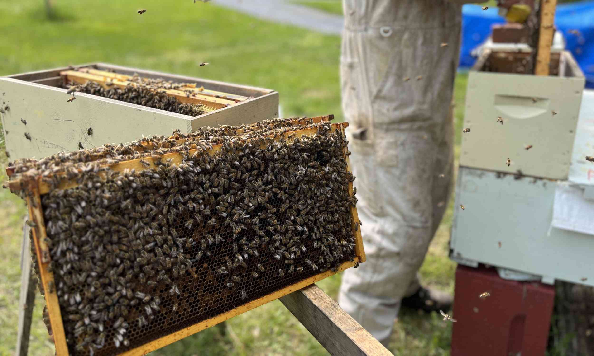
How hygienic bees fight disease could help protect bee colonies
Researchers have found that honey bees from colonies that actively remove sick and dying young, called hygienic colonies, survive infection by the parasite Nosema ceranae at higher rates than other bees.
The finding shows that a trait known for…
Continue Reading
-

Optics / Photonics Information | AZoOptics.com
Terms
While we only use edited and approved content for Azthena
answers, it may on occasions provide incorrect responses.
Please confirm any data provided with the related suppliers or
…Continue Reading
-

100km-wide “hidden giant” discovered beneath Antarctic ice sheet
For decades, pink granite boulders scattered across the volcanic peaks of the Hudson Mountains in West Antarctica have puzzled scientists. However, a study published in October 2025 may have finally solved the mystery surrounding their origin,…
Continue Reading
-

Neanderthals May Have Used the World’s First Antibiotic 50,000 Years Ago
Creating birch tar using Neanderthal methods. Image credits: University of Cologne. If you were a Neanderthal hunter 50,000 years ago, even a small cut could be deadly. Without sterile bandages or antibiotics, a wound was an open…
Continue Reading
-
ALICE sees new sign of primordial plasma in proton collisions – Phys.org
- ALICE sees new sign of primordial plasma in proton collisions Phys.org
- Observation of partonic flow in proton—proton and proton—nucleus collisions Nature
- Physicists Reveal the Big Bang’s “Primordial Soup” Really Flowed Like a Liquid
Continue Reading
-

Human-driven climate change is slowing Earth’s rotation at a rate not seen in 3.6 million years
Human-driven climate change is slowing Earth’s rotation at a rate not seen in 3.6 million years, with sea level rise increasing the length of days by 1.33 milliseconds per century, according to a new study.
Earth spins faster when its mass is…
Continue Reading
-

Humans in The Andes Appear to Have Evolved a Strange Genetic Ability : ScienceAlert
For thousands of years, humans living high in the Argentinian Andes have relied on drinking water that would make most people deathly ill.
There, naturally occurring arsenic in volcanic bedrock leaches into the groundwater, contaminating the…
Continue Reading