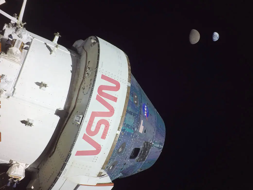
Even most rocket scientists would rather avoid hard math when they don’t have to do it. So when it comes to figuring out orbits in complex three-body systems, like those in Cis-lunar space, which is between the Earth and the Moon,…
Category: 7. Science
-
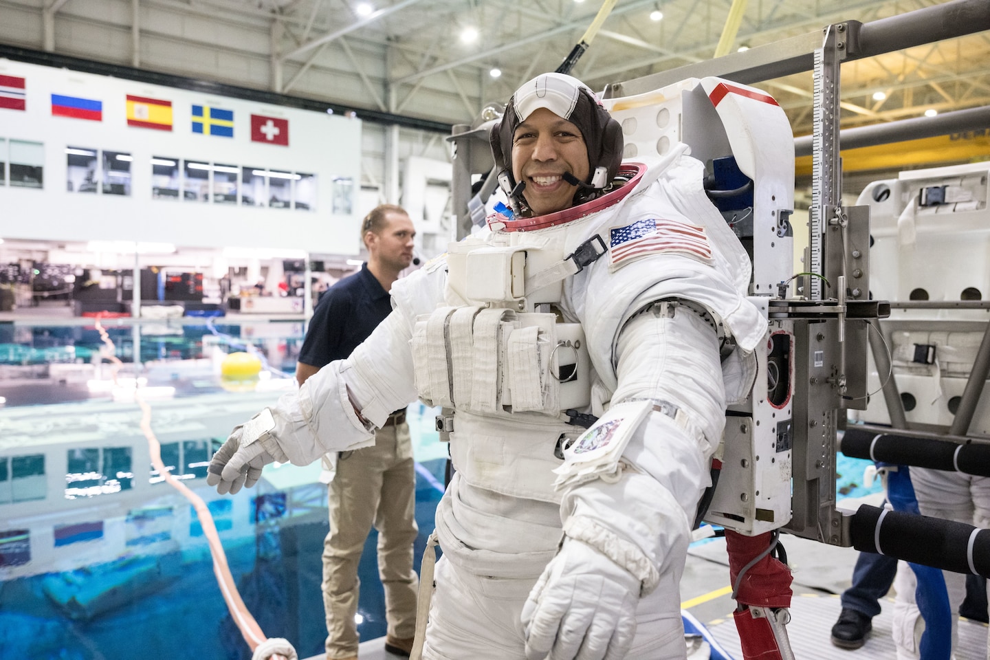
Maryland raised astronaut talks space and fulfilling his childhood dream
While most Americans spent Thanksgiving Day gathered around a table with family and friends, feet firmly planted on the ground, one Maryland-raised man spent the day journeying to the stars.
Chris Williams, who grew up in Potomac, Maryland, has…
Continue Reading
-
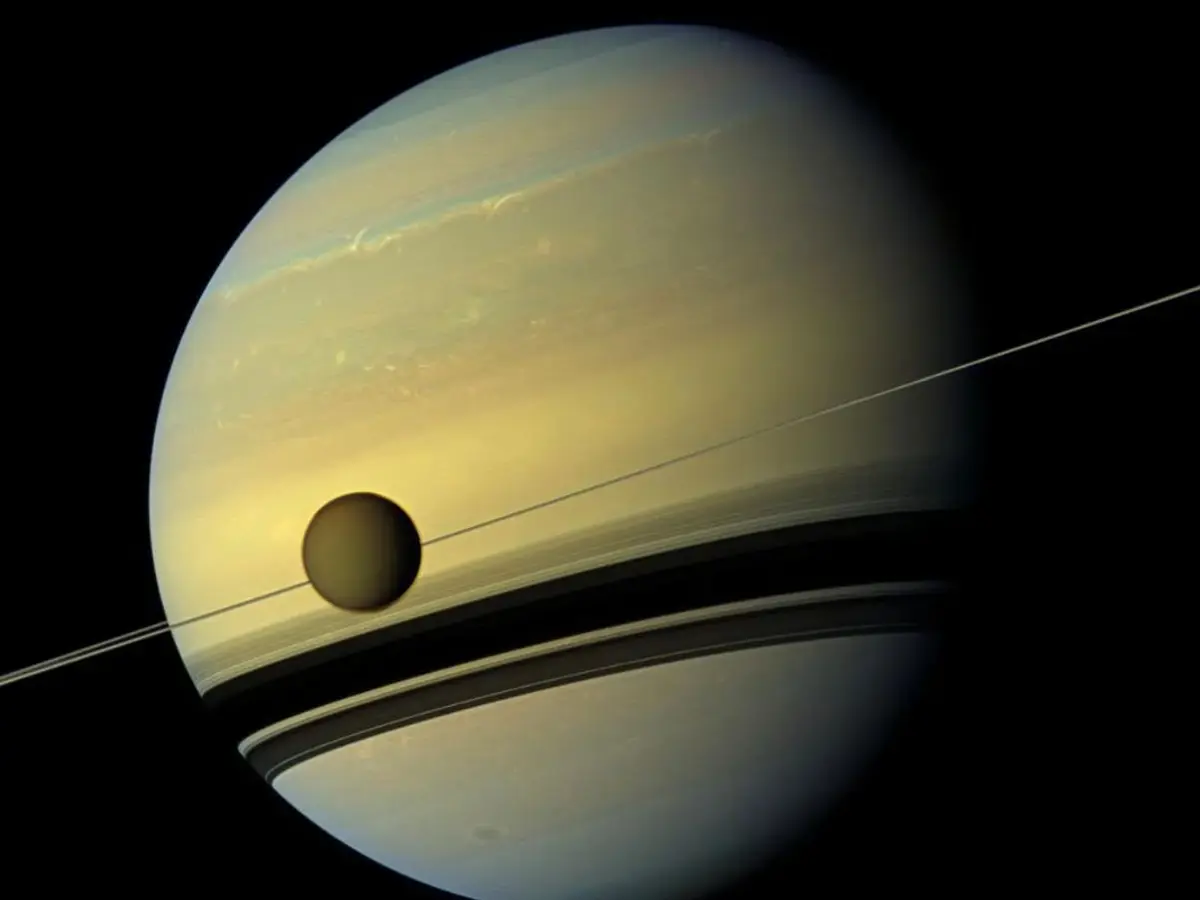
NASA study reveals surprising details about Titan
In the latest study, researchers applied advanced processing techniques to Cassini’s archival data, reducing noise and revealing a strong energy dissipation signature deep within Titan. The research team interpreted this signature as coming…
Continue Reading
-

Deer are secretly lighting up the forest with glowing urine to mate
White-tailed deer use glowing signposts to communicate during mating season, a new study has revealed.
The University of Georgia, US, found that white-tailed deer communication involves a “hidden” visual component: UV-induced…
Continue Reading
-
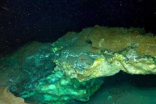
Deepest gas hydrate cold seep ever discovered in the Arctic – Alaska Native News
ROV image of a partially collapsed gas hydrate mound in the Molloy Deep (Freya mounds). The mound hosts frenulate worms and crustaceans.
Photo: UiT / Ocean Census / REV OceanA multinational scientific team led by UiT has uncovered the deepest…
Continue Reading
-
NGC 646: Euclid Mission Captures a Dynamic Galaxy Garland – DIYPhotography
- NGC 646: Euclid Mission Captures a Dynamic Galaxy Garland DIYPhotography
- Euclid’s galaxy garland European Space Agency
- Dark Matter Telescope Captures a Sparkling Galaxy Merger Scientific American
- How the ‘Dark Universe’ telescope Euclid scans…
Continue Reading
-
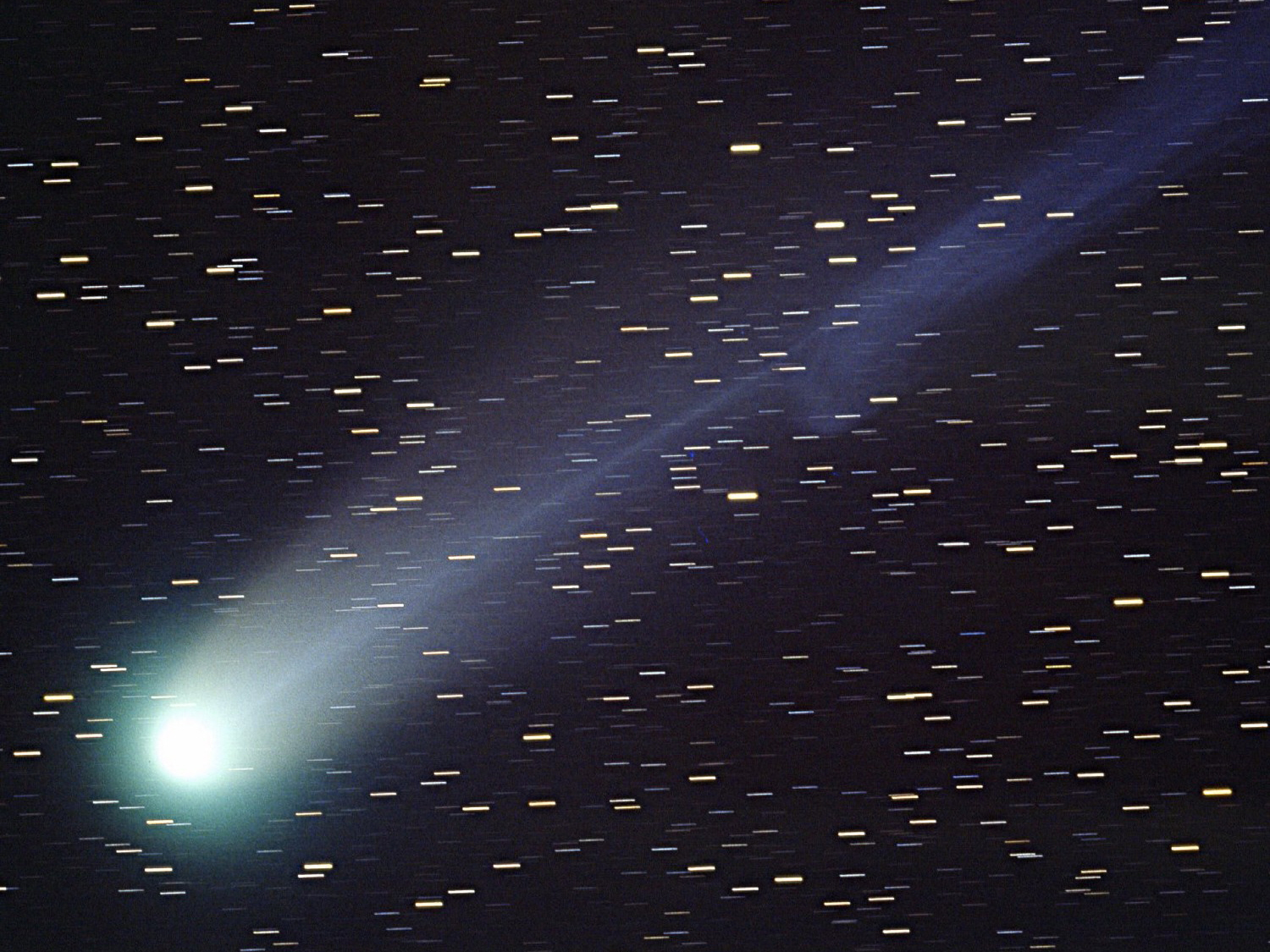
Comet C/1955 Y1 is discovered
Today in the history of astronomy, Yuji Hyakutake finds a comet – but not his most famous one.
Comet Hyakutake, seen here in March 1996, was…
Continue Reading
-
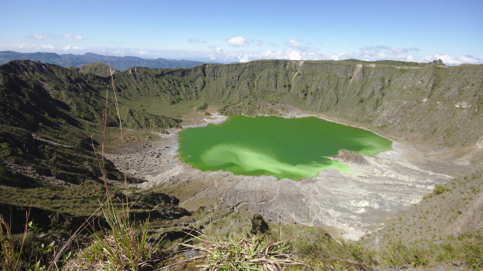
The world’s ‘hidden’ volcanoes pose the greatest risk for global crisis
The next global volcanic disaster is more likely to come from volcanoes that appear dormant and are barely monitored than from the likes of famous volcanoes such as Etna in Sicily or Yellowstone in the US.
Often overlooked, these “hidden”
Continue Reading
-
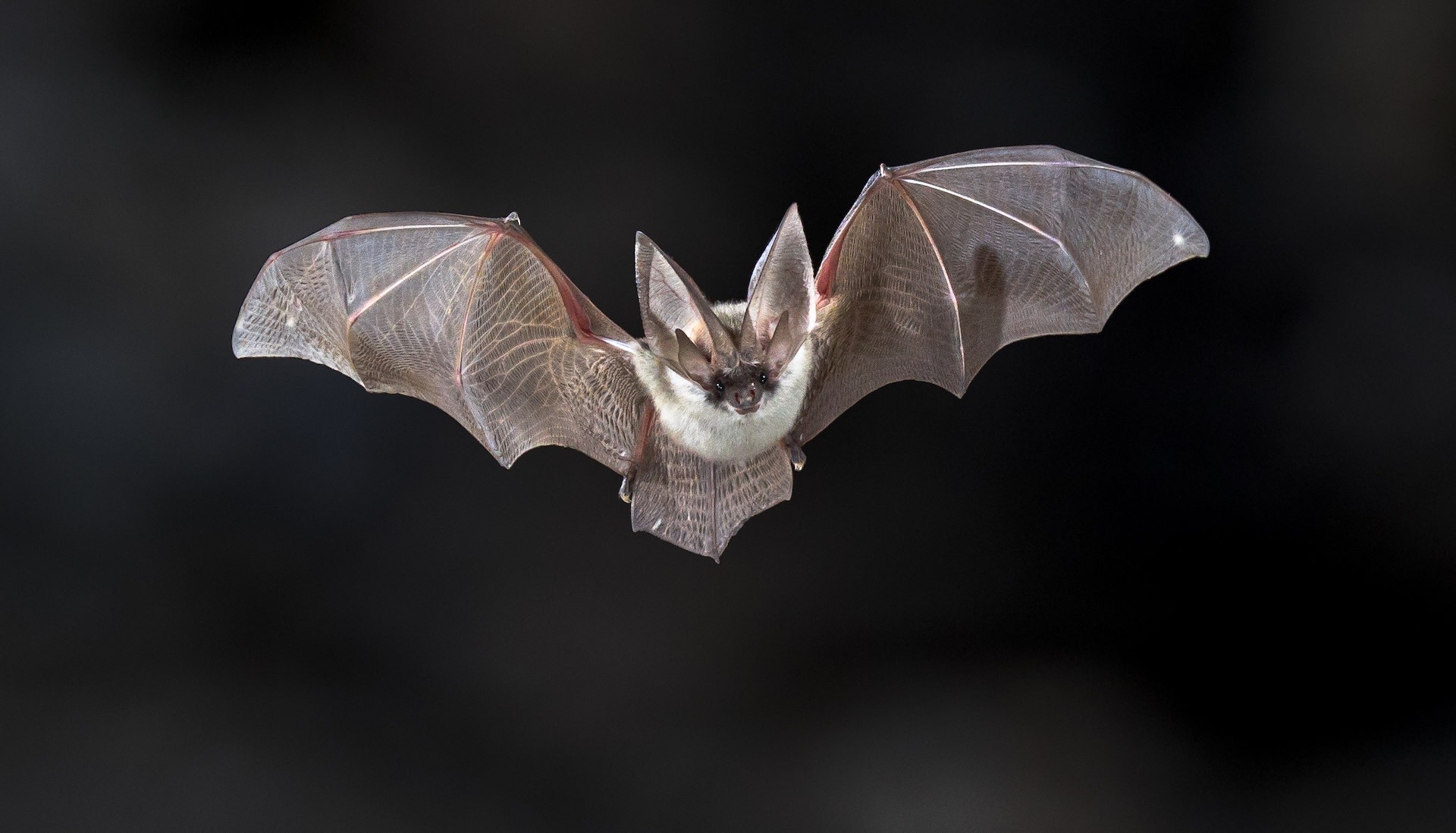
Scientists stunned after discovering unexpected ‘mutation’ in common US creatures: ‘Somehow gets perpetuated ‘
Researchers at the University of Georgia have found that six North American bat species can glow under ultraviolet light.
The findings were published in the journal Ecology and Evolution. Although scientists do not yet understand what…
Continue Reading
-
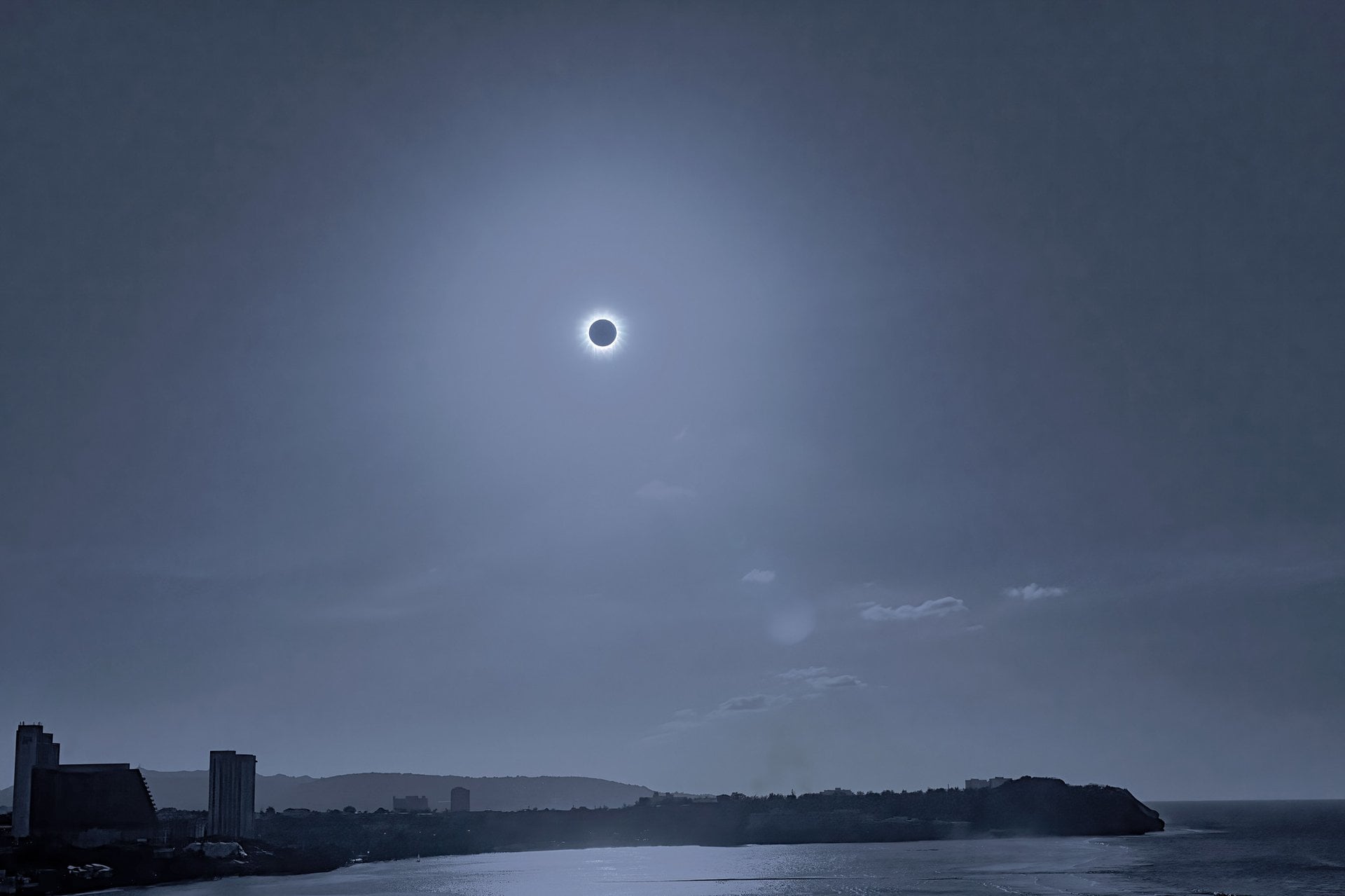
Top Astronomical Events to Watch For in 2026
The coming year offers eclipses, occultations and much more.
Ready for another amazing year of skywatching? 2025 was a wild year with a steady parade of comets knocking on naked eye visibility, and one extra special interstellar…
Continue Reading