Hershey, A. D., Burgi, E. & Ingraham, L. Cohesion of DNA molecules isolated from phage lambda. Proc. Natl. Acad. Sci. USA 49, 748–755 (1963).
Cairns, J. The bacterial chromosome and its…
Hershey, A. D., Burgi, E. & Ingraham, L. Cohesion of DNA molecules isolated from phage lambda. Proc. Natl. Acad. Sci. USA 49, 748–755 (1963).
Cairns, J. The bacterial chromosome and its…

As global temperatures climb, scientists at Cold Spring Harbor Laboratory (CSHL) are striving to develop crops that are stronger and more resilient. This work, however, is far from simple. Many desirable plant traits — such as size or…

Sign up for the Starts With a Bang newsletter
Travel the universe with Dr. Ethan…
Matthew, F., Dixon. Deep learning for spatio-temporal modeling: Dynamic traffic flows and high frequency trading[J]. Applied Stochastic Models in Business & Industry, 35(3):788–807. (2019).
Rui, Z., Yan, R. & Chen, Z. Deep learning and its…

Scientists at the University of Amsterdam have developed a new way to use gravitational waves from black holes to uncover the presence of dark matter and learn more about its behavior. Their approach relies on a detailed theoretical model…

Touchdown airbursts are a form of cosmic impact that may happen more often than the well-known, crater-forming events linked to mass extinctions. Despite their potential for destruction, these explosive encounters remain poorly understood. UC…
Scientists at the University of Amsterdam have developed a new way to use gravitational waves from black holes to uncover the presence of dark matter and learn more about its behavior. Their approach relies on a detailed theoretical model…
Burney, S., Caulfield, J. L., Niles, J. C., Wishnok, J. S. & Tannenbaum, S. R. The chemistry of DNA damage from nitric oxide and peroxynitrite. Mutat. Res. 424, 37–49 (1999).
Tuo, J. et al….
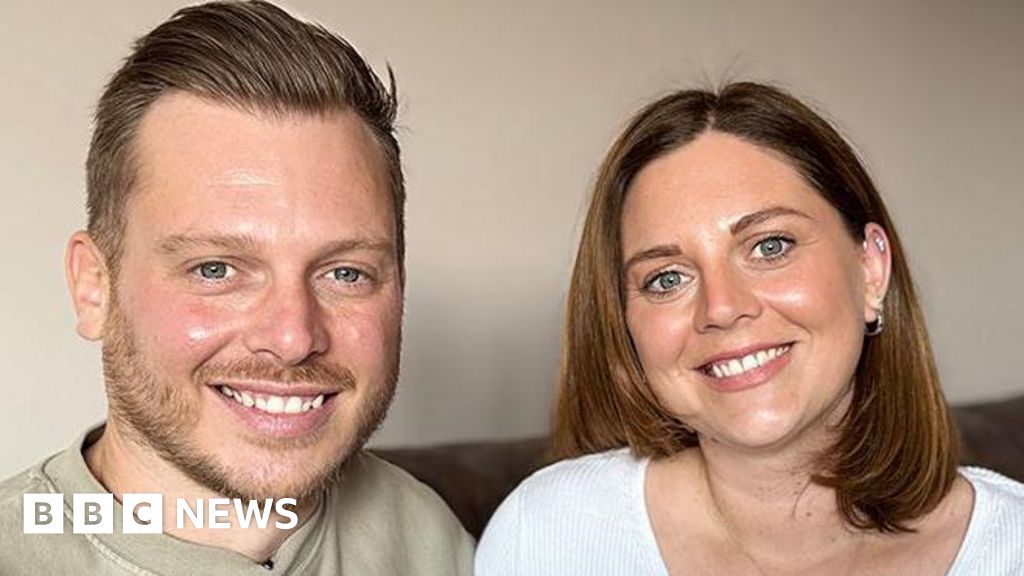
Sharuna SagarNorth East and Cumbria
 Sharuna Sagar/BBC
Siblings only have a 25% chance of being a bone marrow match with each other, but a brother and sister are…
Linden-Carmichael, A. N., Stamates, A. L., Sheehan, B. E. & Lau-Barraco, C. Molly users versus nonusers in a sample of college alcohol drinkers: differences in substance-related harms and sensation seeking. Subst. Abus. 37(3), 474–479 (2016).