Zhang, B., Trapp, A., Kerepesi, C. & Gladyshev, V. N. Emerging rejuvenation strategies—Reducing the biological age. Aging Cell 21, e13538 (2022).
Tarkhov, A. E., Denisov, K. A. & Fedichev, P….
Zhang, B., Trapp, A., Kerepesi, C. & Gladyshev, V. N. Emerging rejuvenation strategies—Reducing the biological age. Aging Cell 21, e13538 (2022).
Tarkhov, A. E., Denisov, K. A. & Fedichev, P….
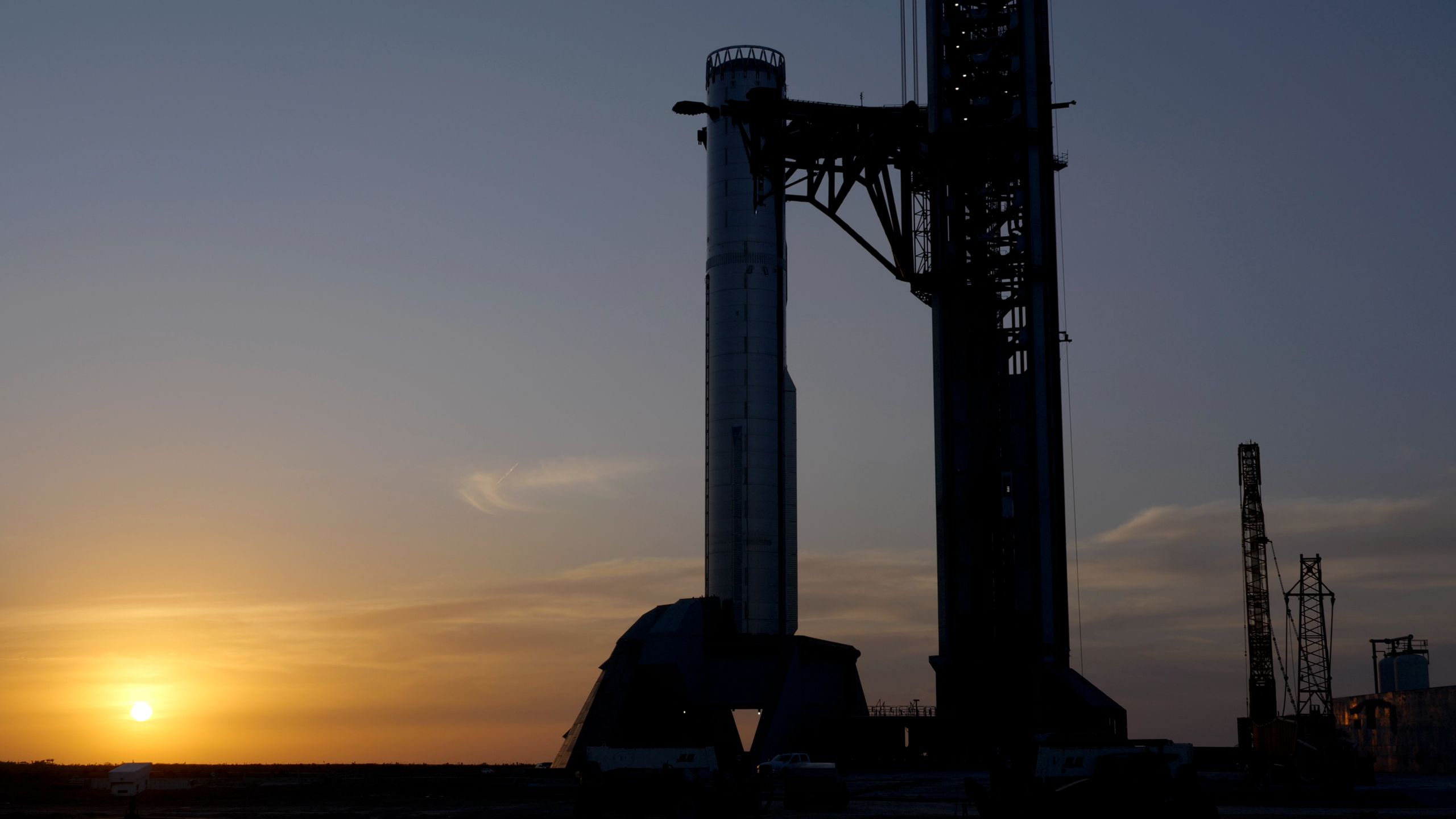
SpaceX’s biggest, most powerful Starship to date just breathed fire for the first time.
On Monday (March 16), the company conducted a static fire test with Starship‘s “Super Heavy” first stage, briefly igniting the booster’s engines while the…
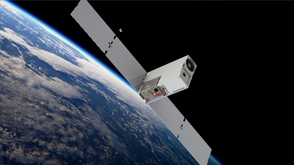
NASA’s Star-Planet Activity Research CubeSat (SPARCS) is a small space telescope that launched to space on January 11th, 2026. Created by NASA and researchers from the School of Earth and Space Exploration (SESE) at the University of…
Neanderthals probably used birch tar for multiple functions, including treating their wounds, according to a study published March 18, 2026 in the open-access journal PLOS One by a team of researchers led by Tjaark Siemssen of the…
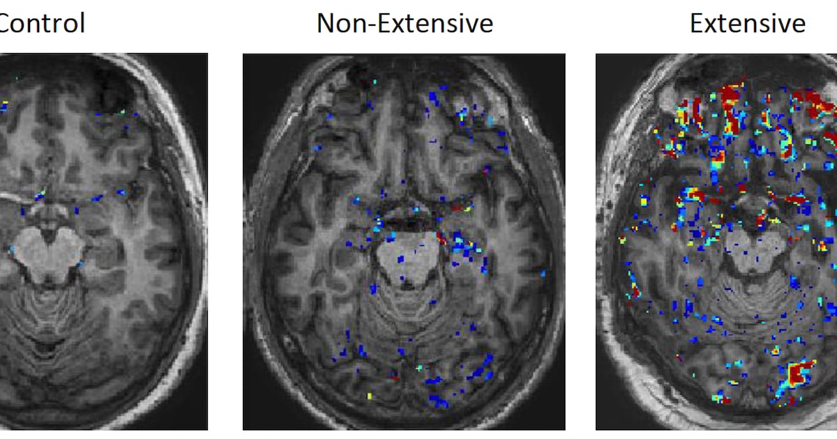
A “leaky” blood-brain barrier is the “key link” between repetitive head injuries and poor long-term brain health in retired athletes, new research from Trinity College Dublin has found.
Sport-related concussion and subconcussive injuries…
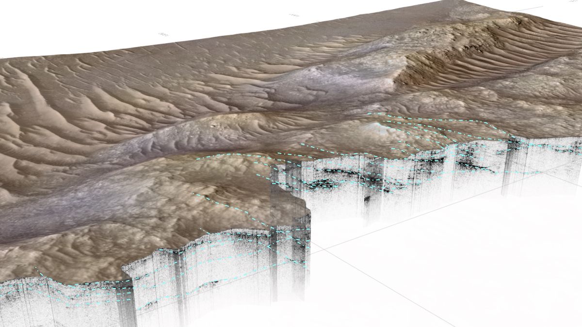
Far away, alone in a crater on a planet inhabited only by robots, NASA’s Perseverance rover explores a dry landscape that was once a river system billions of years ago.
According to a new discovery, however, the Jezero Delta on Mars is not…
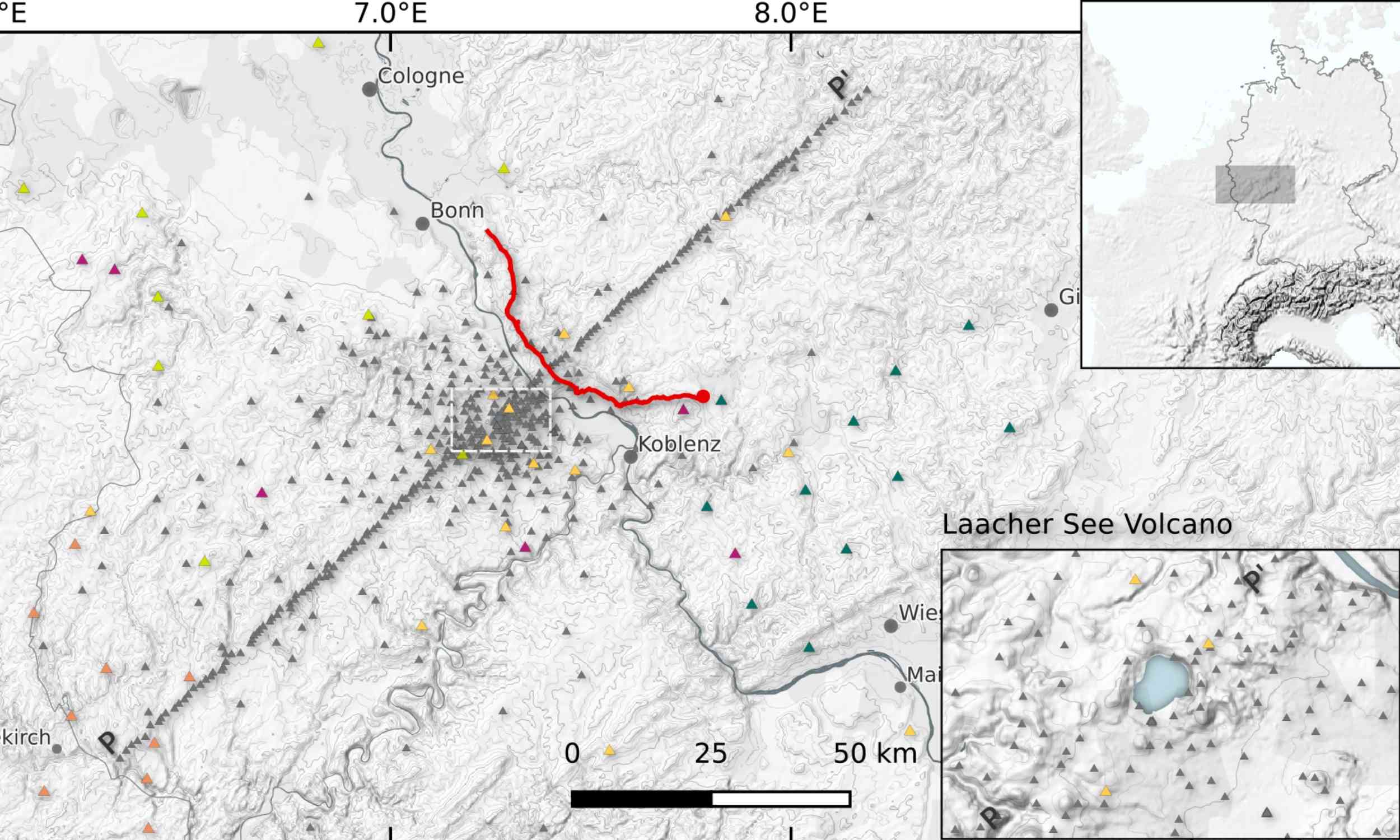
Researchers have revealed that the magma reservoir beneath Germany’s Laacher See volcano lies deeper and tilts southeast instead of extending vertically as long assumed.
The finding connects the volcano’s ancient eruption system with modern…

NASA’s Jet Propulsion Laboratory (JPL) built an ultraviolet camera for NASA’s teeny-tiny SPARCS space telescope, which is about the size of a cereal box. The camera is searching the Milky Way Galaxy for habitable planets.
SPARCS,…
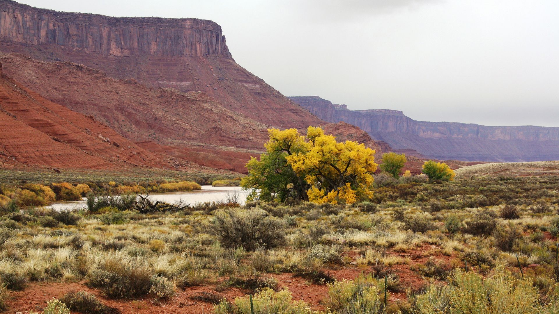
Thirsty plants are sucking up water that would otherwise end up in the Colorado River, according to a new study. The findings could have important implications for water management in regions that rely heavily on snowmelt for their water,…