Category: 7. Science
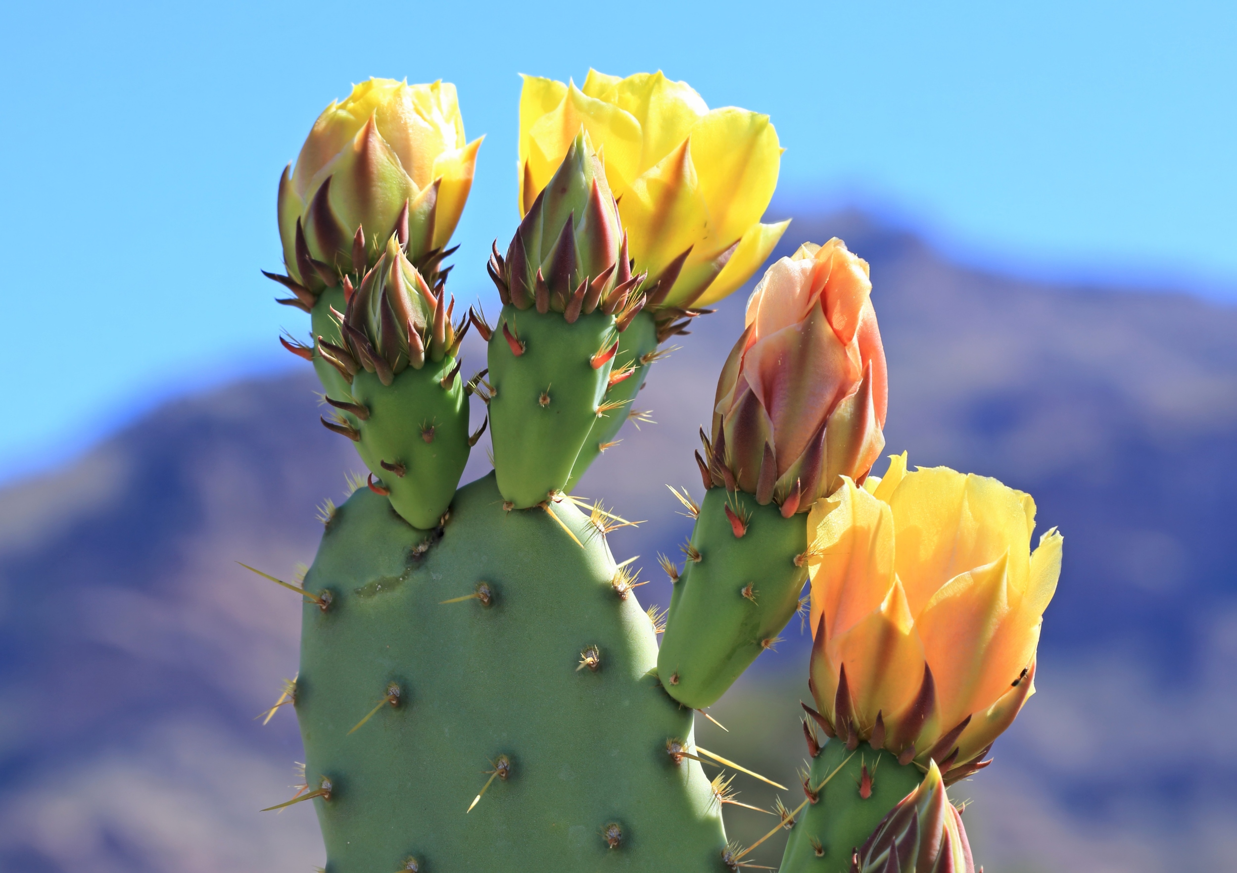
  • Liverworts: Discoveries Through Shot Glass Lens

    Liverworts: Discoveries Through Shot Glass Lens

    Studying plant vegetative reproduction is key to increasing crop yield and for bioengineering. Kobe University research is making progress on studying the genetic regulation of the process in liverworts, which are ideal model plants and even a candidate for space crops.

    Potatoes are tubers, ginger is a rhizome, and both are forms of vegetative plant reproduction, in which plants create structures from which genetically identical individuals can emerge. This mode of reproduction is very important for agriculture and horticulture, but there is very little research on the underlying genetic mechanism. Kobe University plant geneticist ISHIZAKI Kimitsune thinks that the liverwort Marchantia polymorpha is an ideal model organism to study this process and over the past 10 years has been involved in decoding its genome and establishing tools for its convenient genetic manipulation. He says, “Also, the liverwort is so proliferative that it is considered a nuisance to gardeners, growing back quickly no matter how often it is removed.”

    The liverwort spreads through tiny, detachable buds, called “gemmae,” that form in small cups on the upper side of the liverwort’s “leaves” and are dispersed by rain, the wind or animals. Apart from this, the plant also engages in sexual production, switching from vegetative reproduction when the days become longer in summer. “In previous research, we found a gene that seemed to be involved in the formation of both gemma cups and the plant’s sexual reproductive organs. But it was completely unclear what it does, so we wanted to learn more,” says Ishizaki.

    In the journal New Phytologist, the Kobe University team now reports that plants lacking the gene generally don’t form vegetative or sexual reproductive organs, and in rare cases form empty, shot-glass-shaped cups instead of the usually wide and shallow gemma cups, leading them to name the gene “SHOT GLASS.” This shows that the gene is necessary for the development of functioning reproductive structures. Studying the interactions with other genes known to be involved, they found that SHOT GLASS acts by suppressing the development of air chambers in the liverwort’s “leaves” to make space for gemma cup development, and by helping factors needed for the development of sexual reproductive organs to locate to the right place.

    In addition, Ishizaki and his team found something astonishing. They knew that flowering plants, which are much more complex than the simple liverwort, have genes that are related to SHOT GLASS and likely derive from the same gene in the ancestor of all land plants. Interestingly, in flowering plants, those genes are also involved in regulating the development of the secondary meristem that, broadly speaking, makes a plant grow branches. And when they inserted the liverwort’s gene into a flowering plant that lacks one of its own versions, they found that it can even compensate for the gap its more evolved cousin left. Ishizaki explains, “This suggests that the mechanism by which plants create new buds away from the main shoot tip may be common to all land plants.”

    This means that Ishizaki’s liverwort is indeed a convenient model organism to study this agriculturally important process. But the Kobe University researcher has bigger dreams. “Unlike crop plants, liverworts don’t require soil but can be grown with just fog cultivation. We are exploring the development of liverworts where the whole body is directly available as a food resource. This means it could even be used as a food source in space,” Ishizaki explains. He adds: “We are also exploring using the liverwort as an organism for the bioproduction of valuable chemical resources, which has so far practically been restricted to bacteria and yeasts. The engineering technology we are developing and the knowledge we are gathering on the plant’s biology are an important step into that direction.”

    This research was funded by the Ministry of Education, Culture, Sports, Science and Technology Japan (grants 25119711, 15H01233, 17H06472, 21K15125, 19H05673, 19H05670, 20H05780), the Japan Society for the Promotion of Science (grants 21J40092, 15H04391,19H03247, “Program for forming Japan’s peak research universities (J-PEAKS)”), the Japan Science and Technology Agency (grant JPMJGX23B0), the SUNTORY Foundation for Life Sciences, the Yamada Science Foundation, the Asahi Glass Foundation, the Kyoto University Foundation and the Ohsumi Frontier Science Foundation. It was conducted in collaboration with researchers from Kyoto University and Ehime University.

    Kobe University is a national university with roots dating back to the Kobe Higher Commercial School founded in 1902. It is now one of Japan’s leading comprehensive research universities with nearly 16,000 students and nearly 1,700 faculty in 11 faculties and schools and 15 graduate schools. Combining the social and natural sciences to cultivate leaders with an interdisciplinary perspective, Kobe University creates knowledge and fosters innovation to address society’s challenges.

    /Public Release. This material from the originating organization/author(s) might be of the point-in-time nature, and edited for clarity, style and length. Mirage.News does not take institutional positions or sides, and all views, positions, and conclusions expressed herein are solely those of the author(s).View in full here.

    Continue Reading

  • Vesta passes a field star

    Vesta passes a field star

    Vesta is passing an 8th-magnitude field star, offering a great opportunity to see the space rock’s motion on the sky over just a few nights.

    • Asteroid 4 Vesta, a 7th-magnitude object, presents an observable positional shift relative to nearby stars in the constellation Libra, specifically near HD 130193 and Xi1 Librae.
    • Vesta’s apparent motion provides an opportunity for observation and tracking against the static stellar background over several days.
    • The globular cluster NGC 5634 in Virgo, with an apparent magnitude of 9.6, is highlighted as an observable target, suggested to originate from a Milky Way satellite galaxy.
    • Observational timing information is provided for sunrise, sunset, moonrise, moonset, and moon phase (waxing crescent at 33% illumination) at a specified location (40° N 90° W).

    Asteroid 4 Vesta remains an easy-to-spot target in western Libra, just 5° north of Zubenelgenubi, the Balance’s double alpha star. Tonight and tomorrow offer an excellent opportunity to see the 7th-magnitude space rock move relative to the background of stars, as it currently lies less than 0.5° from a slightly fainter 8th-magnitude background star (HD 130193). 

    Tonight, Vesta is 10.5’ northwest of that star. Tomorrow, it is a little less than that distance roughly due east of it. Vesta is also drawing closer to 6th-magnitude Xi11) Librae and will sit due south of this star on August 6, offering another chance to easily catch the asteroid moving against the stationary background for a few days before and after this date. 

    While you’ve got your scope out, swing up 6.7° northwest into Virgo to enjoy NGC 5634, a lovely compact globular cluster that shines at magnitude 9.6. Astronomers believe this cluster was once part of one of the Milky Way’s smaller satellite galaxies, likely the the Sagittarius Dwarf Elliptical Galaxy or the Sagittarius Dwarf Spheroidal Galaxy. Its brightest star, 8th-magnitude SAO 139967, is just southeast of the cluster’s center. The markedly orange-hued sun stands out distinctly against the fuzzy background created by the compact structure of the grouping. 

    Sunrise: 5:57 A.M.
    Sunset: 8:15 P.M.
    Moonrise: 12:00 P.M.
    Moonset: 11:05 P.M.
    Moon Phase: Waxing crescent (33%)
    *Times for sunrise, sunset, moonrise, and moonset are given in local time from 40° N 90° W. The Moon’s illumination is given at 12 P.M. local time from the same location.

    For a look ahead at more upcoming sky events, check out our full Sky This Week column. 

    Continue Reading

  • Australia set for Celestial Spectacle as Southern Delta Aquariids and Alpha Capricornids meteor showers peak this week

    Australia set for Celestial Spectacle as Southern Delta Aquariids and Alpha Capricornids meteor showers peak this week

    Night skies across Australia are set to dazzle as two meteor showers, the Southern Delta Aquariids and Alpha Capricornids, peak this week, offering stargazers a rare celestial double feature.

    The dual showers will be most visible between 11 pm and dawn next week, with peak activity expected around 2 am for the Delta Aquariids and just before midnight for the Alpha Capricornids, according to Professor Jonti Horner, an astrophysicist at the University of Southern Queensland.

    “This is a fantastic opportunity,” Horner said. “You won’t need a telescope or binoculars; in fact, they’ll limit your view. All you need are your eyes, a dark sky, and a bit of patience.”

    Best seats in the house

    Australia is ideally positioned in the southern hemisphere to catch both showers. But location matters; experts recommend escaping city lights. Horner advises scouting a viewing spot during daylight hours to avoid surprises at night.

    “The further you are from artificial light, buildings, streetlamps, car headlights, the better your chances of seeing the meteor trails,” he said. Weather could affect visibility, with clouds forecast in Melbourne and Brisbane, and possible showers in Sydney on Wednesday night.

    What you’ll see

    The Southern Delta Aquariids, the more active of the two showers, are expected to produce up to 20 meteors per hour, entering Earth’s atmosphere at speeds around 40 kilometers per second. These meteors tend to be faint but can leave glowing trails.The Alpha Capricornids, while less prolific, are known for producing bright, slow-moving fireballs, offering a more dramatic viewing experience. Both showers appear to originate from specific points in the sky known as “radiants,” located in the constellations Aquarius and Capricornus, hence their names.

    Stargazing tips

    To maximize your viewing experience, Horner recommends letting your eyes adjust to the dark, ideally for 30 to 45 minutes, and avoiding screens during that time.

    “Bring a blanket, some pillows, maybe a reclining chair,” he said. “Comfort helps. Looking up for long periods can strain your neck.”

    Apps like Sky Map or Star Walk can help locate the radiant points in the sky, but experts caution against getting too distracted.

    For those who miss this week’s display, December promises even more. The Geminid meteor shower, considered the best of the year, will peak on December 14–15 under a new moon, offering dark skies and spectacular viewing conditions.

    Continue Reading

  • China fossil find reveals why ancient marine ‘lamp shells’ practised social distancing

    China fossil find reveals why ancient marine ‘lamp shells’ practised social distancing

    Ancient “lamp shells” – bottom-dwelling invertebrates that once ruled the oceans – may have used hundreds of bristle-like structures to “social distance” in a bid to improve their chances of survival, Chinese palaeontologists have discovered.

    Their findings are based on a fossilised species of brachiopods – or clam-like marine animals – that was discovered in China’s southwestern Guizhou province.

    The fossils, discovered embedded in rocks, were found to be arranged in a “checkerboard-like” pattern across what was once the sea floor.

    Individuals of the extinct species Nucleospira calypta were likely to have used the flexible bristle-like structures – thinner than human hair – to help guide their movement and arrange their population to provide adequate space for filter feeding.

    “We report exceptionally preserved soft, bristle-like structures (“setae”) on 436-million-year-old brachiopods, enabling analysis of ancient behaviour,” the team said in a paper published in the peer-reviewed journal Proceedings of the National Academy of Sciences on July 21.

    Using advanced imaging and spatial analysis, the researchers determined that Nucleospira calypta formed a “highly regular” pattern of spacing that corresponded to about 1.5 to 2 times the length of their setae.

    Continue Reading

  • Moon phase today explained: What the moon will look like on July 30, 2025

    Moon phase today explained: What the moon will look like on July 30, 2025

    The moon is becoming more lit up each night as we progress through the lunar cycle.

    The lunar cycle is a series of eight unique phases of the moon’s visibility. The whole cycle takes about 29.5 days, according to NASA, and these different phases happen as the Sun lights up different parts of the moon whilst it orbits Earth. 

    So, what’s happening tonight, July 30?

    What is today’s moon phase?

    As of Wednesday, July 30, the moon phase is Waxing Crescent. There’s 32% of the moon’s surface visible to us on Earth (according to NASA’s Daily Moon Observation).

    It’s day six of the new lunar cycle, and there’s a lot to see on the moon’s surface.

    With the unaided eye, enjoy a glimpse of the Mare Crisium and the Mare Fecunditatis, an impact basin also known as the “Sea of Fertility.” Add binoculars and you’ll also get a glimpse of the Endymion Crater and the Posidonius Crater, a lava-filled impact crater that’s visible from the fifth to the 19th day of the lunar cycle.

    If you have a telescope, you’ll also see the Apollo 17, the last moon landing of the Apollo program. Use NASA’s interactive moon guide tool to know exactly where to look.

    Mashable Light Speed

    When is the next full moon?

    The next full moon will be on August 9. The last full moon was on July 10.

    What are moon phases?

    According to NASA, moon phases are caused by the 29.5-day cycle of the moon’s orbit, which changes the angles between the Sun, Moon, and Earth. Moon phases are how the moon looks from Earth as it goes around us. We always see the same side of the moon, but how much of it is lit up by the Sun changes depending on where it is in its orbit. This is how we get full moons, half moons, and moons that appear completely invisible. There are eight main moon phases, and they follow a repeating cycle:

    New Moon – The moon is between Earth and the sun, so the side we see is dark (in other words, it’s invisible to the eye).

    Waxing Crescent – A small sliver of light appears on the right side (Northern Hemisphere).

    First Quarter – Half of the moon is lit on the right side. It looks like a half-moon.

    Waxing Gibbous – More than half is lit up, but it’s not quite full yet.

    Full Moon – The whole face of the moon is illuminated and fully visible.

    Waning Gibbous – The moon starts losing light on the right side.

    Last Quarter (or Third Quarter) – Another half-moon, but now the left side is lit.

    Waning Crescent – A thin sliver of light remains on the left side before going dark again.

    Continue Reading

  • Developing Next-Generation Analytical Technique for Gene and Cell Doping and Ensuring Ethics and Fairness in Sports

    Developing Next-Generation Analytical Technique for Gene and Cell Doping and Ensuring Ethics and Fairness in Sports

    Newswise — Changmin Sung, a principal researcher at the Doping Control Center at the Korea Institute of Science and Technology (KIST), announced that he and his collaborators at the Department of Biomedical Engineering at Korea University have developed a high-throughput multiplexed gene and cell doping analysis (HiMDA) based on gene scissors (CRISPR-Cas).

    Unethical doping practices to enhance athletic performance is becoming more sophisticated with the use of advanced technology, and gene and cell doping – the use of gene or cell therapies to manipulate body functions – poses a serious threat to fairness in sports. Gene-based drugs such as insulin-like growth factor (IGF-I) and erythropoietin (EPO), which can maximize strength and endurance, are likely to be abused by athletes in some sports as a means of performance enhancement. The World Anti-Doping Agency (WADA) has prohibited this practice since 2003, but diagnostic techniques that can accurately identify gene and cell doping are still in early stage.

    Quantitative polymerase chain reaction (qPCR)-based gene testing has been piloted at the Tokyo Summer Olympics since the World Anti-Doping Agency first published guidelines for genetic doping in 2021. Current protein-level doping analytical methods cannot clearly distinguish between exogenous genetic targets that produces proteins structurally identical to endogenous proteins. This has led to the need for new analytical platforms that can distinguish exogenous genes at the DNA level.

    The HiMDA directly amplifies the target gene from the blood without complex sample preparation, and then applies CRISPR-Cas, the Nobel Prize-winning gene editing technology to determine the presence of the exogenous gene rapidly and precisely. By injecting representative gene doping substances such as hGH, EPO, IGF-I into an experimental mouse model and applying the assay platform, the researchers were able to accurately detect exogenous genes at the 2.5 copies within 90 minutes using as little as 5 μL (microliters) of blood sample, less than half the size of a fingertip drop. This demonstrated superior performance in both sensitivity and specificity compared to existing assays.

    The developed assay is not limited to doping tests, but is considered to be a platform-based diagnostic technology that can be applied to early diagnosis of infectious diseases, detection of antibiotic resistance genes, genetic disease testing, evaluation of cell therapy drug adaptability, and precision medicine. Currently, the technology is undergoing the certification process to be adopted as a World Anti-Doping Agency-approved method, and is attracting attention as a next-generation anti-doping testing platform that can respond to various new doping methods based on genes and proteins.

    “By applying gene editing technology to doping tests, this study provides a practical solution that can overcome the limitations of existing techniques and contribute to protecting sports ethics and fairness,” said Changmin Sung, a principal researcher at KIST. “It has the potential to develop into a core foundation for precision medicine and genetic diagnostic technologies in the future.”

    ###

    KIST was established in 1966 as the first government-funded research institute in Korea. KIST now strives to solve national and social challenges and secure growth engines through leading and innovative research. For more information, please visit KIST’s website at https://www.kist.re.kr/eng/index.do

    This research was supported by the Ministry of Science and ICT (MSIT) under the KIST Institutional Program and the World Anti-Doping Agency Research Support Program (241E07CS). The findings were published in the latest issue of the international journal Science Advances (IF 12.5, JCR top 8.5%).


    Continue Reading

  • New algorithms enable efficient machine learning with symmetric data | MIT News

    New algorithms enable efficient machine learning with symmetric data | MIT News

    If you rotate an image of a molecular structure, a human can tell the rotated image is still the same molecule, but a machine-learning model might think it is a new data point. In computer science parlance, the molecule is “symmetric,” meaning the fundamental structure of that molecule remains the same if it undergoes certain transformations, like rotation.

    If a drug discovery model doesn’t understand symmetry, it could make inaccurate predictions about molecular properties. But despite some empirical successes, it’s been unclear whether there is a computationally efficient method to train a good model that is guaranteed to respect symmetry.

    A new study by MIT researchers answers this question, and shows the first method for machine learning with symmetry that is provably efficient in terms of both the amount of computation and data needed.

    These results clarify a foundational question, and they could aid researchers in the development of more powerful machine-learning models that are designed to handle symmetry. Such models would be useful in a variety of applications, from discovering new materials to identifying astronomical anomalies to unraveling complex climate patterns.

    “These symmetries are important because they are some sort of information that nature is telling us about the data, and we should take it into account in our machine-learning models. We’ve now shown that it is possible to do machine-learning with symmetric data in an efficient way,” says Behrooz Tahmasebi, an MIT graduate student and co-lead author of this study.

    He is joined on the paper by co-lead author and MIT graduate student Ashkan Soleymani; Stefanie Jegelka, an associate professor of electrical engineering and computer science (EECS) and a member of the Institute for Data, Systems, and Society (IDSS) and the Computer Science and Artificial Intelligence Laboratory (CSAIL); and senior author Patrick Jaillet, the Dugald C. Jackson Professor of Electrical Engineering and Computer Science and a principal investigator in the Laboratory for Information and Decision Systems (LIDS). The research was recently presented at the International Conference on Machine Learning.

    Studying symmetry

    Symmetric data appear in many domains, especially the natural sciences and physics. A model that recognizes symmetries is able to identify an object, like a car, no matter where that object is placed in an image, for example.

    Unless a machine-learning model is designed to handle symmetry, it could be less accurate and prone to failure when faced with new symmetric data in real-world situations. On the flip side, models that take advantage of symmetry could be faster and require fewer data for training.

    But training a model to process symmetric data is no easy task.

    One common approach is called data augmentation, where researchers transform each symmetric data point into multiple data points to help the model generalize better to new data. For instance, one could rotate a molecular structure many times to produce new training data, but if researchers want the model to be guaranteed to respect symmetry, this can be computationally prohibitive.

    An alternative approach is to encode symmetry into the model’s architecture. A well-known example of this is a graph neural network (GNN), which inherently handles symmetric data because of how it is designed.

    “Graph neural networks are fast and efficient, and they take care of symmetry quite well, but nobody really knows what these models are learning or why they work. Understanding GNNs is a main motivation of our work, so we started with a theoretical evaluation of what happens when data are symmetric,” Tahmasebi says.

    They explored the statistical-computational tradeoff in machine learning with symmetric data. This tradeoff means methods that require fewer data can be more computationally expensive, so researchers need to find the right balance.

    Building on this theoretical evaluation, the researchers designed an efficient algorithm for machine learning with symmetric data.

    Mathematical combinations

    To do this, they borrowed ideas from algebra to shrink and simplify the problem. Then, they reformulated the problem using ideas from geometry that effectively capture symmetry.

    Finally, they combined the algebra and the geometry into an optimization problem that can be solved efficiently, resulting in their new algorithm.

    “Most of the theory and applications were focusing on either algebra or geometry. Here we just combined them,” Tahmasebi says.

    The algorithm requires fewer data samples for training than classical approaches, which would improve a model’s accuracy and ability to adapt to new applications.

    By proving that scientists can develop efficient algorithms for machine learning with symmetry, and demonstrating how it can be done, these results could lead to the development of new neural network architectures that could be more accurate and less resource-intensive than current models.

    Scientists could also use this analysis as a starting point to examine the inner workings of GNNs, and how their operations differ from the algorithm the MIT researchers developed.

    “Once we know that better, we can design more interpretable, more robust, and more efficient neural network architectures,” adds Soleymani.

    This research is funded, in part, by the National Research Foundation of Singapore, DSO National Laboratories of Singapore, the U.S. Office of Naval Research, the U.S. National Science Foundation, and an Alexander von Humboldt Professorship.

    Continue Reading

  • SpaceX launches 28 Starlink satellites on a Falcon 9 rocket from Florida

    SpaceX launches 28 Starlink satellites on a Falcon 9 rocket from Florida

    Twenty-eight more Starlink satellites are now in Earth orbit after launching from Florida on Tuesday night (July 29).

    A SpaceX Falcon 9 rocket lifted off at 11:37 p.m. EDT (0337 GMT on July 30) rom Space Launch Complex 40 (SLC-40) at Cape Canaveral Space Force Station in Florida. After a nine minute climb into space, the 28 Starlink broadband internet satellites (group 10-29) were on track to be deployed into their intended orbit.

    The first stage of a SpaceX Falcon 9 rocket lands atop an ocean-based droneship in the Atlantic Ocean on Tuesday, July 29, 2025. (Image credit: SpaceX)

    Continue Reading

  • Isolation and characterization of phages ΦZC2 and ΦZC3 against carbapenem-resistant Acinetobacter baumannii, and efficacy of ΦZC3 on A549 cells | Virology Journal

    Isolation and characterization of phages ΦZC2 and ΦZC3 against carbapenem-resistant Acinetobacter baumannii, and efficacy of ΦZC3 on A549 cells | Virology Journal

    Bacterial isolates and growth conditions

    Forty-one MDR A. baumannii clinical isolates were obtained from sputum and respiratory secretions of hospitalized patients in Egypt. These isolates were provided by Mabaret Elasafra clinical microbiology laboratory serving multiple hospitals in Alexandria, Egypt, between August 2020 and April 2021, where they had been pre-identified and characterized, with species-level identification conducted using the VITEK2 Compact system (bioMérieux, Marcy-l’Étoile, France). The presence of carbapenemase-encoding genes was further confirmed by PCR with specific primers [34].

    The collected bacterial isolates were sub-cultured on MacConkey agar (Oxoid, England), and they were preserved in tryptic soy broth (TSB; Merck, USA) supplemented with 25% glycerol for long-term storage at -80 °C.

    Bacteriophage isolation and characterization

    Isolation and purification of bacteriophages

    The protocol described by Clokie and Kropinski (2009) was used for phage isolation with minor modifications [35]. The wastewater samples were centrifuged in a 50 ml centrifuge tube at 5,000 × g for 20 min at 4 °C. For enrichment, 5 ml supernatant of wastewater was transferred to a sterile centrifuge tube with an equal volume of the TSB 2X in a sterile flask with 100 µl of mixed culture (AB20, AB21, AB22, AB23, AB24, AB25, AB26, AB27, AB28, and AB29) of A. baumannii isolates for overnight in a shaking incubator at 37 °C. The enrichment was transferred to 50 ml centrifuge tube and centrifuged at 5,000 ×g for 20 min at 4 °C. The supernatant was filtered by a 0.45 μm syringe membrane filter (Membrane Solution, USA). Double agar overlay plaque assay was used to evaluate bacteriophage activity; an overnight bacterial culture was combined with molten soft nutrient agar with a concentration of 0.75% agar and poured onto a 1.5% agar plate. 10 µl of supernatant was dropped onto the plate and incubated overnight at 37°C [36].

    The formed plaques were purified by picking the single plaques from the plate with a sterile pipette tip and transferred to sterile SM buffer (100 mM NaCl, 8 mM MgSO4 • 7H2O, 50 mM Tris-Cl; pH 7.5) to elute the phage overnight at 4°C [4]. The eluted phage was diluted tenfold in SM buffer to be spotted on the overlay of bacterial lawn to pick a single plaque, and this procedure was repeated seven times to ensure that a single phage type was isolated from the plaques. The isolated phages were amplified in small volumes (5 ml) by mixing TSB of log-phase bacterial culture 108 CFU/ml and 50 µl of isolated phages with titer 109 PFU/ml.

    Host range assay

    The phages’ host range was investigated using the spot assay method against forty-one MDR A. baumannii isolates, as previously described [37]. In brief, 10 µL of each phage lysate was added on overlays of bacterial lawns from each isolate and followed by overnight incubation at 37 °C. The phage lysis intensity to the host was assessed based on the clarity of the formed plaques. This assay was carried out in triplicate.

    Relative efficiency of platting (EOP)

    As detailed previously [27]the relative efficacy of plating (EOP) of the phages was determined by counting the number of clear lysis plaques formed after applying 10-fold successive dilutions of the phage onto freshly prepared lawns of each susceptible bacterial isolate. The spotting assay was performed on a double agar overlay, and the plaques were enumerated. The EOP was determined by calculating the ratio of the phage titer on each tested clinical isolate to the titer of host isolate. The EOP ratio was categorized into four levels based on defined quantitative thresholds. Isolates with EOP values ≥ 0.5 were classified as highly efficient. Moderate efficiency was assigned to isolates with 0.1 ≤ EOP < 0.5, while low efficiency corresponded to values in the range of 0.001 ≤ EOP < 0.1. Isolates with EOP values < 0.001 were considered inefficient or resistant.

    Morphological characterization

    The phages were serially diluted to obtain a plaques per plate, when plating 0.1 ml of the diluted stock. Dilutions were prepared in TSB, for plaque formation, 0.1 ml of bacterial culture was mixed with 3.0 ml of molten soft nutrient agar (0.75% agar), followed by the addition of 0.1 ml of the diluted phage. The mixture was poured onto a tryptic soy agar (TSA; Merck, USA) plate. Plates incubated at 37 °C for 18 h. Plaque formation was observed, with the potential for plaque lysis [38]. Plaque size measurement was measured by image j software.

    The structure of phages was analyzed using transmission electron microscopy (TEM) in the Faculty of Science, University of Alexandria, Egypt. Initially, 10 µL of the phage, containing 1010 PFU/ml was stained with 2.5% uranyl acetate. Subsequently, the sample was attached to a carbon-coated Cu-grid and incubated for 10 min before being analyzed using TEM. The stained phage was imaged using a TEM (1230 JEOL, Tokyo, Japan). The captured images from a JEOL 1230 TEM were measured using ImageJ software version 1.53n [39].

    Phage production by bioreactor

    Phage production was carried out in the BioFlo 120 bioreactor = (Eppendorf, Germany), as previously described, with minor modification [40],1.6 L of TSB was autoclaved in a vessel (Borosilicate glass, 316 L stainless steel). The temperature was 37 °C, and pH 7 was optimized through two front-mounted fixed-speed pumps connected to NaOH and HCL bottles. A single colony of the A. baumannii host isolate was resuspended in 200 ml TSB and incubated at 37 °C for overnight culture. After cultivation, 200 ml of the overnight culture was subcultured.

    in the bioreactor vessel. The bacterial culture was cultivated at 37 °C and 100 rpm for 4 h until reaching the early log phase (OD600 = 0.1). The multiplicity of infection (MOI) of 0.1 was achieved with 0.8 ml of phage (8 × 109 PFU/ml) to the bioreactor vessel by syringe. The agitation was set to 100 rpm, and the gas flow was 2 barg for 5 h. Aliquots were collected in triplicate for phage enumeration and bacterial OD measurement from sample ports using syringes at each time point.

    Phage stability assay

    Phage stability assays were carried out as previously described, with minor modifications [41]. Phage lysates were serially diluted and spotted on their hosts of bacterial culture to evaluate their pH, temperature, and UV stability. The phages’ titers were measured after incubating 1 h with different pH values (2, 3, 4, 7, 10, 11, and 12). Likewise, the temperature stability was assessed by incubating tubes containing phages separately at temperature points − 80, -20, 4, 37, 50, 60, 70, 80 and 90 °C for 1 h. The phage’s tolerance to UV exposure was assessed at 15, 30, 45, 60, 80, and 90. minutes. The stability of the phage was evaluated based on the reduction in phage titer compared to the initial phage titer in SM buffer. For the UV stability assay, the phages were exposed to UV-C light at a wavelength of 254 nm using the built-in UV lamp of the biosafety cabinet (ESCO, Singapore). The distance from the lamp to the sample surface was approximately 60 cm. The lamp’s intensity at this distance was estimated to be 40µW/cm² [42].

    Time kill kinetics assay

    Kinetics of bacterial killing by the isolated phages was investigated at different phage MOIs (0.01, 0.1, 1, 10, 100) using the previously established method of time-killing curve assay [27]. The bacterial strains AB23 and AB24 were used as bacterial host for the isolated phages. Briefly, bacterial culture at 106 CFU/ml was treated with the phage at different MOIs in a 96-well microtiter plate. The bacterial O.D. at a wavelength of 600 nm was determined at 1 h intervals for a duration of 11 h by using FLUOstar® Omega plate reader, BMG LABTECH, Germany. A control of bacterial culture without phage treatment and a blank containing only the culture media were used.

    Bacteriophage insensitive mutants (BIM)

    The BIM frequency was estimated using the previously described method [43]. Briefly, 1 ml of bacterial culture at a concentration 107 CFU/ml was mixed with the phage to achieve MOI of 100. The bacterial strains AB23 and AB24 were used as hosts for isolated phages to assess phage-resistant mutant bacterial cells. After 24 h of incubation at 37 °C, 100 µl of the mixture (phage with its bacterial host) was serially diluted, spotted on TSA, and incubated at 37 °C for 24 h. Phage-resistant mutant bacterial cells were calculated by dividing viable bacterial counts after phage infection by the initial viable count [44].

    Phage genome sequencing

    Bacteriophage genomic DNA extraction

    The genomic DNA of the phage was extracted using phenol-chloroform–isoamyl alcohol method with a minor modification [45]. The phage genomic DNA was extracted from 10 ml of high-titer (1010 PFU/ml) and filter-sterilized in a sterile tube. In brief, 1 ml of lysis buffer was added to the phage and then incubated at 56 °C for 1 h. After incubation, 10 ml of phenol/chloroform isoamyl alcohol was added and then centrifuged at 18,000 × g for 10 min to separate the organic phase and aqueous phase; the DNA was collected from the top of the aqueous phase while the organic phase, which contains proteins was discarded. The DNA from the aqueous phase was precipitated overnight at -20 °C by adding a 1:10 volume ratio of 3 M sodium acetate (pH 5.2) and a 2:1 volume ratio of ice-cold isopropanol. By centrifugation at 18,000 × g for 10 min, the precipitated DNA was formed in a pellet. The supernatant was removed, and the pellet was re-suspended in 90% ice-cold ethanol before being transferred to a new 1.5 mL tube. The DNA pellet was washed twice with 70% ethanol, allowed to dry, and then resuspended in 100 ml of nuclease-free water. DNA concentration and quality were evaluated using the FLUOstar Omega Microplate reader (BMG LABTECH, Germany).

    Genome sequencing and bioinformatics analysis

    The Illumina MiSeq platform was used to perform nucleotide sequencing. Library preparation was carried out using the Illumina Nextera tagmentation protocol (Illumina, Cambridge, UK). Quality control of the sequence reads was performed with FastQC (v0.11.9), and de novo assembly was carried out using Unicycle (v0.4.8) via the BV-BRC portal [46]. Genomic visualization, comparison, and orientation were performed using ProgressiveMauve and Ugene software, v43.0 [47, 48]. The single assembled contig was then annotated with the Rapid Annotation using Subsystem Technology Toolkit (RASTtk) pipeline [49]. The annotation process was customized, beginning with the ‘annotate-proteins-phage’ step, followed by ‘annotate-proteins kmer-v2 [50]. Following the RASTtk annotation, a second round of annotation was performed to confirm the assigned functions and to assign functions to proteins that were previously unassigned, using NCBI BLASTp, HHPred, and InterProScan. ARAGORN v1.2.41 was used to identify tRNA genes [51]. A phage genomic map was generated using the Proksee web-based tool [52] by CGView family tools [53].

    The phage lifestyle prediction, virulent factor and antimicrobial resistance gene detection, and anti-CRISPR protein were analyzed by PhageScope [54]. In addition, the therapeutic potential of the phage was assessed using PhageLeads [55]which analyzes the phage genome for temperate genetic markers, antimicrobial resistance (AMR), and virulence factors. The transmembrane domains of the phage-predicted proteins were examined using DeepTMHMM, a tool that employs a deep learning algorithm based on a protein language model [56]. The genomic sequence was analyzed with the Phage Depolymerase Finder (PhageDPO, Galaxy Version 0.1.0) prediction tool to identify genes potentially encoding depolymerase functions [57].

    Phylogenetic analysis

    Genome-genome distancing was assessed by ViPtree (proteome-based) [58] and VIRIDIC (nucleotide-based) [59]. VIRIDIC assigned taxa using ICTV species and genus thresholds based on average nucleotide identity (ANI) at 95% and 70%, respectively. Additionally, a phylogenetic tree was constructed on NGPhylogeny.fr [60]using MUSCLE for amino acid sequence alignment [61] and PhyML 3.3.2 with the LG model for amino acid substitution [62]and a BioNJ initial tree to infer the evolutionary relationships of the terminase large subunit protein (encoded by a core gene). A gamma distribution with 4 rate categories was used to handle different mutation rates across sites, and SH-like branch supports were calculated to assess the reliability of the tree branches [63].

    Cell cytotoxicity assay

    The phage with a broader host range and better killing capacity was further evaluated for its safety on the human alveolar basal epithelial cell line A549 (ATCC Cat No. CCL-185) by using an MTT assay [27]. A549 cells were cultured in 96-well plates with a seeding density of 104 cells/well, in RPMI-1640 medium supplemented with 10% fetal bovine serum 100 I.U./ml penicillin, and 100 µg/ml streptomycin and incubated for 24 h at 37 °C/ 5% CO2. Then, serially diluted phage lysate was added to the cells with different concentrations (109, 108, 107, and 106) and incubated for another 24 h at 37 °C/ 5% CO2. After incubation, the medium was removed, and 100 µl of MTT solution (10 µl MTT and 90 µl RPMI) was added and incubated for 4 h at 37 °C and 5% CO2. 100 µl of DMSO as a solvent was added and incubated for 15 min in the dark, following OD measurement at 590 nm by using FLUOstar® Omega plate reader, BMG LABTECH, Germany, and the images was taken by an inverted microscope camera (Optika IM-3, Italy).

    Dynamic of phage-bacteria interaction with A549 cells

    To assess the effectiveness of the phage in the cell culture line, the bacteria were co-cultured with A549 cell lines and exposed to phage with different MOIs [27]. Bacterial and phage titer were assessed throughout the experiment at different time points, A549 cell viability was assessed only at the end of the assay time (6 h). In brief, human alveolar basal epithelial cell line A549 was used as a model for the in vitro study of phage-bacteria interaction. The cells were cultured in RPMI-1640 medium supplemented with 10% fetal bovine serum, 100 I.U./ml penicillin and 100 (µg/ml) streptomycin at 37 °C/ 5% CO2. When it reached confluency, the cells were trypsinzed and cultured in 96-well plates with seeding density 5 × 104 cells/well, incubated at 37 °C/ 5% CO2 for 24 h.

    At the same time, an overnight culture of AB24 was prepared and incubated at 37 °C. After 24 h, the bacterial culture was centrifuged at 8000 × g for 1 min, resuspended in phosphate buffer saline (PBS) (MP Biomedicals, LLC, Ohio), and OD was measured to obtain a final concentration of 105 CFU/ml. Then, the bacterial culture was centrifuged again and resuspended into complete RPMI without antibiotics.

    After checking the cell adhesion on a 96-well plate, the old medium was removed and washed twice with PBS, and 100 µl/well of bacteria suspension in RPMI was added to reach a final concentration of 105 CFU/well. The cells were incubated for 1 h at 37 °C/ 5% CO2. Then, the planktonic bacteria were removed, and 100 µl of phage was added with several MOI (0.1, 1, 10, and 100); then, 100 µL of RPMI without antibiotics was added to all the wells. these wells were compared to the control with bacteria and the free cells without bacteria.

    The plate count technique was used to enumerate the viable bacterial count and phage titer at (zero, 2, and 6 h). After 6 h, the cells were counted by removing the supernatant, washed twice with PBS, and 40 µl of trypsin was added and incubated for 15 min. Then, 60 µl of complete RPMI was added and mixed well, and cells were counted with trypan blue under a hemocytometer.

    Statistical analysis

    Each experiment was carried out in triplicate, with results expressed as the mean ± standard deviation (SD). GraphPad Prism 9.5.1 software was used for data analysis, including generating graphs and conducting one-way ANOVA.

    Continue Reading

  • This tiny lung-on-a-chip could predict—and fight—the next pandemic

    This tiny lung-on-a-chip could predict—and fight—the next pandemic

    Respiratory infections such as COVID-19 have been responsible for numerous pandemics and have placed a substantial burden on healthcare systems. Such viruses can cause significant damage to our lungs, especially to the proximal region, or airway, and distal region, also known as the alveoli.

    The responses of different lung regions to such infections are varying and complex, so accurately replicating them using traditional models, such as animals and simple in vitro systems, poses a challenge.

    To solve this problem, a team of researchers at Kyoto University has developed a micro physiological system, or MPS, capable of emulating different regions of human lungs. Specifically, their device can simulate the airway and alveoli to investigate viral pathologies. Coupled with isogenic iPSCs, the team is preparing for more personalized and accurate treatment of respiratory diseases.

    “Our iPSC-derived lung chips enable us to model the distinct responses of proximal and distal lung regions, derived from an isogenic source to respiratory virus infections,” states the lead author Sachin Yadav, a PhD student at Kyoto University.

    The team’s research provides a more accurate platform for studying tissue- and virus-specific disease mechanisms, as well as for evaluating drug effectiveness. Moreover, their approach holds significant promise for addressing future pandemics.

    “The insights gained can be used to develop models of other human organs and multi-organ systems, facilitating the study of organ interactions,” notes team leader Ryuji Yokokawa.

    “The ability to accurately replicate the variations in host responses to different viruses and lung regions can significantly enhance our understanding of emerging viruses and facilitate early drug screening,” says senior researcher Takeshi Noda.

    “Integrating iPSCs into our MPS offers unparalleled advantages, with the cells providing benefits such as personalized medicine and isogenic models,” adds Shimpei Gotoh, another senior team member.

    This chip system has potential applications beyond viral infections, and could be used to enhance personalized medicine by enabling the use of patient-specific iPSCs to study other conditions as well.

    Continue Reading