- IBM Fellow and Quantum Pioneer Charles H. Bennett Receives A.M. Turing Award, Computing’s Highest Honor Financial Times
- Turing Award Goes to Inventors of Quantum Cryptography The New York Times
- A Quantum Leap for the Turing Award WIRED
- Quantum…
Category: 7. Science
-
IBM Fellow and Quantum Pioneer Charles H. Bennett Receives A.M. Turing Award, Computing's Highest Honor – Financial Times
-
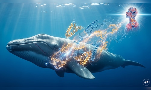
Whale protein discovery may unlock human lifespans of 200 years, scientists say
A protein discovered in exceptional abundance in bowhead whales can significantly improve DNA repair in human cells, according to a new study. This finding raises the possibility that human lifespans may eventually equal those of the…Continue Reading
-

German Researchers Identify Bacterial Consortium Capable of Degrading Persistent Phthalate Ester Plasticizers – geneonline.com
- German Researchers Identify Bacterial Consortium Capable of Degrading Persistent Phthalate Ester Plasticizers geneonline.com
- Ocean bacteria team up to break down biodegradable plastic MIT News
- A ‘consortium’ of bacteria cooperates to eat…
Continue Reading
-
Update, translation, cross-cultural adaptation, and validation of Nottingham Stroke Dressing Assessment into Spanish
Feigin, V. L. & Owolabi, M. O. Pragmatic solutions to reduce the global burden of stroke: a world stroke organization-Lancet Neurology Commission. Lancet Neurol. 22(12), 1160–1206 (2023).
Hou, S. et al. Global, regional, and national…
Continue Reading
-
A 'consortium' of bacteria cooperates to eat phthalate plasticizers that single microbes can't stomach – Phys.org
- A ‘consortium’ of bacteria cooperates to eat phthalate plasticizers that single microbes can’t stomach Phys.org
- EarthTalk: Scientists seek better ways to battle plastic with bacteria Arizona Daily Sun
- Ocean microbes are key to breaking down…
Continue Reading
-
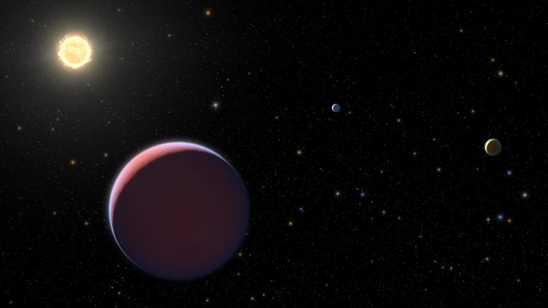
Even JWST can’t see through this planet’s massive haze
A newly studied exoplanet, Kepler-51d, is wrapped in an unusually dense layer of haze that may be hiding both what it is made of and how it formed. Using NASA’s James Webb Space Telescope (JWST), a team led by Penn State researchers took a closer…
Continue Reading
-
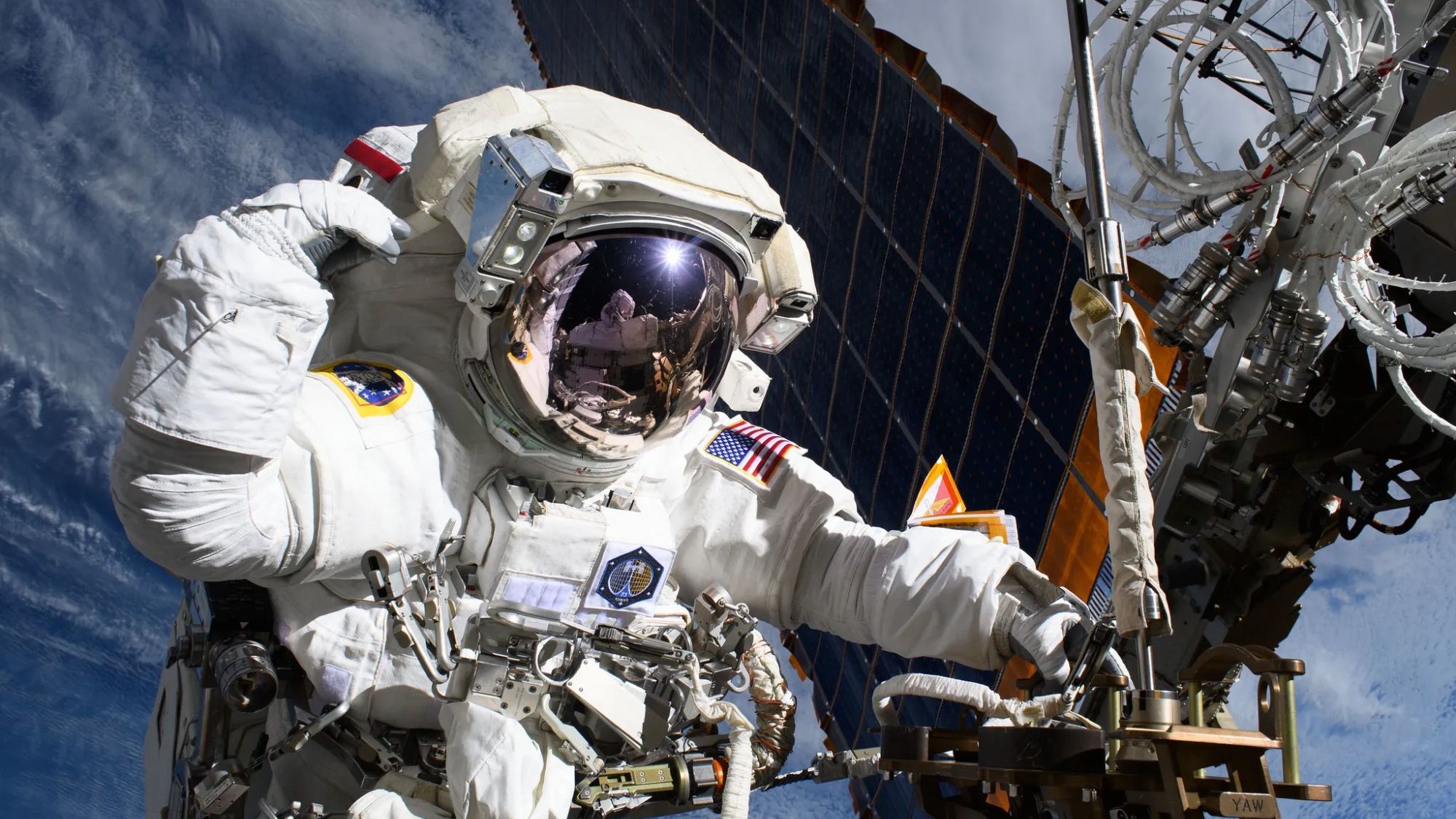
Watch live today: NASA astronauts conducting spacewalk delayed by ISS medical evacuation
Two NASA astronauts will conduct a long-delayed spacewalk today (March 18), and you can watch the action live.
Continue Reading
-
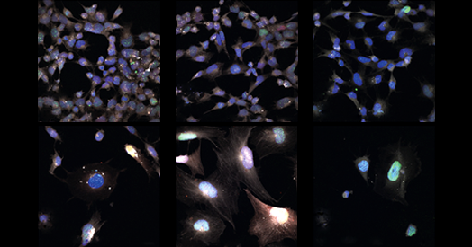
Signs of aging vary across brain cells
As cells age and acquire damage, they stop dividing and enter a comatose-like state. This natural process, called senescence, has several classic hallmarks, including the expression of cell cycle arrest genes and enlarged nuclei, and…
Continue Reading
-
Large-scale multi-omics profiling reveals environmental and evolutionary drivers of fungal phylogeographic and metabolic diversity
Case, N. T. et al. Fungal impacts on Earth’s ecosystems. Nature 638, 49–57 (2025).
Iliev, I. D. et al. Focus on fungi. Cell 187, 5121–5127 (2024).
Continue Reading
-
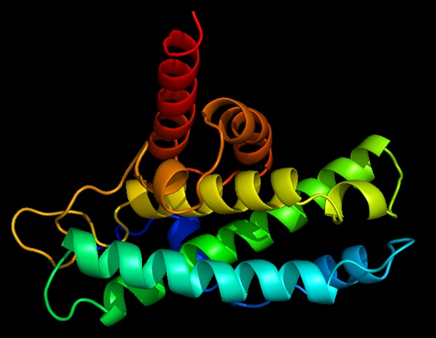
AlphaFold Database expands with millions of predicted protein complexes
A new collaboration between EMBL’s European Bioinformatics Institute (EMBL-EBI), Google DeepMind, NVIDIA, and Seoul National University has made millions of AI-predicted protein complex structures openly available through the…
Continue Reading