- Huge rotating structure of galaxies and dark matter is detected Reuters
- Astronomers spot one of the largest spinning structures ever found in the Universe EurekAlert!
- This Might Be the Biggest Thing in the Universe That Spins Gizmodo
- Science news…
Category: 7. Science
-
Huge rotating structure of galaxies and dark matter is detected – Reuters
-
Webb reveals double helium tails escaping from a 'hot Jupiter' – Phys.org
- Webb reveals double helium tails escaping from a ‘hot Jupiter’ Phys.org
- A ‘super-puff’ exoplanet is losing its atmosphere, and the James Webb Space Telescope had a look Space
- A Giant Helium Cloud Is Escaping From a Distant Planet, And JWST Just…
Continue Reading
-

From Artificial Intelligence to Mirror Molecules • The Revelator
The proliferation of artificial intelligence technologies, molecular manipulation, and literal sea changes are among the top issues a team of conservation experts anticipate will affect biodiversity in the year ahead and beyond, according to a…
Continue Reading
-
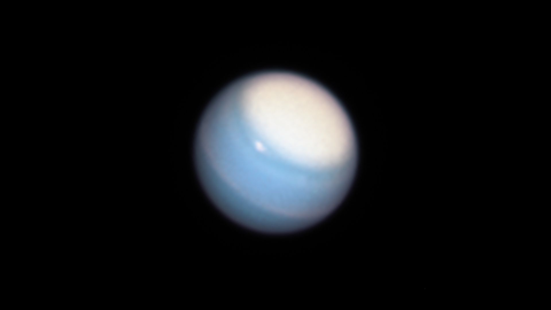
Uranus’s small moons are dark, red, and water-poor
This article was originally published at Eos. The publication contributed the article to Space.com’s Expert Voices: Op-Ed & Insights.
The solar system’s oddball planet has some pretty odd moons, too. The first infrared spectra of Uranus’s small…
Continue Reading
-

Swiss startup turns urine into plant fertilizer
When most people need to go number one, they find the nearest bathroom and don’t give half a thought to what happens to their pee once it disappears down the toilet or urinal. It turns out that the nitrogen in human urine can be used in…
Continue Reading
-

3I/ATLAS Is Carrying Ingredients for Life, NASA Finds
Methanol has long been considered a basic building block of life as we know it: the molecule plays a crucial role in producing the proteins and amino acids that make up DNA and RNA, upon which all known life is based.
Its discovery in…
Continue Reading
-
Young M Dwarfs Flare Activity Model: Towards Better Exoplanetary Atmospheric Characterisation
Space Weather & Heliophysics
…Continue Reading