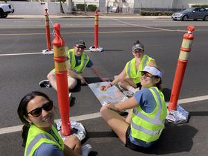
Astronomers are up in arms, protesting against a proposed constellation of tens of thousands of orbiting mirrors intended to reflect light onto ground-based solar power plants and SpaceX’s envisioned one million orbiting data centers.
The…
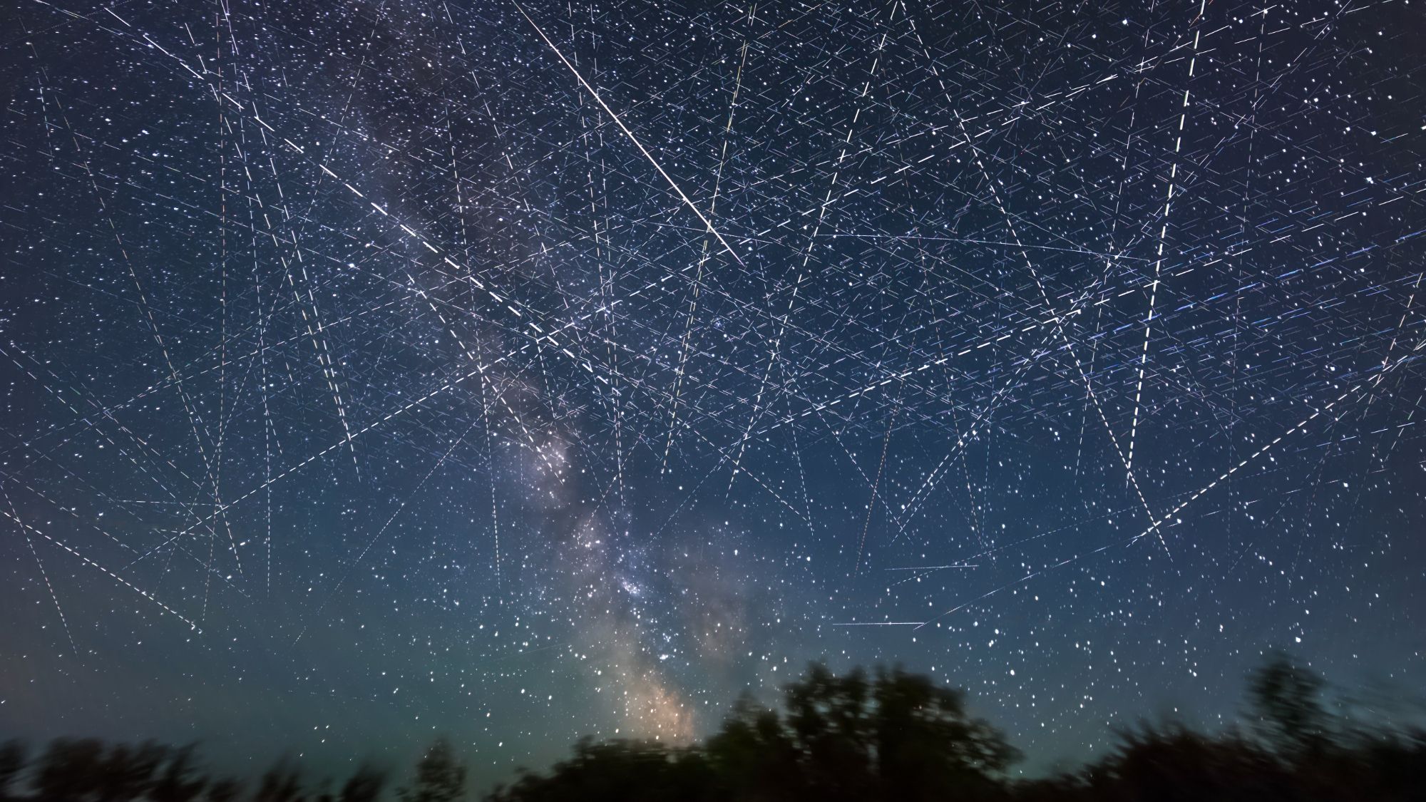
Astronomers are up in arms, protesting against a proposed constellation of tens of thousands of orbiting mirrors intended to reflect light onto ground-based solar power plants and SpaceX’s envisioned one million orbiting data centers.
The…
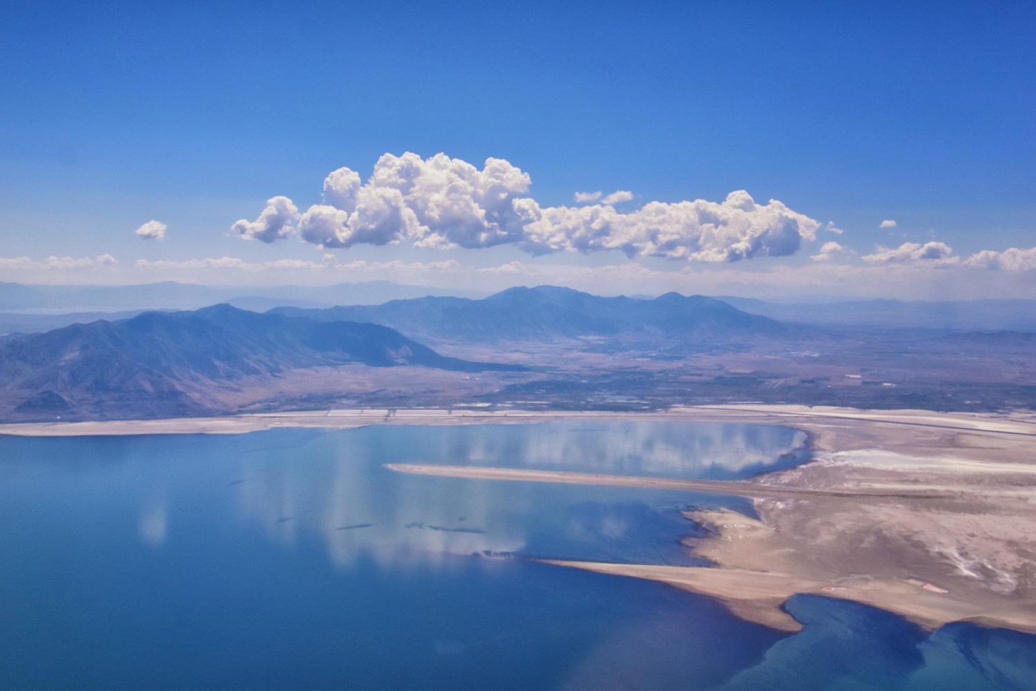
The Great Salt Lake in Utah is the largest inland saltwater body in the Western Hemisphere. However, beneath its shimmering, salty surface, scientists have discovered something surprising. There is a large layer of freshwater that reaches depths…

A team of astronomers led by Carnegie has uncovered the clearest evidence yet that a rocky planet outside our Solar System has an atmosphere. Using NASA’s James Webb Space Telescope (JWST), the researchers identified signs of gas surrounding an…
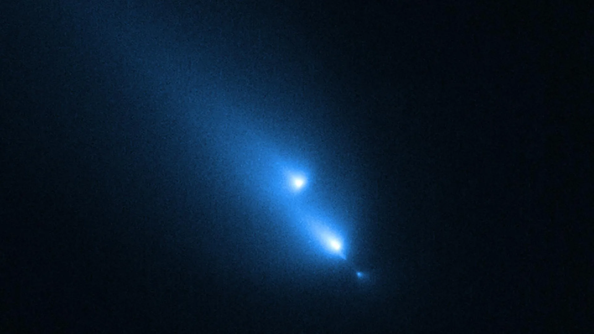
In a remarkable stroke of luck, NASA’s Hubble Space Telescope observed a comet in the middle of breaking apart. The odds of witnessing such an event at exactly the right moment are extremely low. The findings were published in the journal Icarus.

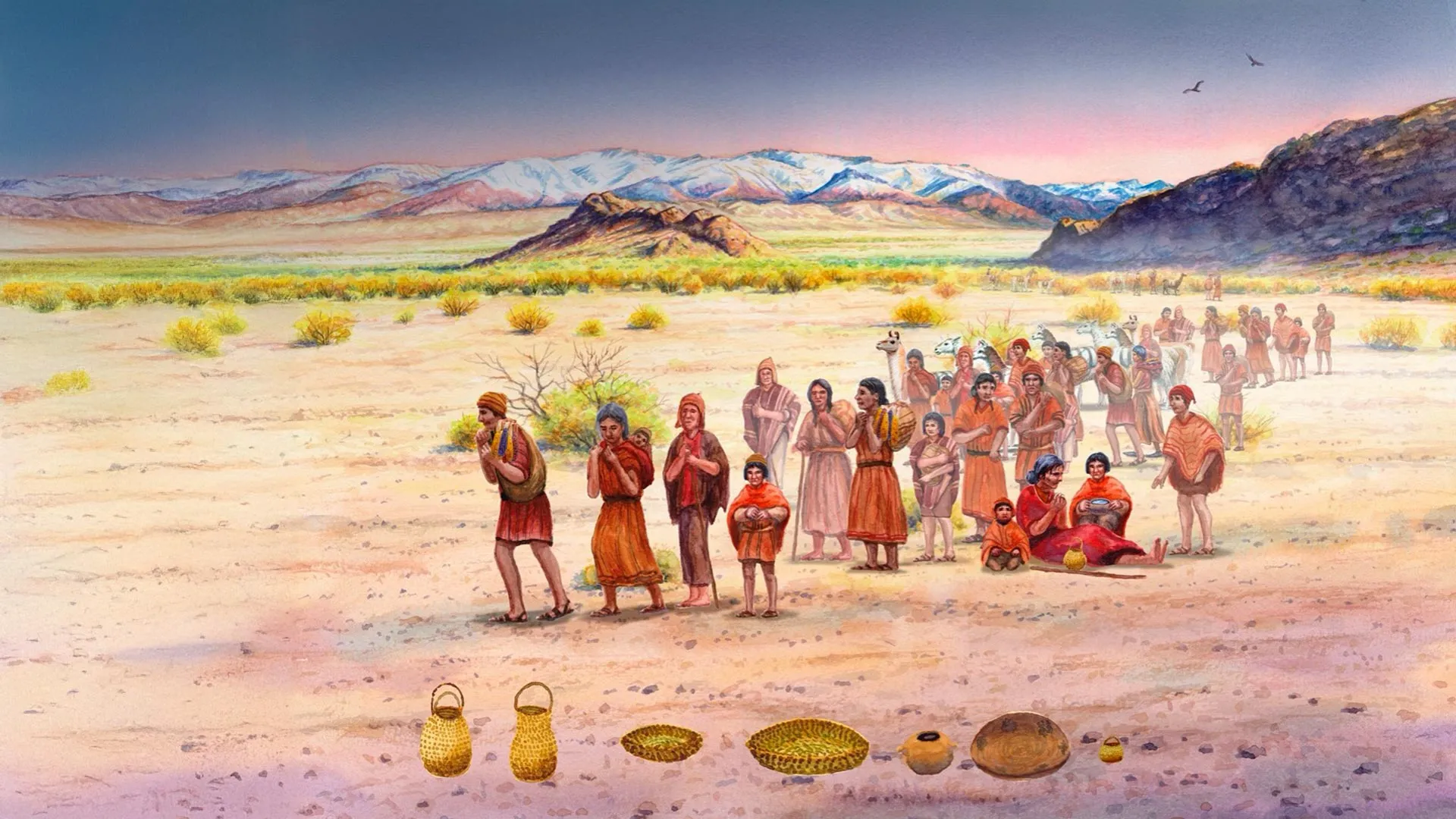
A new interdisciplinary study published in Nature traces more than 2,000 years of population history in Argentina’s Uspallata Valley (UV), a key southern edge of ancient Andean farming. The research offers new insight into how agriculture…
BEIJING, March 21 (Xinhua) — A global research team has used a new technique to capture minute-scale structural changes in farmland soil, revealing how farming practices influence soil water…
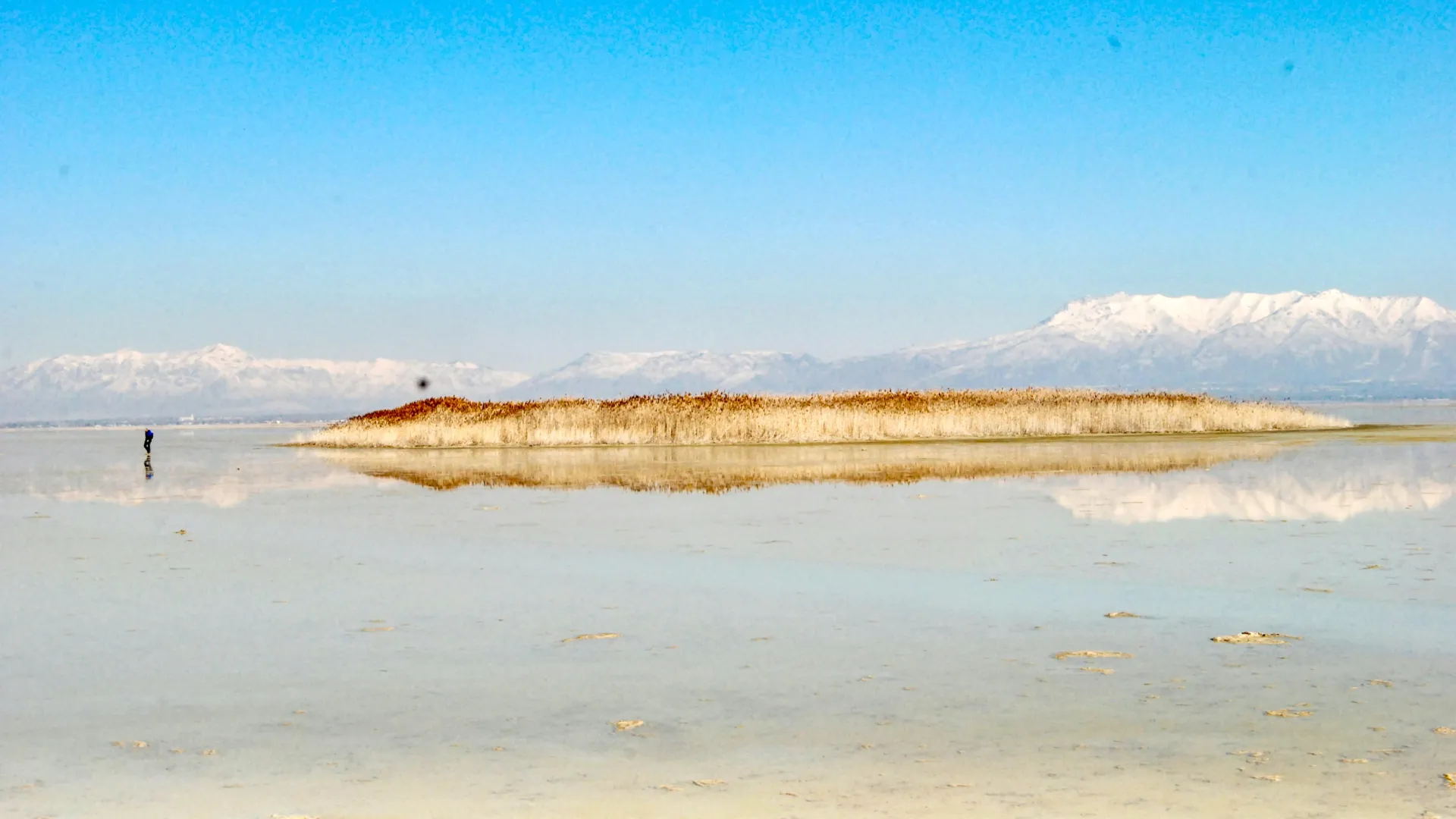
A newly identified underground freshwater system beneath the Great Salt Lake is becoming clearer thanks to a study that used airborne electromagnetic (AEM) surveys to map geologic formations below Farmington Bay and Antelope Island along the…
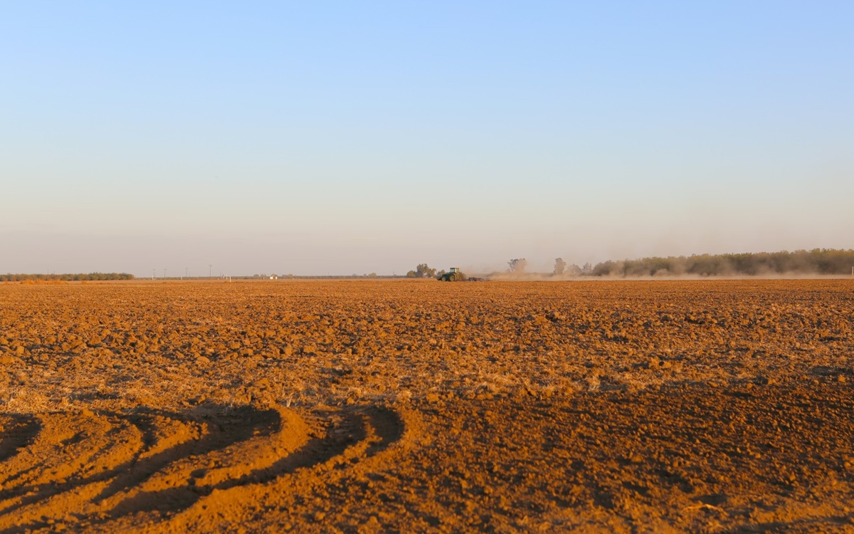
The team, led by the Institute of Geology and Geophysics at the Chinese Academy of Sciences, employed distributed fiber-optic sensing, installed across an experimental farm at Harper Adams University in the United Kingdom, to…