- Earth’s Days Are Getting Longer At Unprecedented Rate Not Seen In 3.6 Million Years – Here’s Why IFLScience
- Climate change slows Earth’s spin: Day lengthening unprecedented in 3.6 million years | Newswise Newswise
- Rising sea levels are…
Category: 7. Science
-
Earth's Days Are Getting Longer At Unprecedented Rate Not Seen In 3.6 Million Years – Here's Why – IFLScience
-

NASA begins building nuclear-powered Dragonfly drone for 2028 launch to Saturn moon Titan
NASA is one step closer to sending a drone mission to another world.
Technicians at the Johns Hopkins Applied Physics Laboratory (APL) in Maryland have begun building and testing the nuclear-powered Dragonfly rotorcraft, which will launch toward…
Continue Reading
-

Robotics Information | AZoRobotics.com – Page not found
Terms
While we only use edited and approved content for Azthena
answers, it may on occasions provide incorrect responses.
Please confirm any data provided with the related suppliers or
…Continue Reading
-

Column | New tech helps us hear these nature sounds beyond normal human range
AMHERST, Mass. — Brian House, in long beard and muck boots, leads me through a pine forest on a cold afternoon…
Continue Reading
-
Astronomers Detect a Vast Hidden Web of Galaxies in the Early Universe – SciTechDaily
- Astronomers Detect a Vast Hidden Web of Galaxies in the Early Universe SciTechDaily
- Enormous 3D map of the universe shows brilliant ‘sea of light’ near the cosmic dawn Live Science
- HETDEX data reveal a vast ‘sea of light’ between early galaxies
Continue Reading
-
NASA’s DART Impact Actually Changed an Asteroid System’s Orbit Around the Sun – SciTechDaily
- NASA’s DART Impact Actually Changed an Asteroid System’s Orbit Around the Sun SciTechDaily
- NASA images show binary asteroids passing dust and debris to each other Earth.com
- How a Tiny Nudge From NASA’s DART Spacecraft Showed We Can Actually…
Continue Reading
-
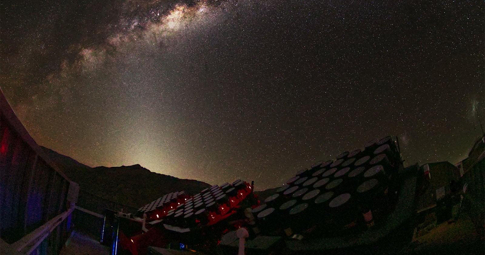
1,140 Canon 400mm f/2.8 Lenses Will Search Space for Dark Matter
The partially completed MOTHRA array below the Milky Way at El Sauce Observatory in Chile. Several of the telescope’s mounts are working now. The full array is expected to be complete by the end of the year. The world’s largest…
Continue Reading
-
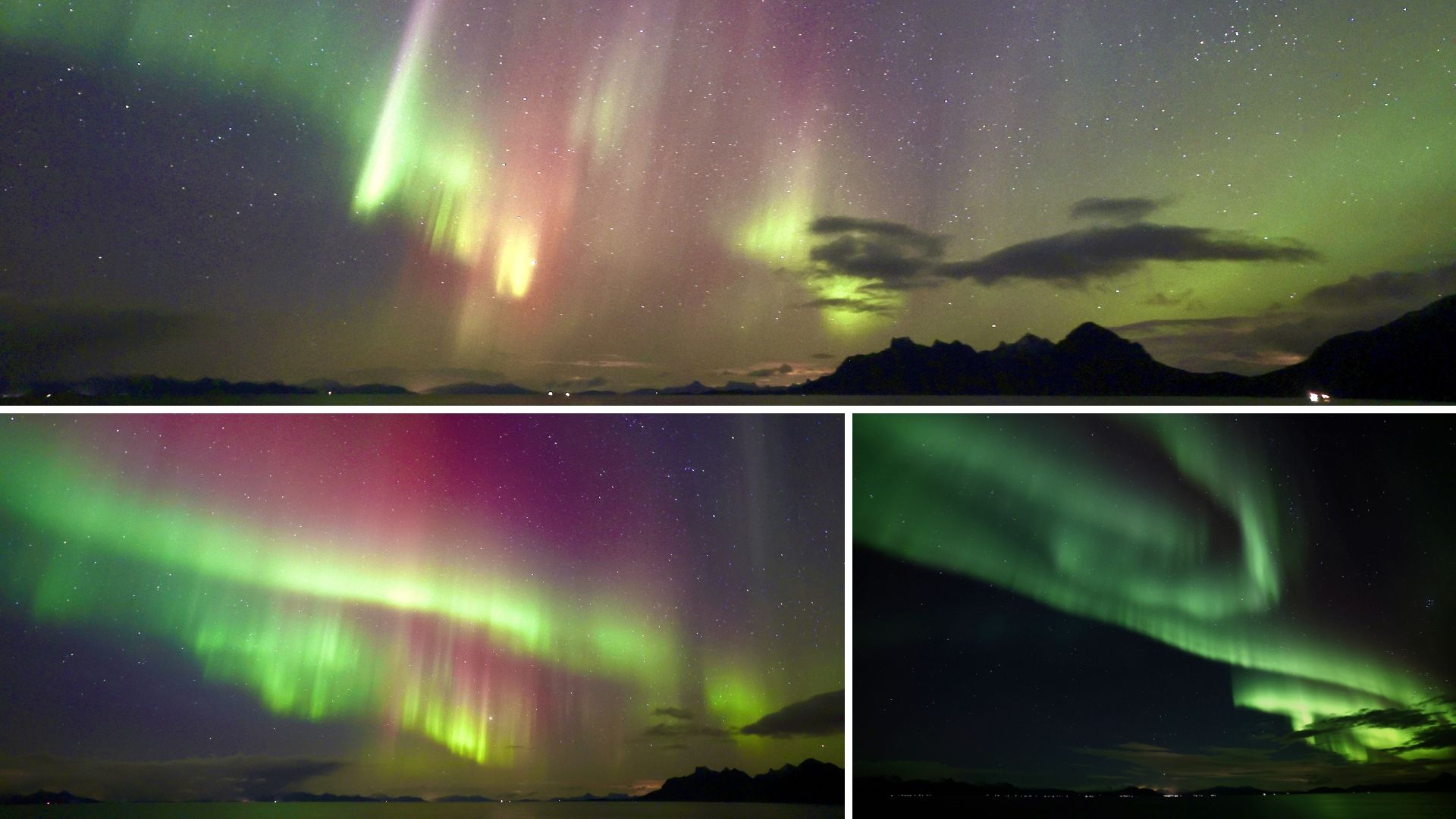
Why March is the best month to see the northern lights
March is widely considered one of the best times of year to see the northern lights, thanks to a seasonal boost in geomagnetic activity around the spring equinox.
The northern lights, or aurora borealis, occur when energetic particles from the…
Continue Reading
-
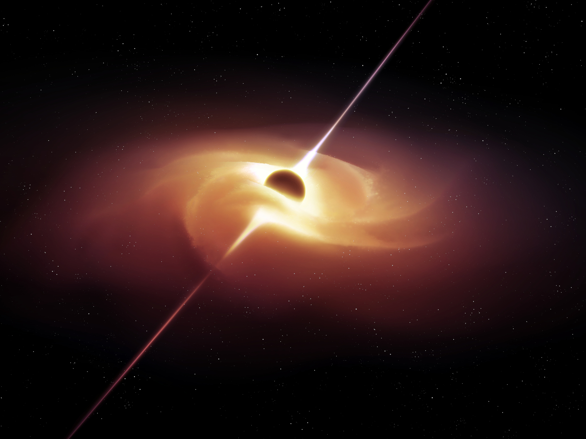
Oval orbit reveals new clues about black hole-neutron star collisions
image: ©Nazarii Neshcherenskyi | iStock Scientists have found the first strong evidence that a neutron star and a black hole merged while travelling in an oval orbit rather than the near-perfect circle scientists long…
Continue Reading
-
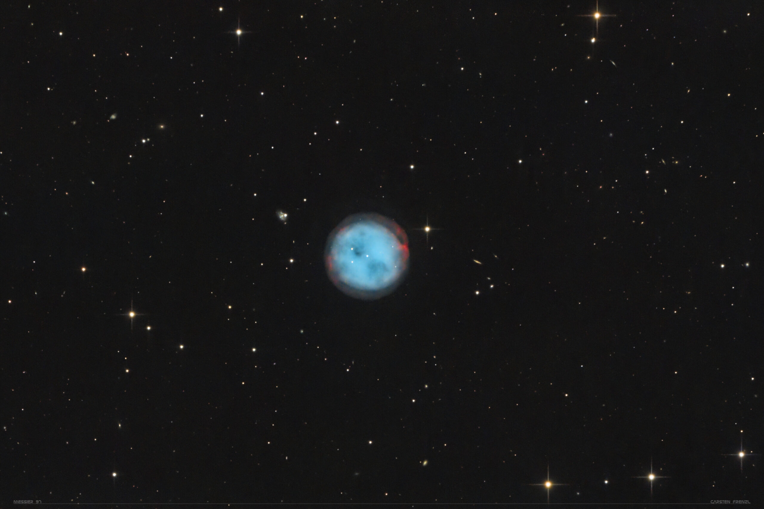
Look into the eyes of the Owl
The Owl Nebula, located in Ursa Major the Great Bear, is one of four planetary nebulae on Messier’s list.
Looking for a sky event…
Continue Reading