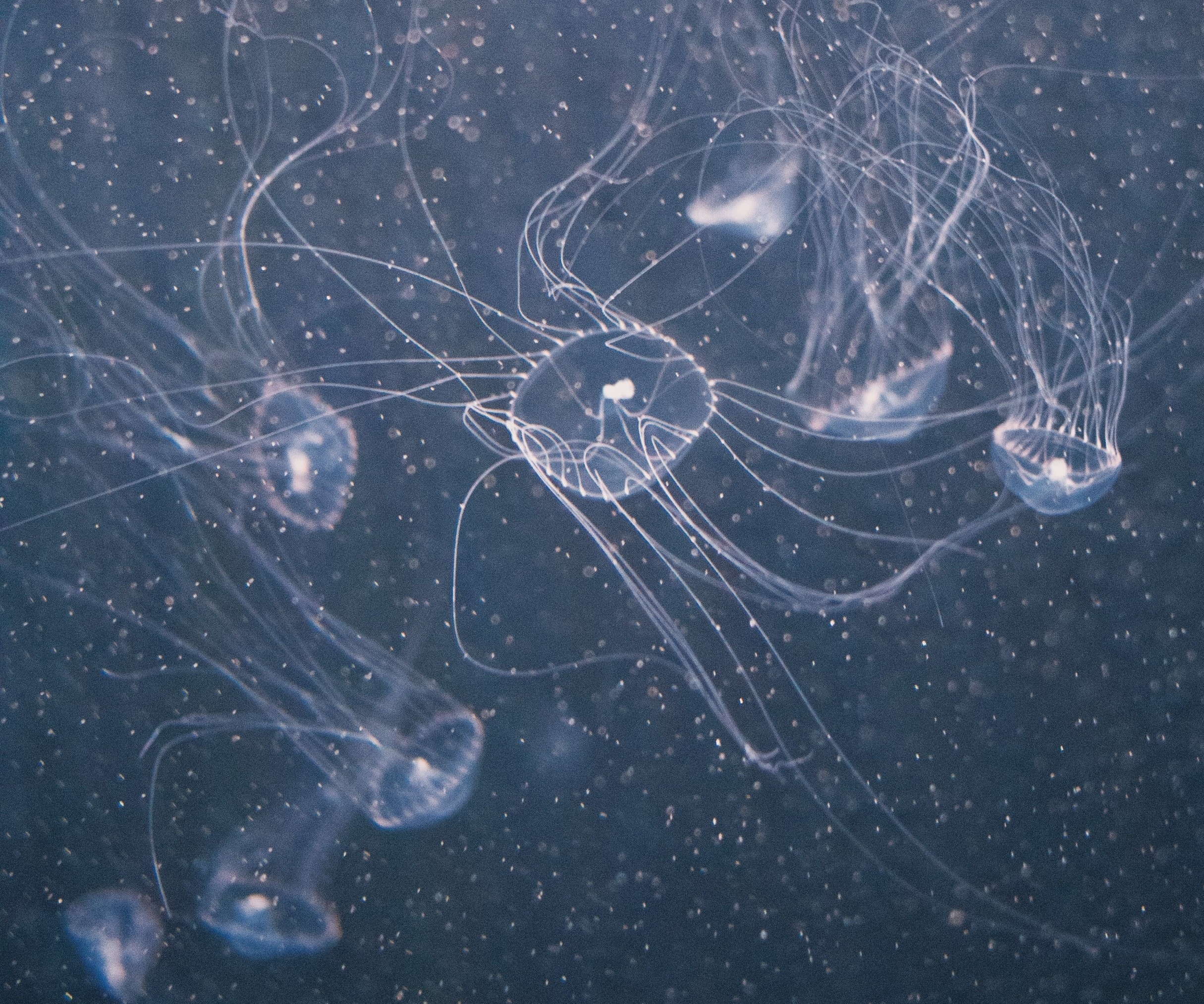
Imagine snow falling in the deep ocean. It sounds strange, but something very similar happens every day beneath the waves. Tiny particles drift slowly downward through the water like snowflakes.
Scientists call this marine snow. This quiet fall…
Imagine snow falling in the deep ocean. It sounds strange, but something very similar happens every day beneath the waves. Tiny particles drift slowly downward through the water like snowflakes.
Scientists call this marine snow. This quiet fall…

Researchers at Cardiff University have uncovered how a particularly severe form of DNA damage arises – shedding new light on mutation processes that contribute to cancer and inherited genetic conditions.
The study, led by Dr Greg Ngo and…

“When lahars are significantly large, the signal can be detected up to 20 minutes before the mudslide reaches the monitoring station,” says Béjar. This all depends on the AI’s ability to correctly distinguish a lahar from another type of…

Researchers have shed new light on how a unusual rock formation in Oman was created, which could reveal new details about the Earth’s ability to store carbon dioxide (CO2) for millions of years.
The study, led by Keele University, in…
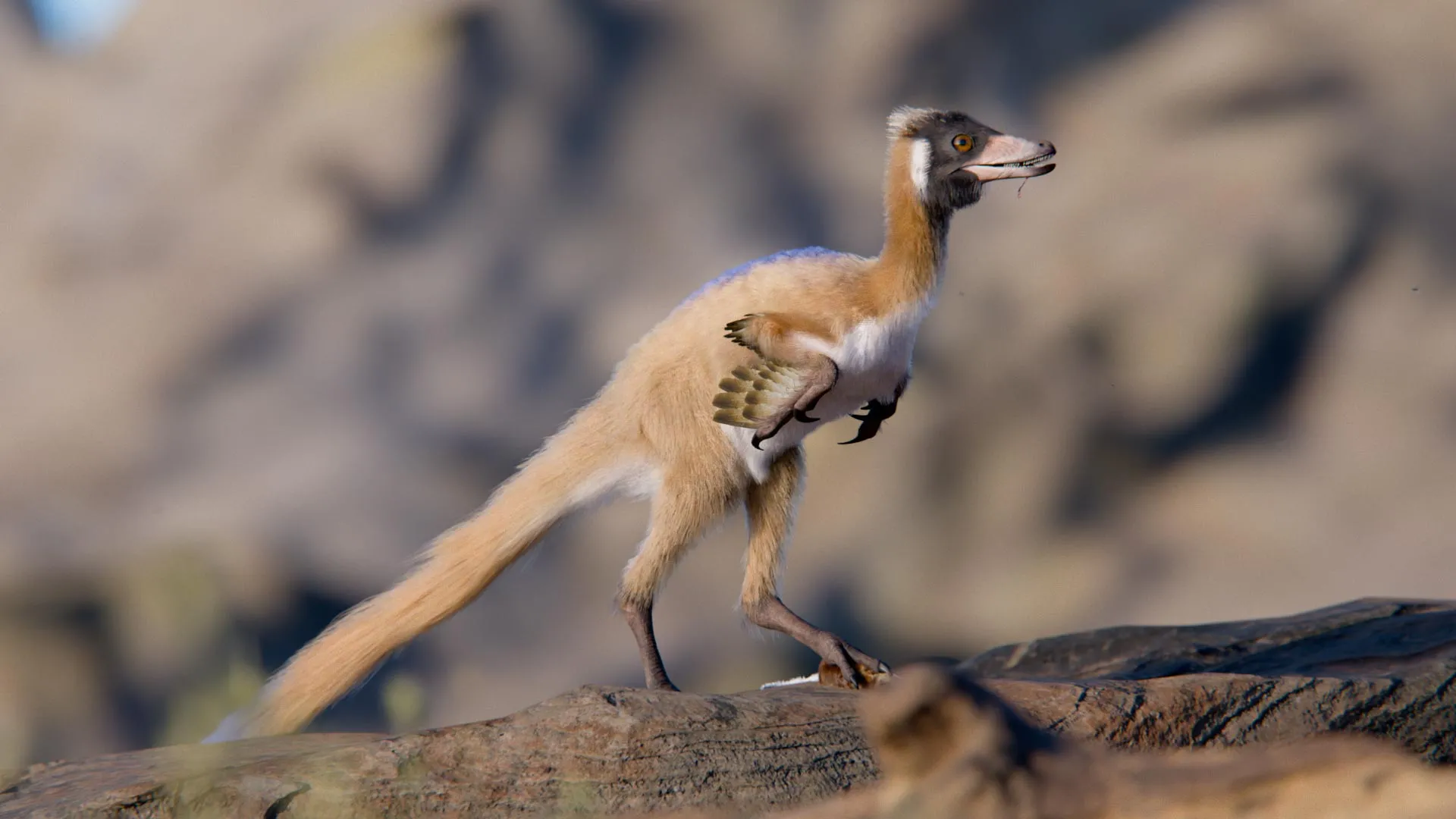
Researchers have identified a 90 million year old fossil that helps solve a long standing mystery about a strange group of prehistoric animals. The discovery was led by University of Minnesota Twin Cities scientist Peter Makovicky along with…
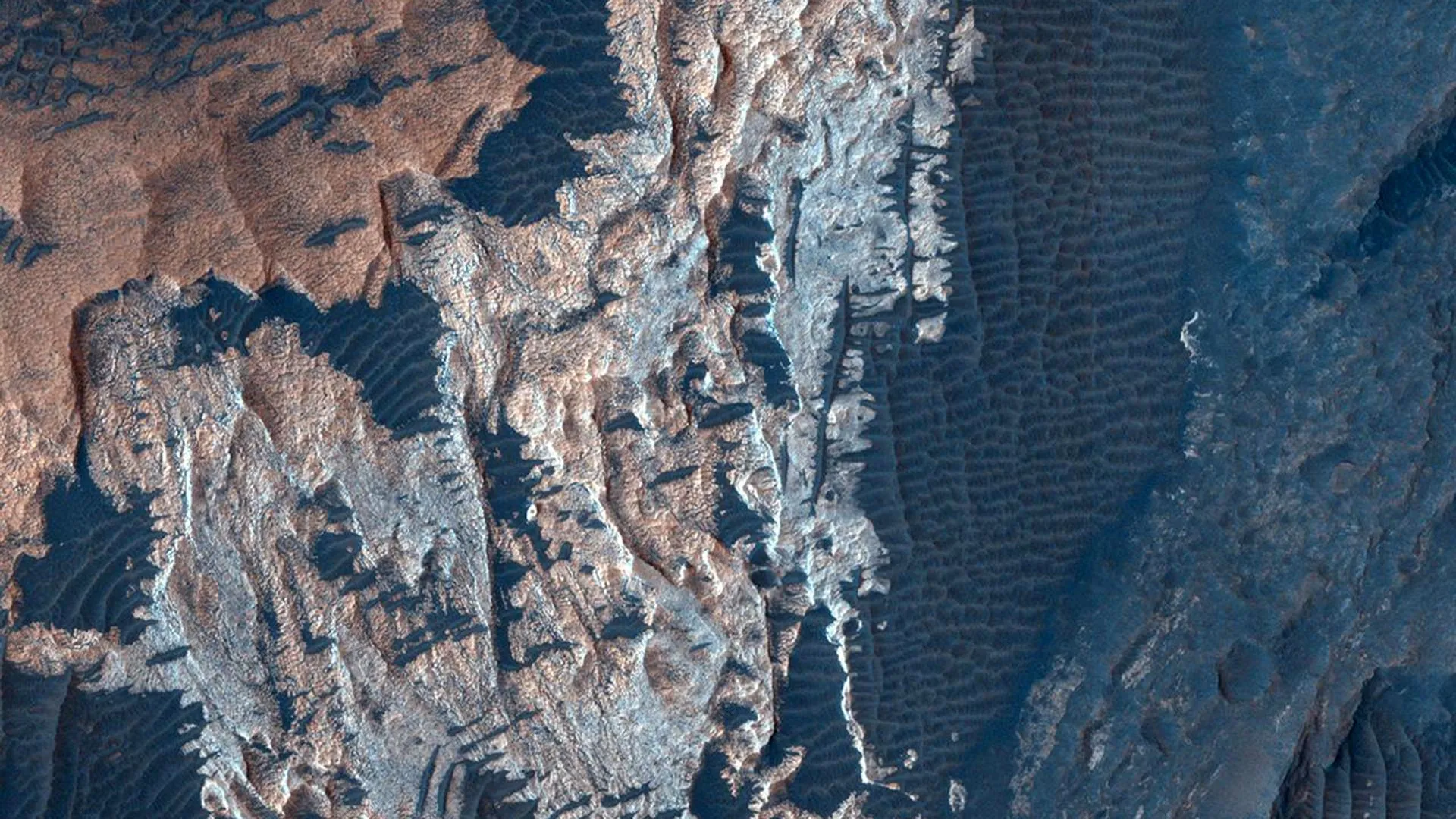
A new study published in Nature Communications reports the detection of an iron sulfate on Mars that may represent a previously unknown mineral. Sulfur is abundant on Mars and commonly combines with other elements to create sulfate minerals. On…

Long before humans cultivated crops or sailed between continents, a group of plant viruses was already evolving among wild plants in Eurasia. According to a new international study published in Plant Disease, the ancestors of modern…
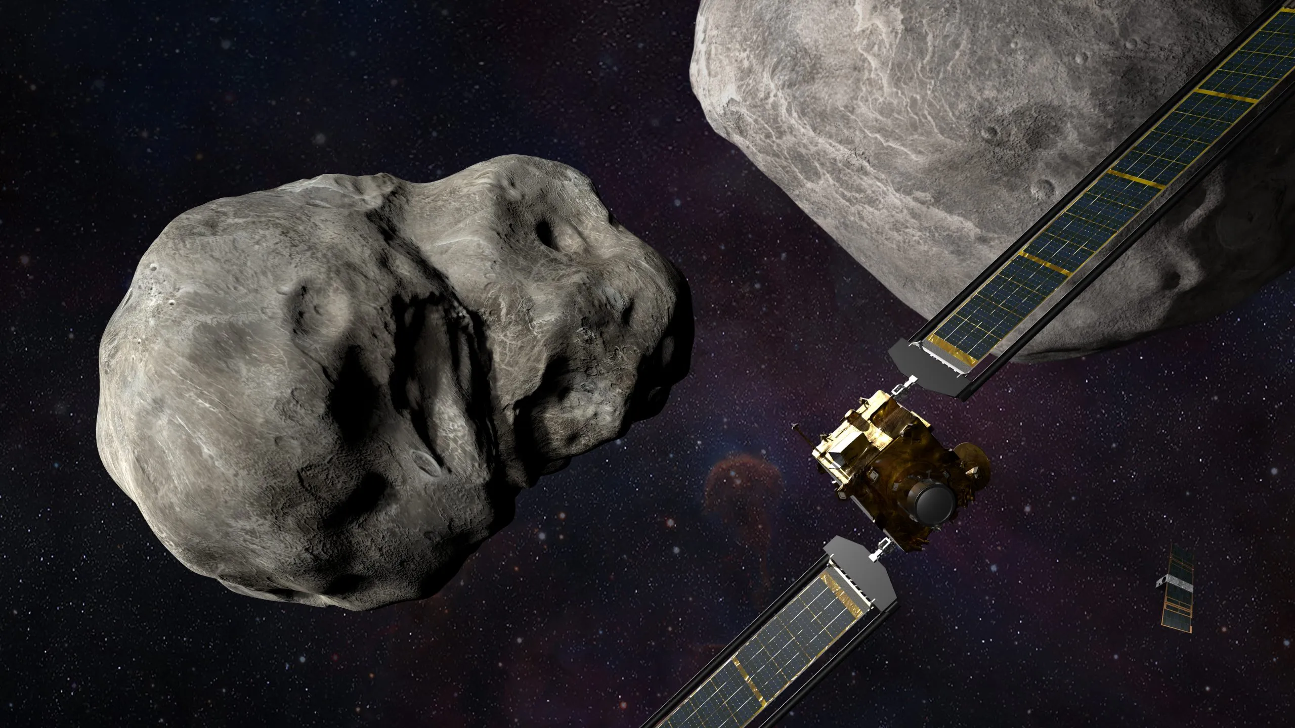
Around 15 percent of near-Earth asteroids have small moons, which suggests binary systems are fairly common. Many of the larger asteroid pairs have likely formed via collisions in the main belt; however, smaller ones, such as near-Earth binaries…
…