Long before humans cultivated crops or sailed between continents, a group of plant viruses was already evolving among wild plants in Eurasia. According to a new international study published in Plant Disease, the ancestors of modern…
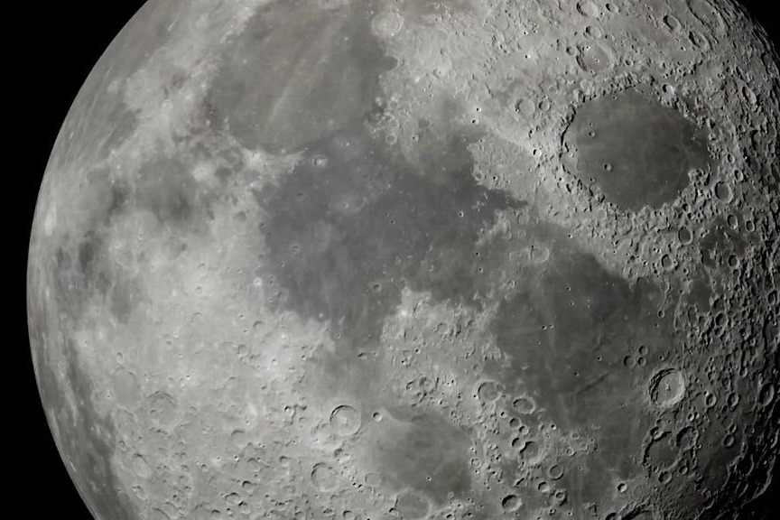


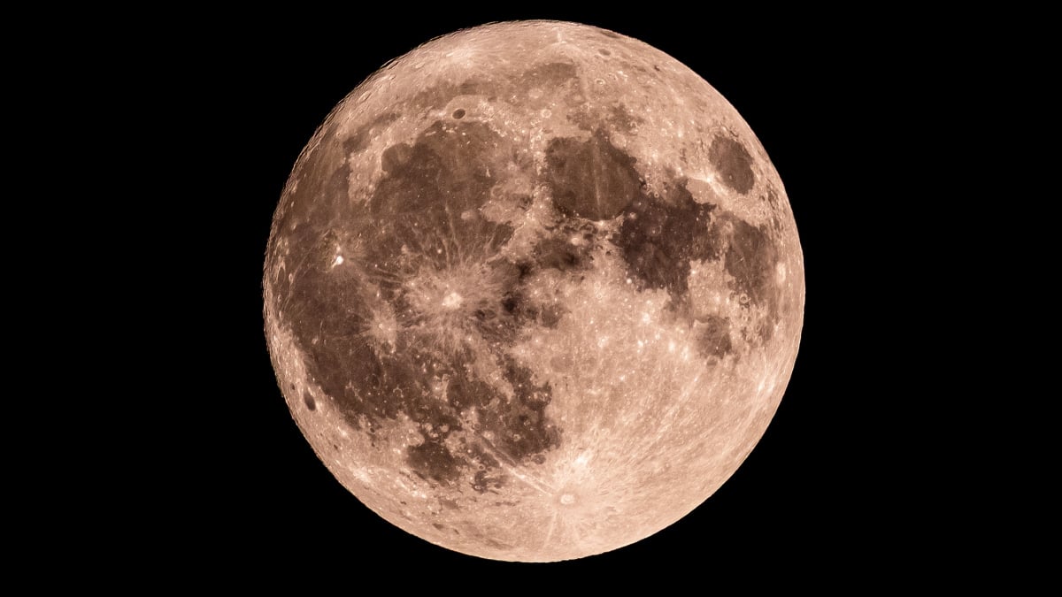
Can you make out any features on the Moon’s surface tonight? There’s plenty to see, so here’s what to look for when you look up.
What is today’s Moon phase?
As of Tuesday, March…