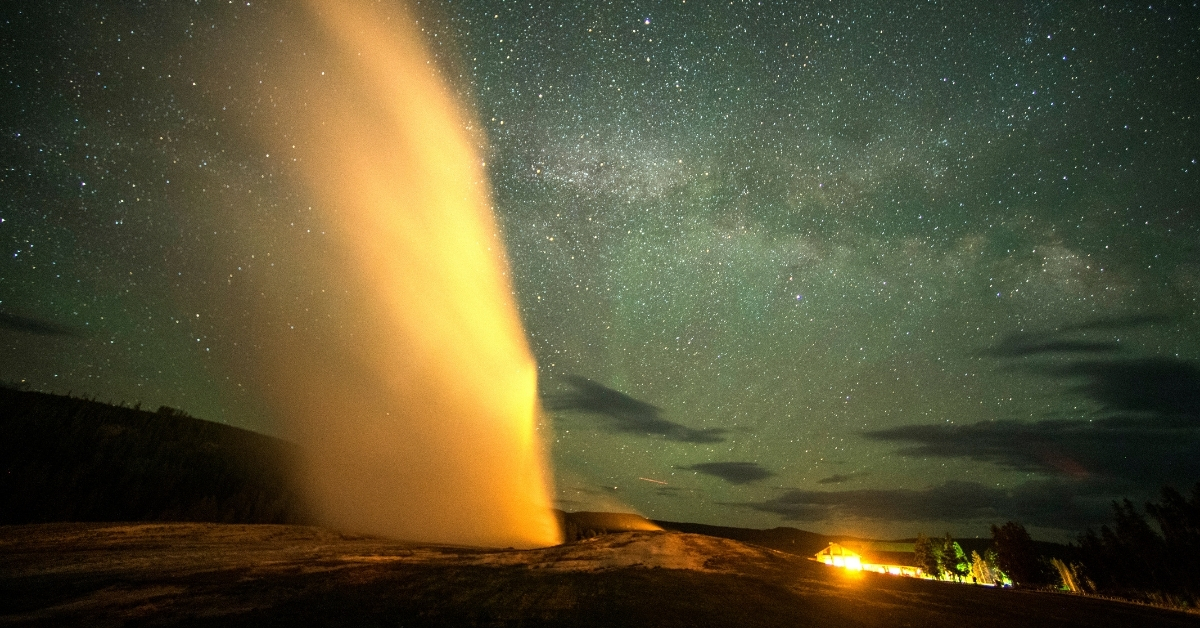
The geysers at Yellowstone National Park are still very much alive, and one of them just reminded us. The Echinus Geyser is the largest acidic geyser on earth, and it just started erupting again for the first time in years, reports ABC…
Category: 7. Science
-

US unveils bizarre-legged robots that self-repair and survive damage
Engineers at Northwestern University have developed a new class of modular robots called “legged metamachines.”
These machines possess built-in athletic intelligence that adapts on the go.
The robots are composed of autonomous,…
Continue Reading
-
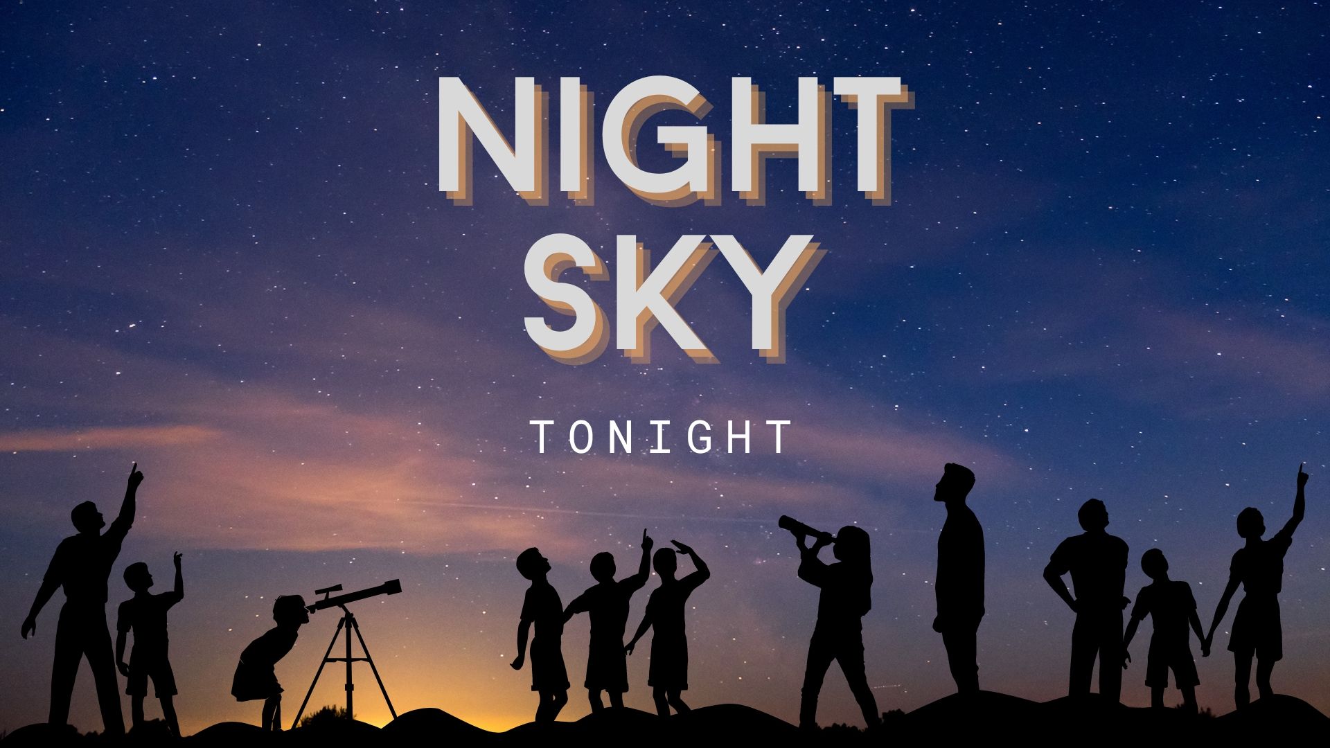
Night sky tonight (March 9) — The moon meets Antares in the predawn sky
Refresh
Monday, March 9: Northern lights season peaks (after dark)
Auroras become more likely in mid-March. (Image credit: Steven Robinson Pictures via Getty Images) With the fading of the full moon’s light and…
Continue Reading
-
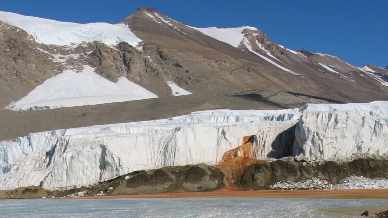
Study explains Antarctica’s mystery Blood Falls
Scientists say they have resolved a long-standing Antarctic mystery with new research in the journal Antarctic Science that identifies the physical engine driving Blood Falls, the rust-red outflow from Taylor Glacier. Measuring devices caught a…
Continue Reading
-
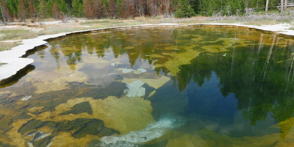
Mushroom Pool: The subtle spring with the spectacular story
Yellowstone Caldera Chronicles is a weekly column written by scientists and collaborators of the Yellowstone Volcano Observatory. This week’s contribution is from Michael Poland, geophysicist with the U.S. Geological Survey and…
Continue Reading
-
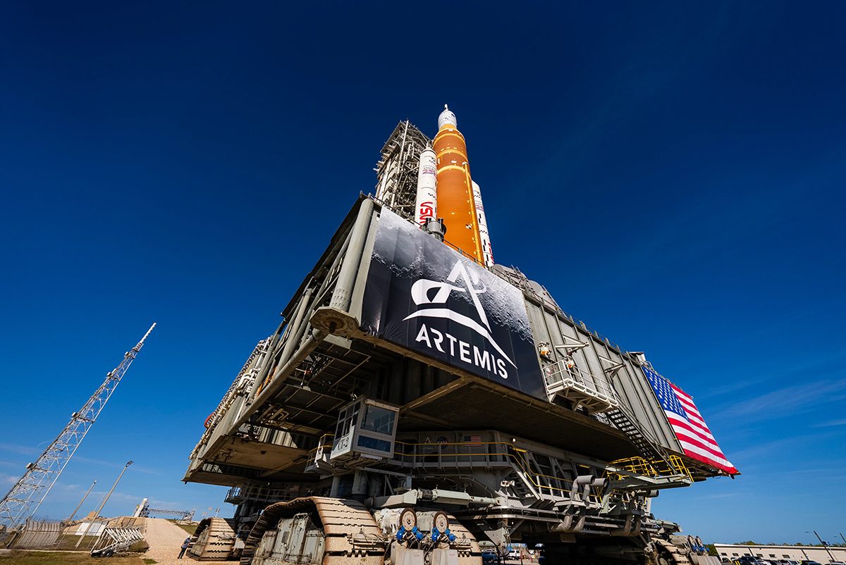
Is anyone else bored of Artemis already? Why I’m not over the Moon about NASA’s new lunar missions
What is the point of sending humans to the Moon, when we could carry out the same lunar science but with rovers and robots instead? Why risk lives?
Following Artemis I’s successful dry run in 2022, NASA’s next mission in the Artemis programme…
Continue Reading
-

Rare porpoise mating behaviour captured by drones off Shetland
Rare harbour porpoise mating behaviour has been captured by drones off Shetland’s coast.
The footage, taken between 2019 and 2023, provides one of the most detailed records of…
Continue Reading
-
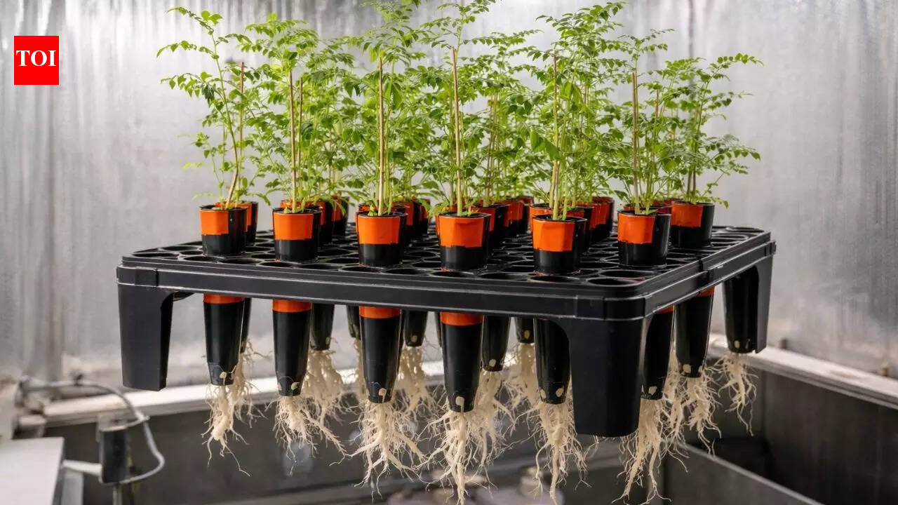
Chickpeas Grow In Moon Soil: Can chickpeas grow in Moon soil? New study reveals surprising results for space farming |
As the US plans a long-duration mission to the Moon under the Artemis Programme, one of the biggest challenges that the scientists face is what astronauts will eat in space. This challenge has led researchers to explore whether crops can be…
Continue Reading
-
Utilization of machine learning to identify lower extremity biomechanical predictors of rupture in a validated cadaveric model of ACL injury
Awan, M. J., Rahim, M. S. M., Salim, N., Rehman, A. & Garcia-Zapirain, B. Automated knee mr images segmentation of anterior cruciate ligament tears. Sensors 22, 1552 (2022).
Putera, K. H. et…
Continue Reading
-
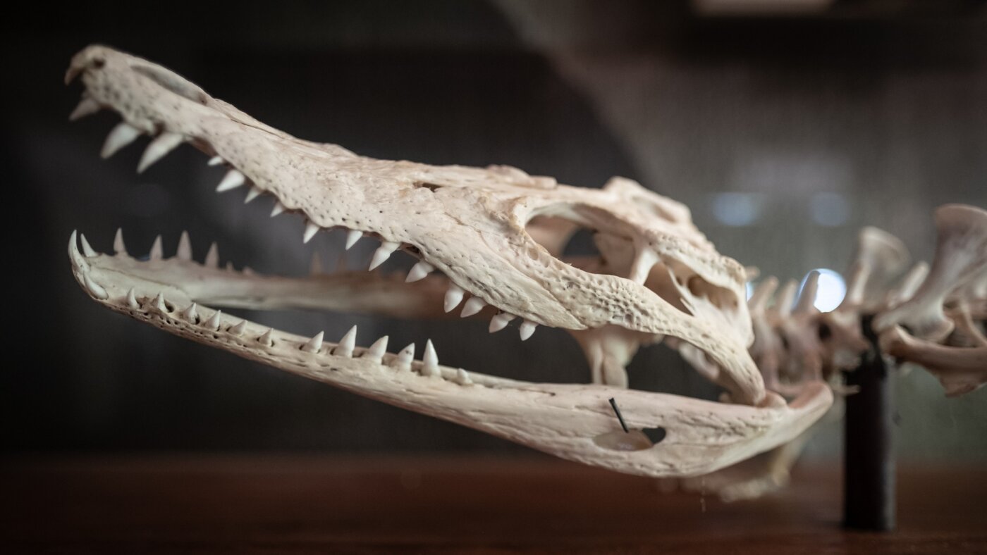
What crocodile bones teach us about dinosaurs : Short Wave : NPR
Historically, dinosaur ages have been estimated using the growth rings in their bones — one ring per year. But new…
Continue Reading