New scientific analysis from the Antarctic Peninsula warns that decisions made in this decade will shape the southern continent’s future “for centuries to come” and have worldwide impacts, as a university report detailed.
What’s happening?
The
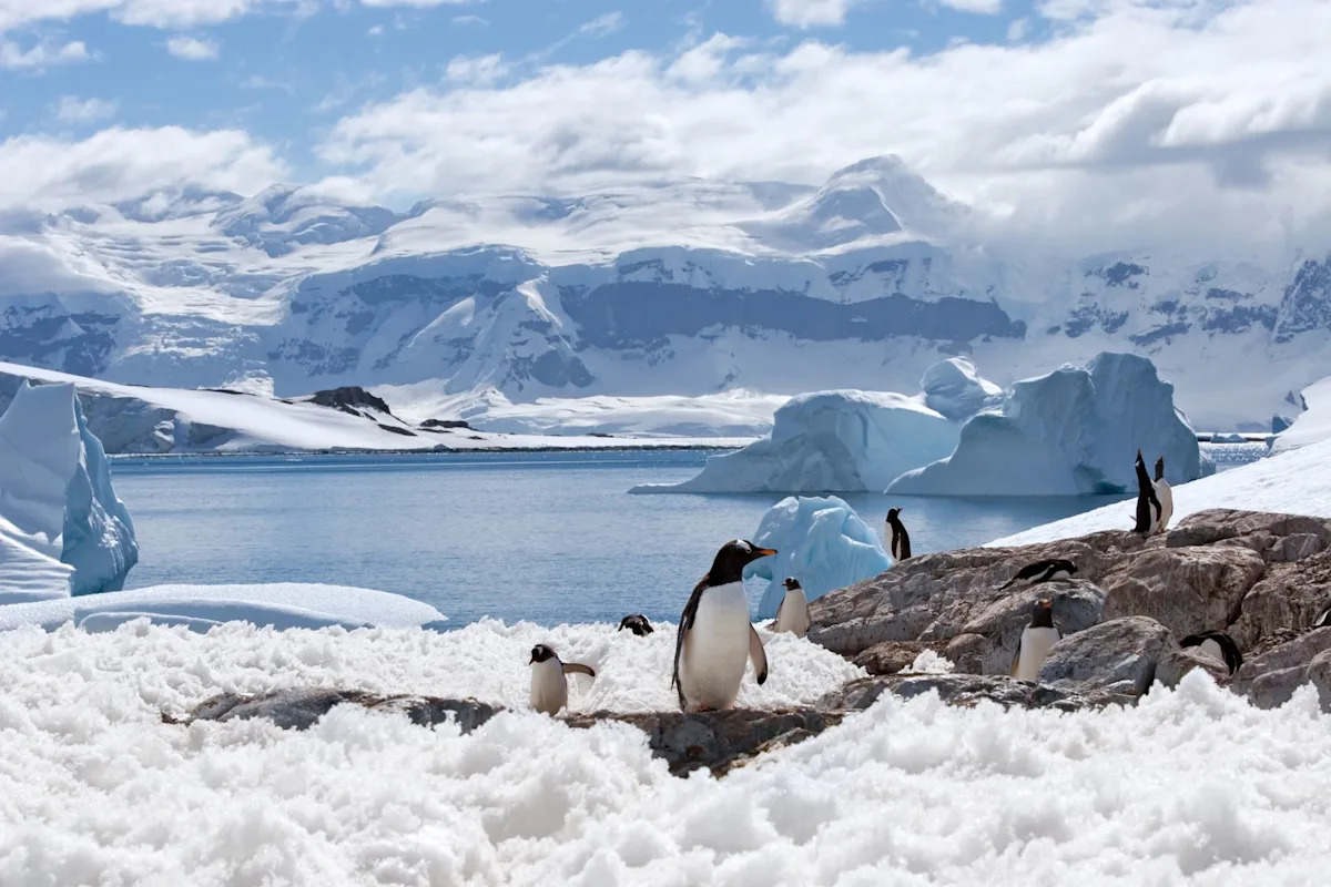
New scientific analysis from the Antarctic Peninsula warns that decisions made in this decade will shape the southern continent’s future “for centuries to come” and have worldwide impacts, as a university report detailed.
The

NASA’s DART actually changed the orbit of an Asteroid around the Sun.
We knew that DART changed the orbit of Dimorphos, but that was orbiting another larger asteroid called Didymos. Now, scientists have…
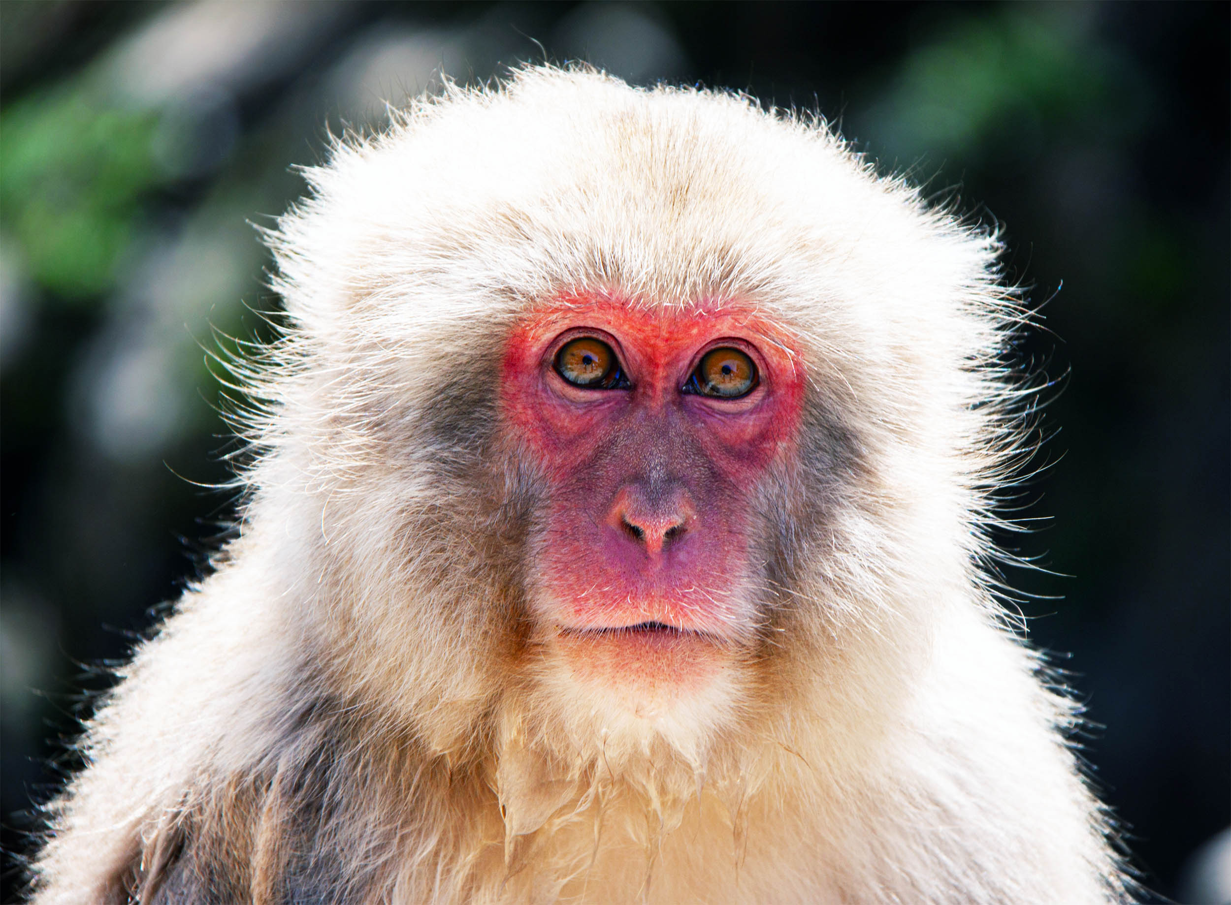
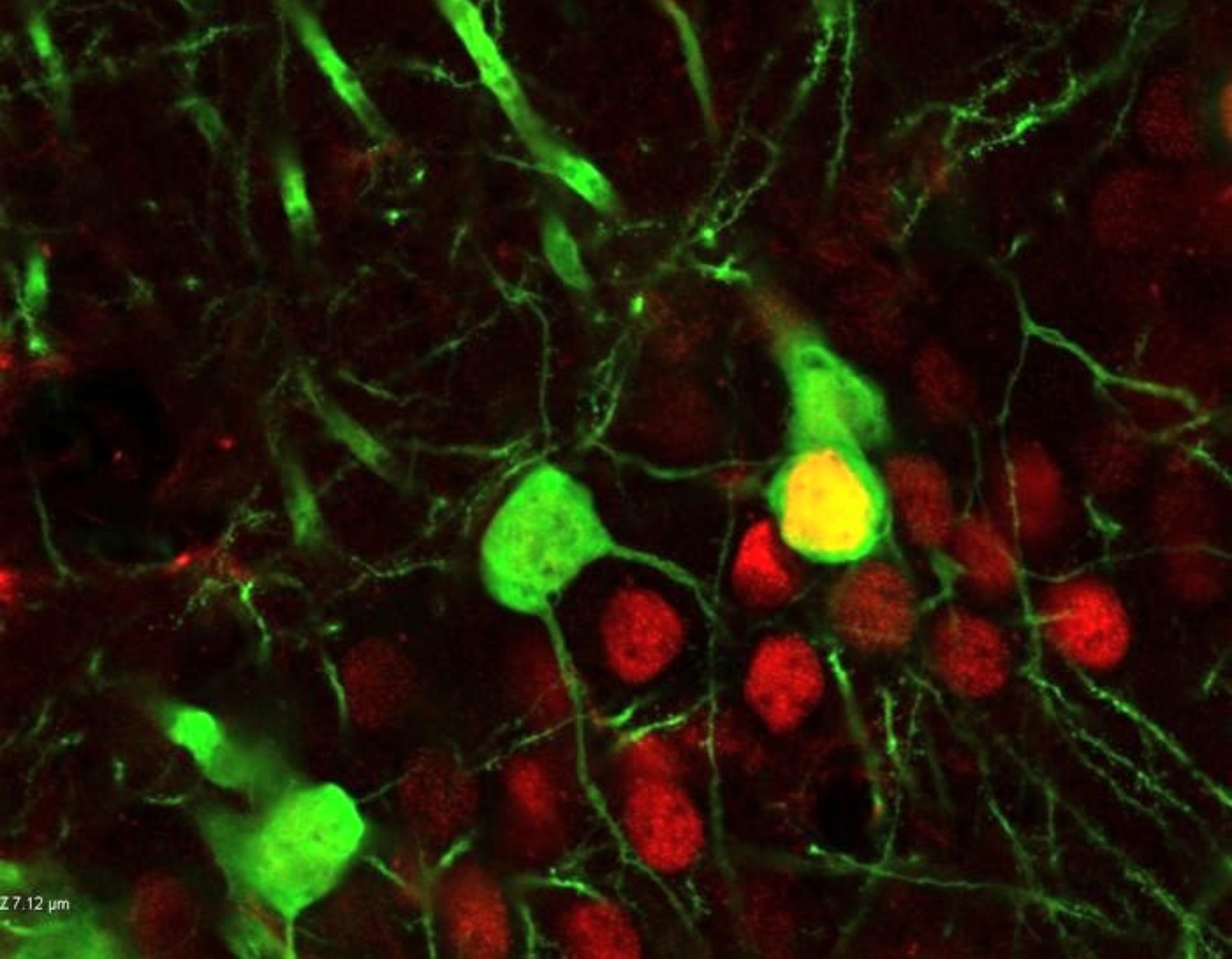
Two macaque monkeys have demonstrated the ability to tap in time with musical rhythms and maintain that timing even when rewards disappear.
The result places a key element of musical timing inside a primate brain, complicating the long-held belief…
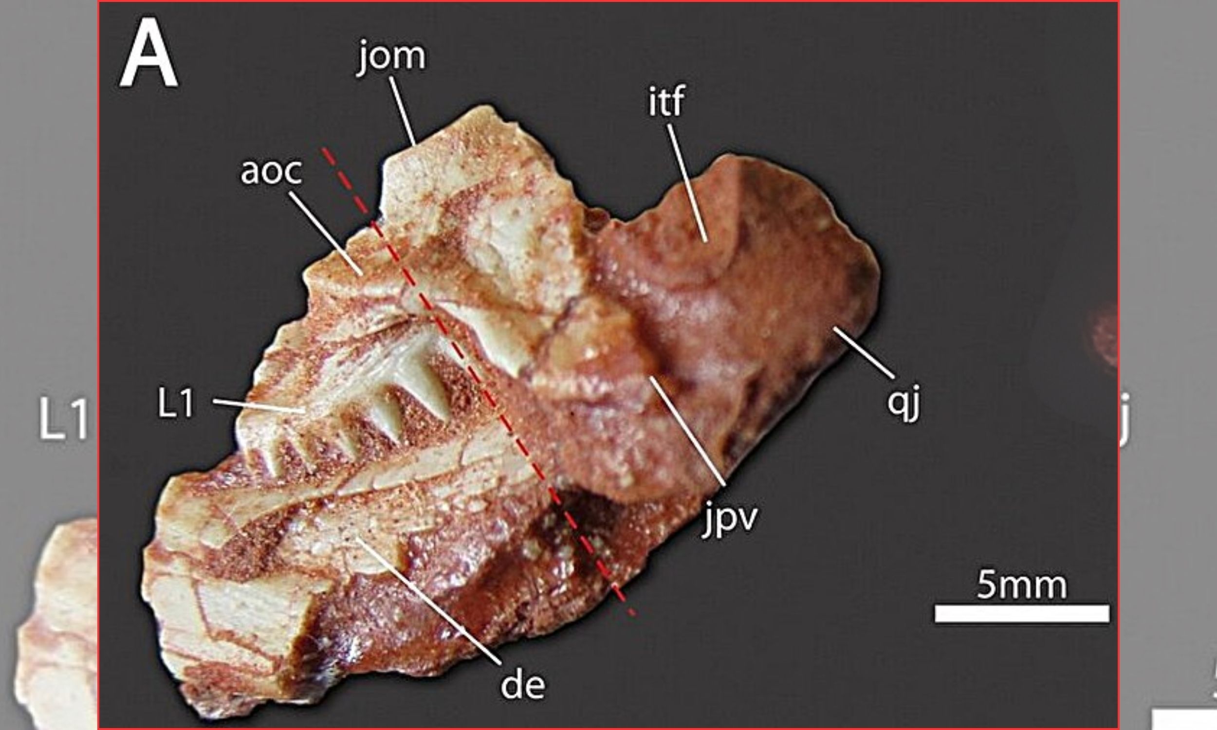
A fossil skull barely 1 inch long has proved to be a newborn reptile from Brazil’s Late Triassic, a period more than 230 million years ago when early reptiles dominated land ecosystems.
Its tiny jaw already carries the cutting surfaces seen in…
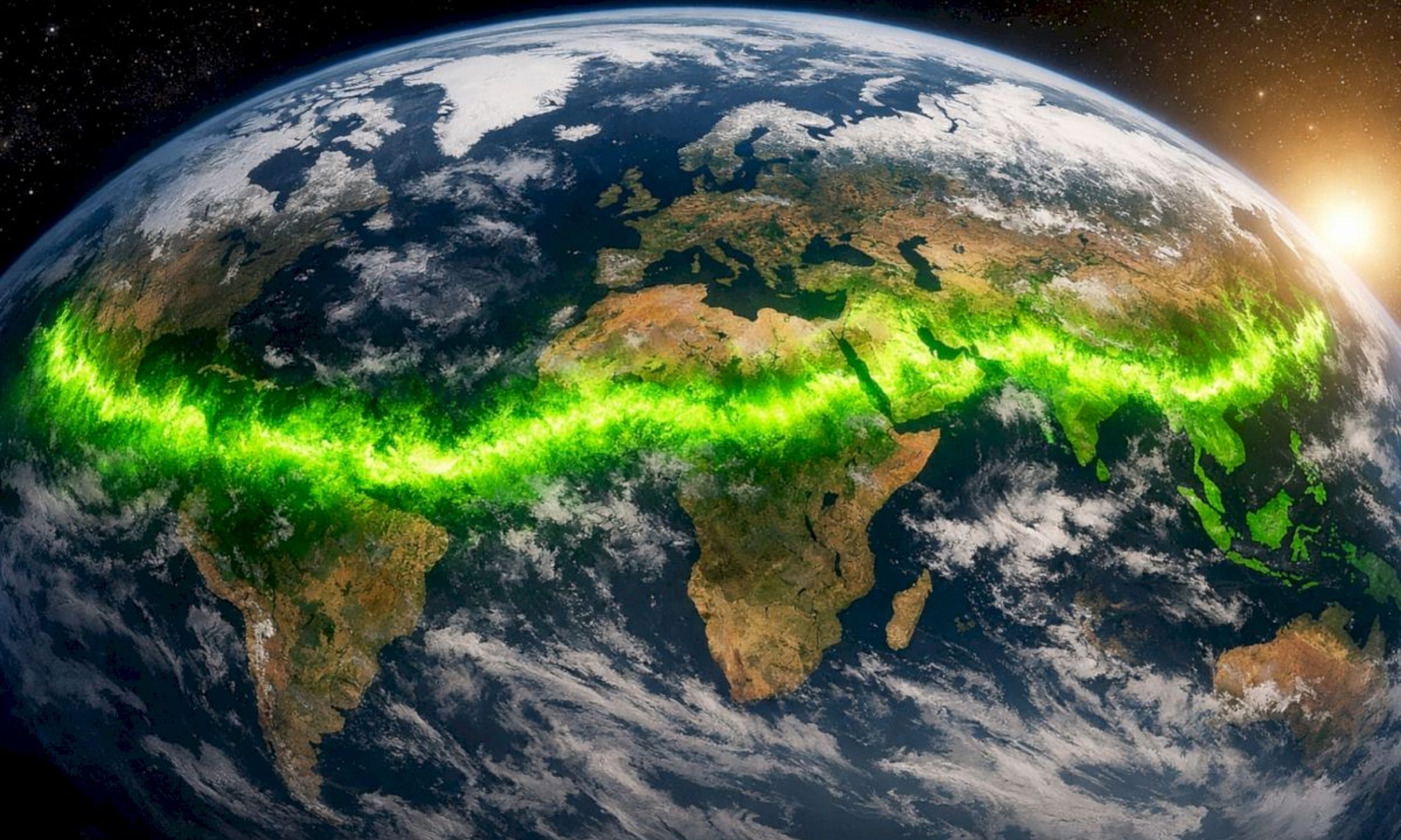
Earth’s vegetation has shifted measurably northeast over recent decades, relocating the global center of the planet’s seasonal green growth.
The finding reveals that the living surface of the planet is reorganizing in response to…
Quitting cocaine does not always silence the urge to use it again. Even after long periods of abstinence, memories tied to the drug can suddenly reignite powerful cravings.
New research shows that repeated cocaine exposure may rewire a…
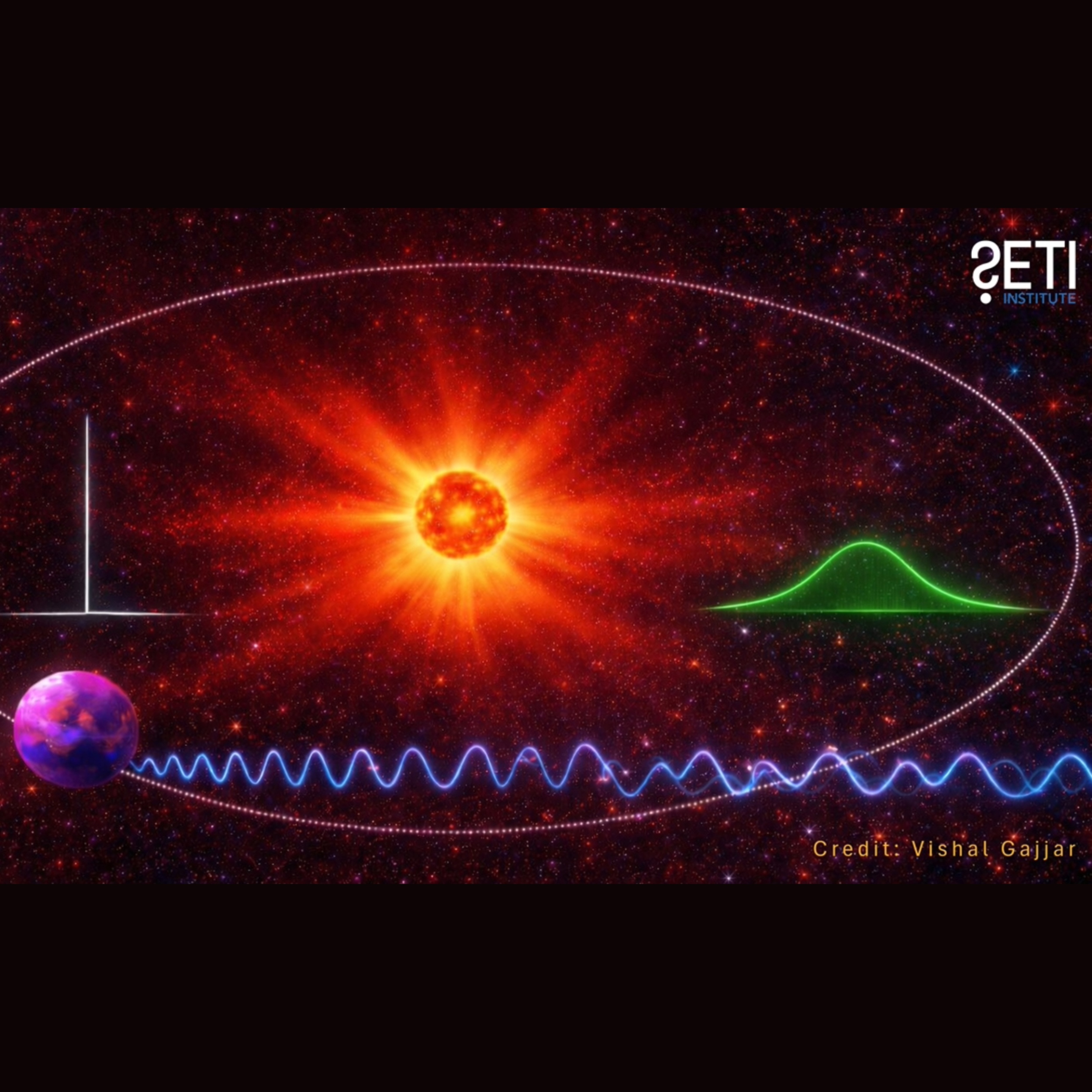
For decades, astronomers searching for extraterrestrial technology have focused on extremely narrow radio signals. Such signals rarely occur naturally, making them attractive targets in the hunt for alien transmissions.
But a new study suggests…
You’ve probably heard of Albert Einstein, Charles Darwin, and Thomas Edison, some of the most famous scientists and…
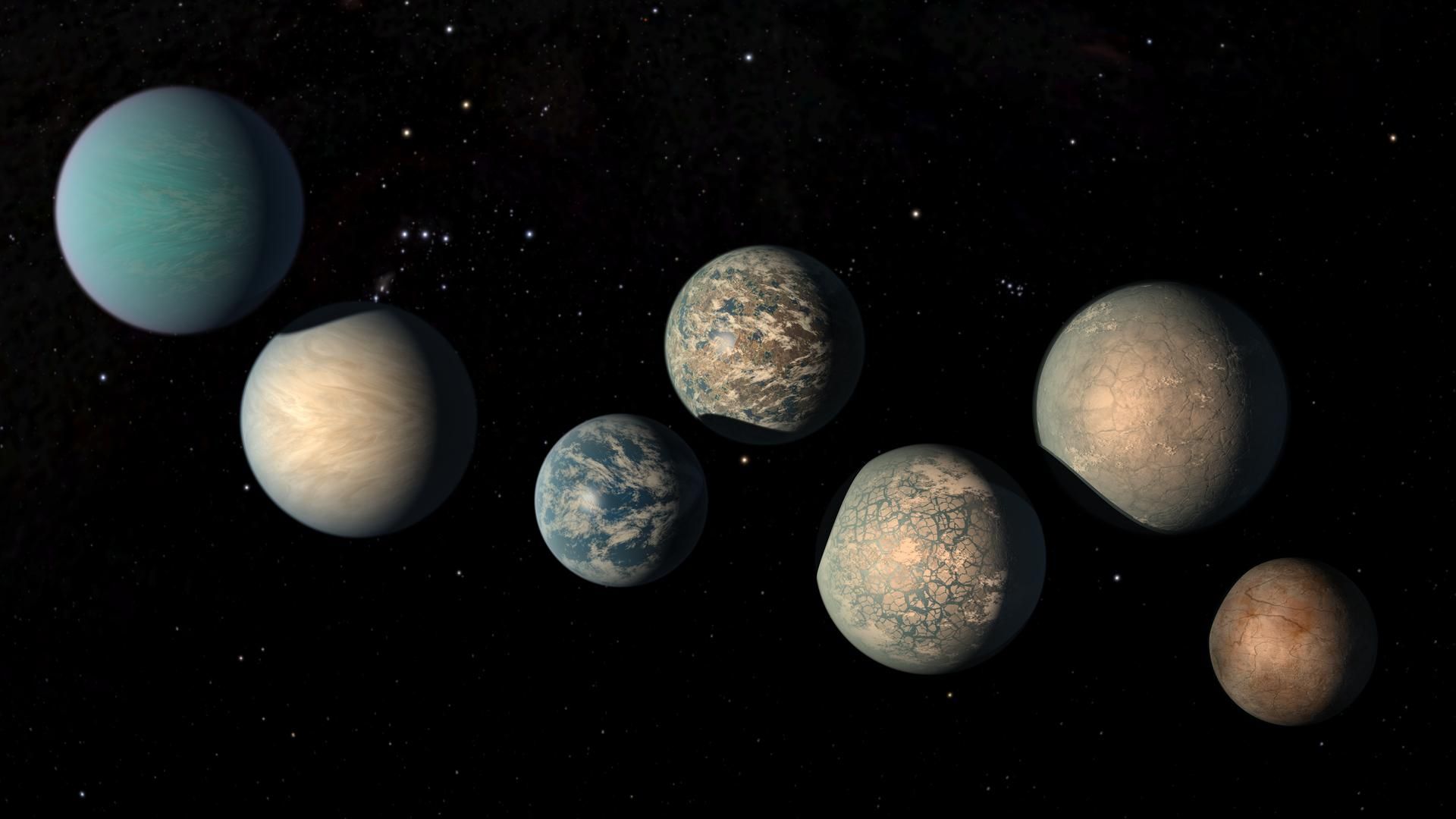
Scientists have found a potential shortcut for identifying stars that host planets. The technique, based on specific signals in starlight, could make it easier to search for exoplanets, according to a new study.
The team has already used their…
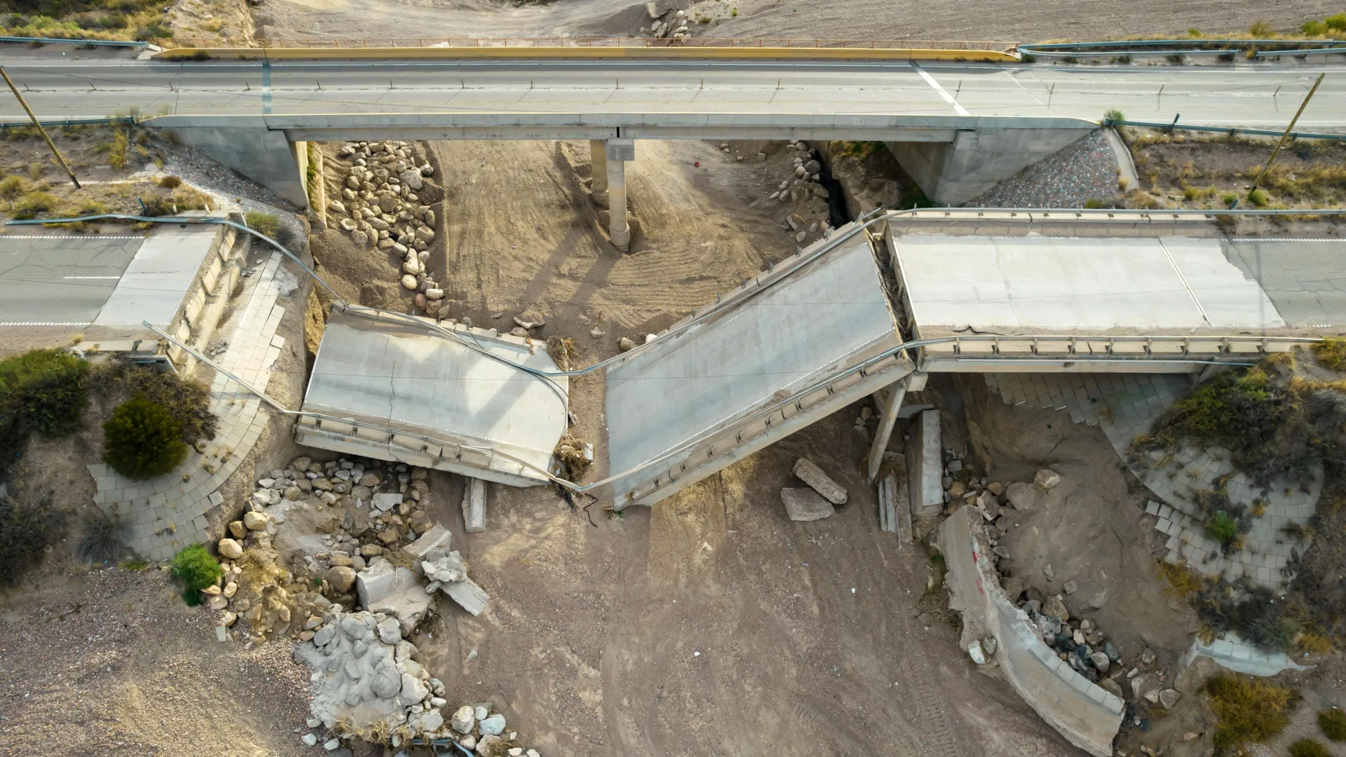
Scientists are using satellites to reveal which bridges around the world may be at risk of failure — and how to catch problems before disaster strikes.