Qaasid News
Download Our App
Latest News from Pakistan
‘I’ve Been Misinterpreted.’ Timothée Chalamet Sets The Record Straight On His ‘Quest’ For An Oscar
February 3, 2026
China unveils humanoid 'Bolt' – video shows it's earned its name – news.cgtn.com
February 3, 2026
Best Workouts for Longevity: 4 Weekly Training Schedules
February 3, 2026
Will Japanese Stocks Rally to Fresh Record Highs?
February 3, 2026
China condemns Balochistan attacks, says it will always support Pakistan in combatting terrorism – Dawn
February 3, 2026
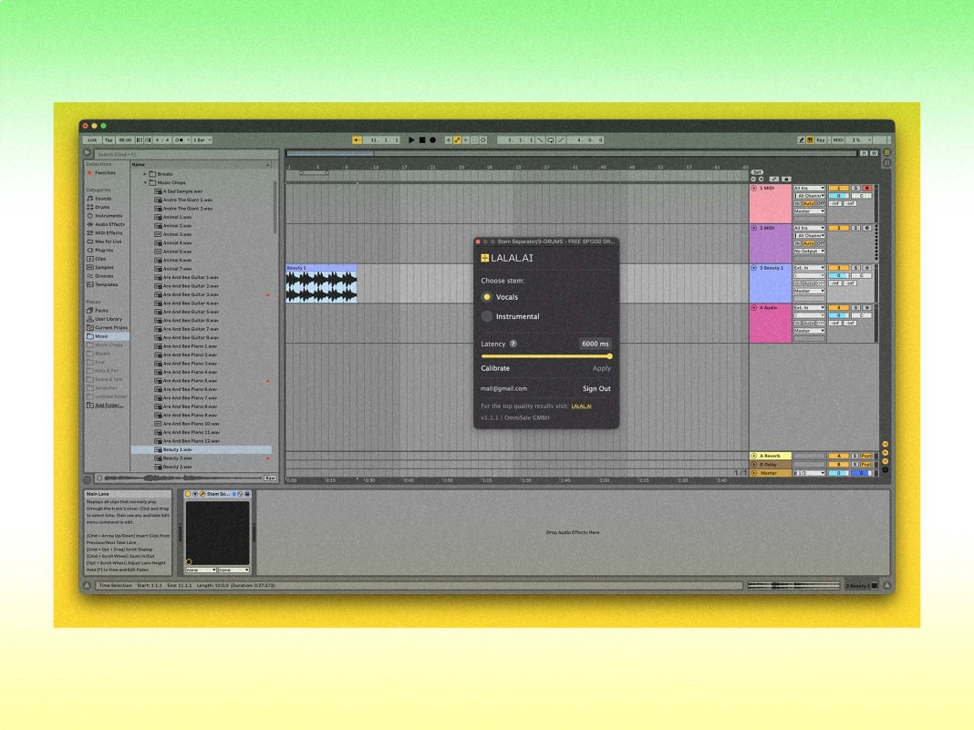
LALAL.AI launches its first stem-splitting VST plugin
February 3, 2026
Crude Picks Up As U.S.-India Trade Deal to Affect Russian Oil – The Wall Street Journal
February 3, 2026
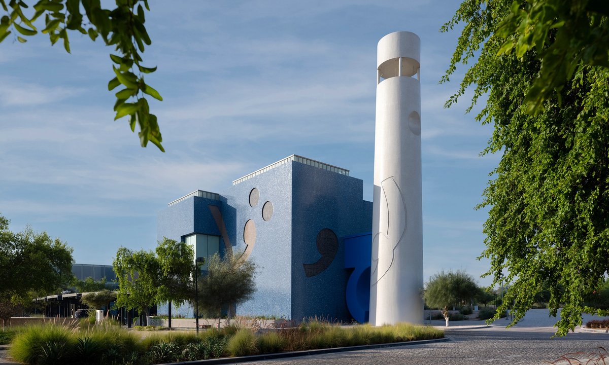
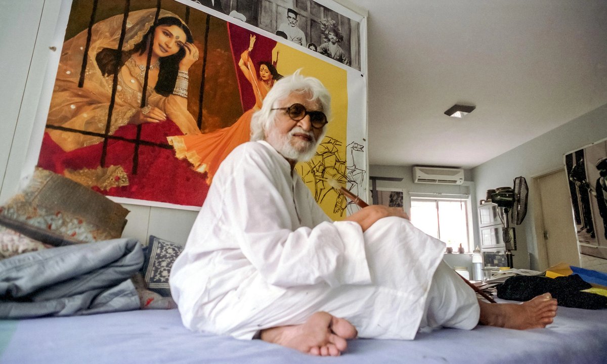
New Doha museum celebrates M.F. Husain’s global legacy – The Art Newspaper
February 3, 2026
M.F. Husain in Qatar: bridging Asia and the Arab world – The Art Newspaper
February 3, 2026
Special control room set up for Basant safety: CM Punjab – RADIO PAKISTAN
February 3, 2026