Qaasid News
Download Our App
Latest News from Pakistan
Microsoft’s AI Efforts Are Faceplanting
February 7, 2026
Access Denied
February 7, 2026
Kon Knueppel To Compete In 2026 KIA Shooting Stars Competition – NBA
February 7, 2026
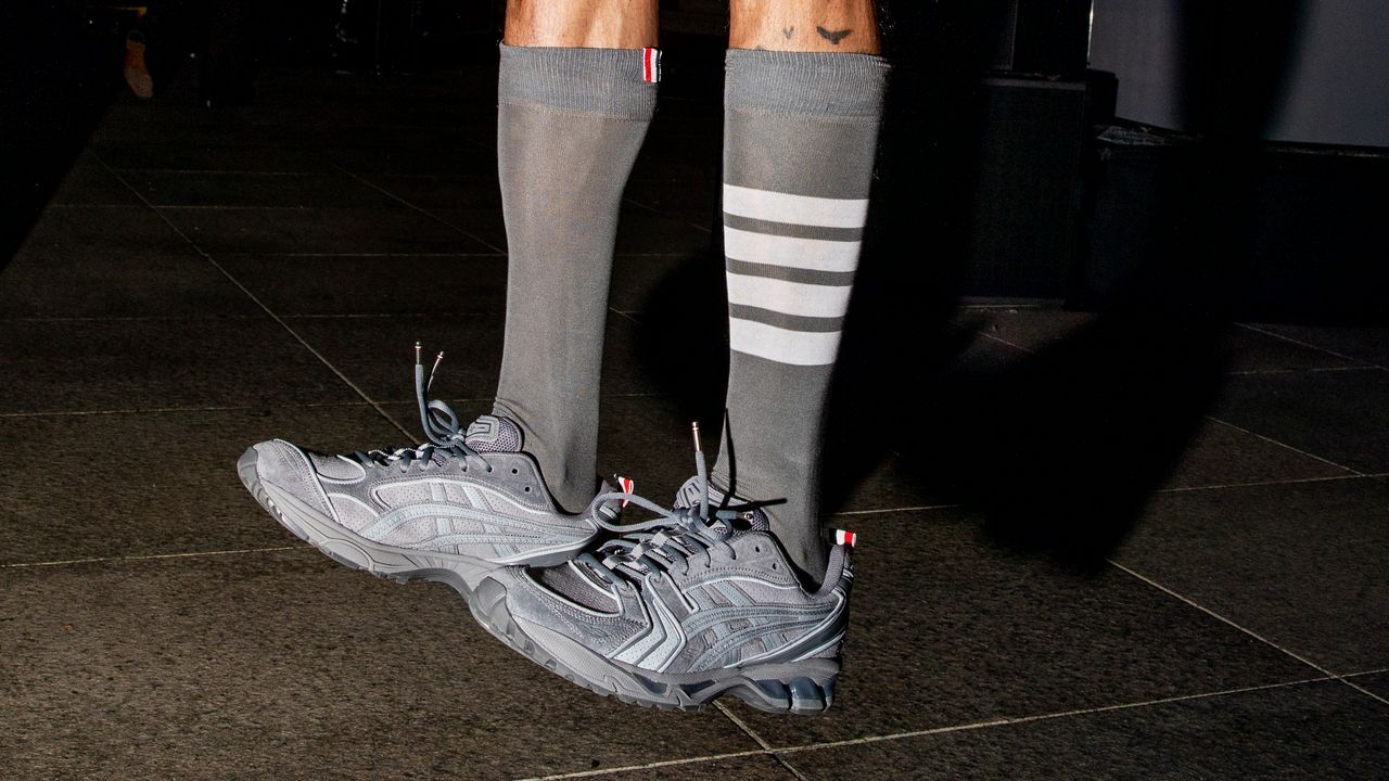
Thom Browne Sent a Surprise Asics Collab Down the GQ Bowl Runway
February 7, 2026
Bournemouth 1-1 Aston Villa (Feb 7, 2026) Game Analysis
February 7, 2026
Scientists show how to narrow the hunt for merging giant black holes
February 7, 2026
Brendan Behan ‘took the language of the streets and proved it belonged on the page’ – The Irish Times
February 7, 2026
Suspected RSF drone attack on aid convoy kills dozens
February 7, 2026
That dry, bitter taste may be waking up your brain
February 7, 2026
NBA announces participants for 2026 AT&T Slam Dunk Contest
February 7, 2026