Qaasid News
Download Our App
Latest News from Pakistan
Amy Yang Finishes Runner-Up to Nelly Korda at 2026 Hilton Grand Vacations Tournament of Champions – LPGA
February 2, 2026
New Punjab police chief on the cards – Dawn
February 2, 2026
Firefox will soon let you block all of its generative AI features
February 2, 2026
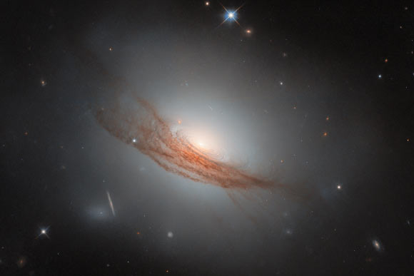
Hubble Space Telescope Spies Beautiful Lenticular Galaxy: NGC 7722
February 2, 2026
Requiem for a film-maker: Darren Aronofsky’s AI revolutionary war series is a horror | Darren Aronofsky
February 2, 2026
MTG Arena Announcements – February 2, 2026
February 2, 2026
Jack Schlossberg Says Donald Trump Might ‘Demolish’ the Kennedy Center, but His Grandpa JFK Can Be ‘Kept Alive’ Another Way
February 2, 2026
Traffic alert: Key capital roads to face diversions tomorrow evening
February 2, 2026
Trump administration sued over pause on immigrant visa processing – Reuters
February 2, 2026
Access Denied
February 2, 2026