Qaasid News
Download Our App
Latest News from Pakistan
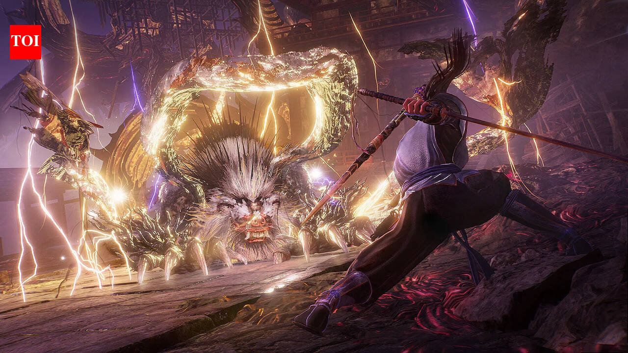
Nioh 3 Trophy List (2026): Full achievement guide and unlock requirements | Esports News
February 7, 2026
Human redheads and orange birds share a cellular ‘superpower’
February 7, 2026
‘Can Mette-Marit be queen after this?’: Rape trial and Epstein files bring double crisis for Norway’s royals | Norway
February 7, 2026
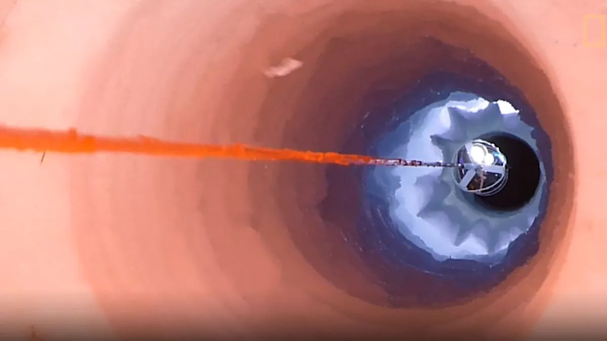
Scientists make concerning discovery while studying ocean hidden under ice — here are the details
February 7, 2026
NASA discovers a massive ‘starless’ cloud in deep space
February 7, 2026
Experts Say These 4 Supplements May Support Gut Health
February 7, 2026
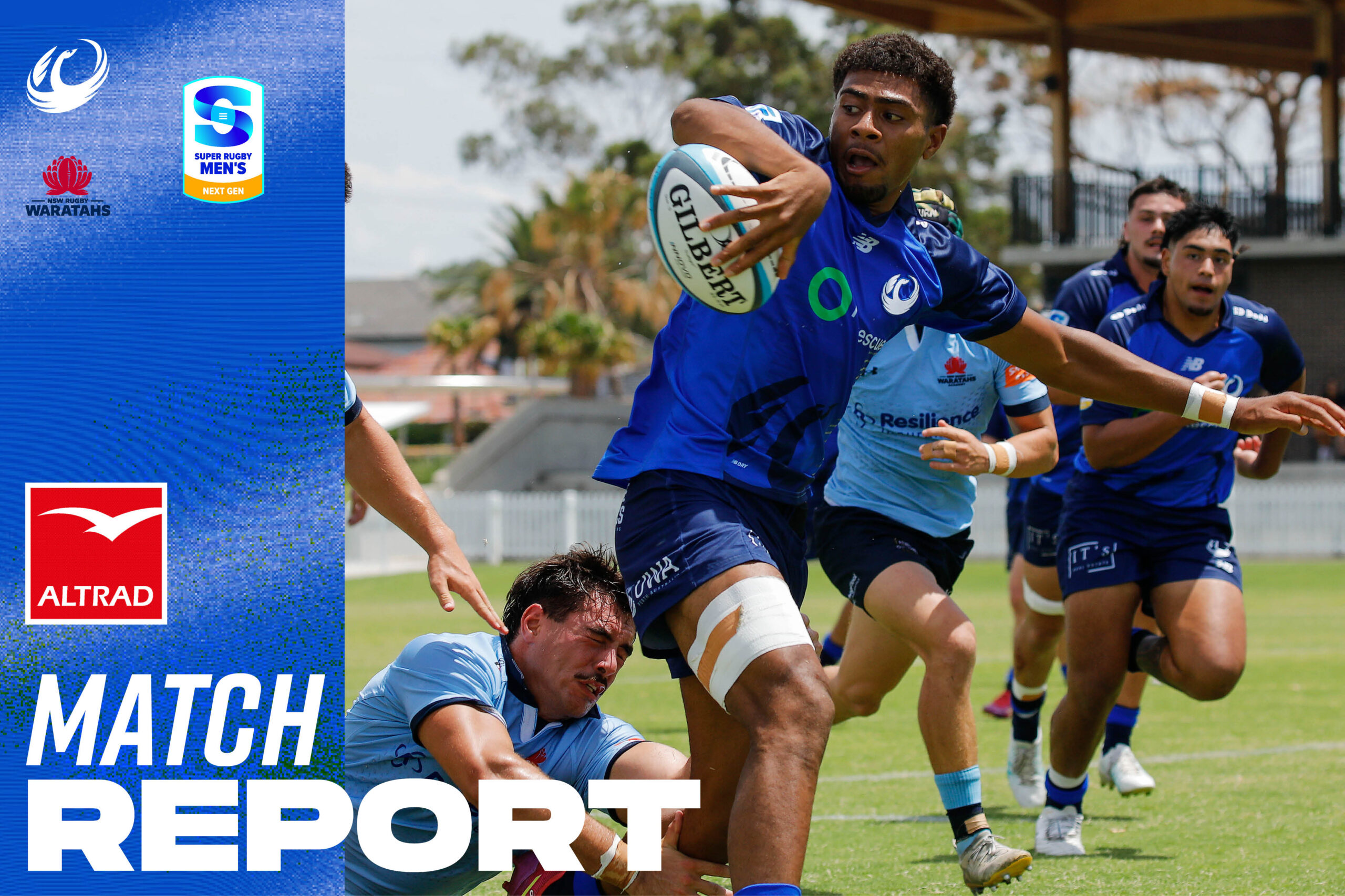
Force downed by ruthless Waratahs in Next Gen opener
February 7, 2026
New forecasts offer early warning of Arctic sea ice loss
February 7, 2026
New forecasts offer early warning of Arctic sea ice loss
February 7, 2026
Heterogeneous multicopy of blaCTX-M variants on the same plasmid enhances evolutionary adaptability in clinical Klebsiella pneumoniae
February 7, 2026