Qaasid News
Download Our App
Latest News from Pakistan
Petroleum company begins payments after Rs47 billion fraud recovery case
February 2, 2026
Lahore Sehari, Iftari timings as Ramadan 2026 approaches
February 2, 2026
PM Shehbaz meets KP CM Sohail Afridi in Islamabad – Dawn
February 2, 2026
PM Shehbaz meets KP CM Sohail Afridi in Islamabad – Dawn
February 2, 2026
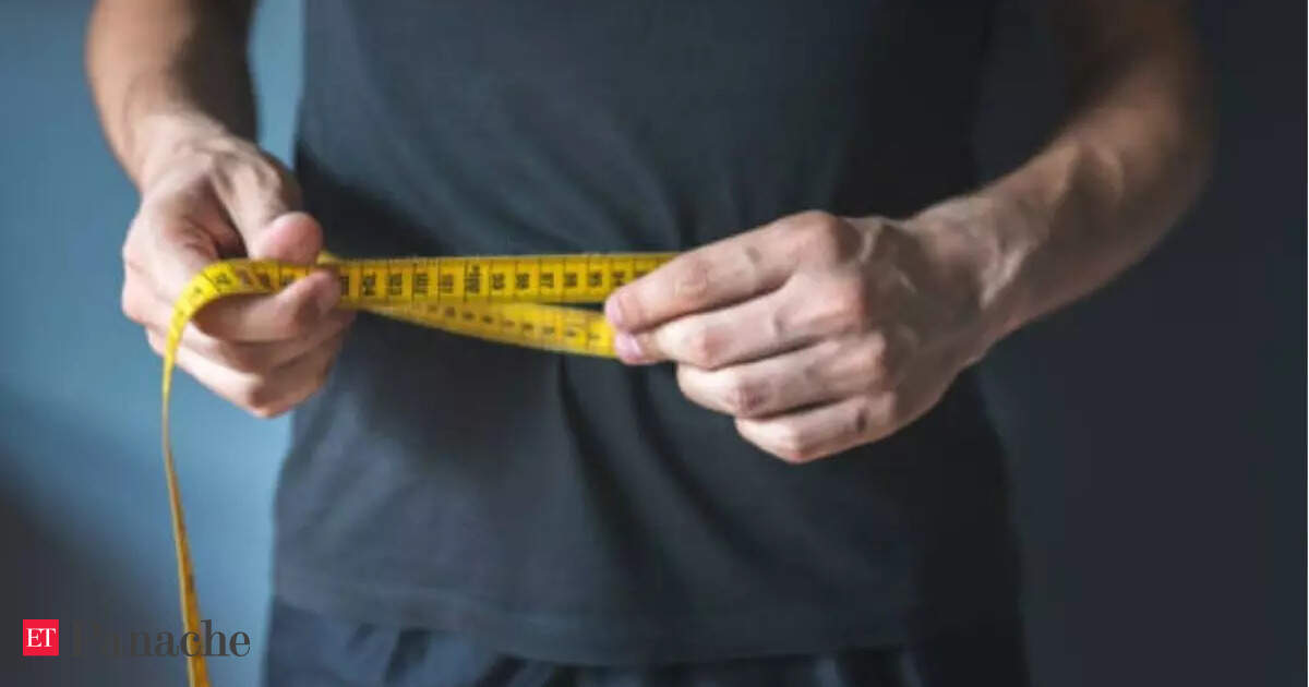
Man who once weighed 144 kilos, shares how he lost 63 kg in two years without fancy diet
February 2, 2026
Forces carrying out proactive operations against terrorists in Balochistan: Defense Analyst – RADIO PAKISTAN
February 2, 2026
Anahat Singh wins maiden PSA Bronze title after beating top seed in Squash On Fire Open 2026
February 2, 2026
Israel reopens Gaza’s Rafah border crossing to Egypt, with tight limits – Arab News
February 2, 2026
Google expands Gemini Ai in Maps navigation
February 2, 2026
her snowboard athletes to watch at Milano Cortina 2026
February 2, 2026