Qaasid News
Download Our App
Latest News from Pakistan
Tharparkar: child deaths rise to 50 as malnutrition crisis deepens
January 31, 2026
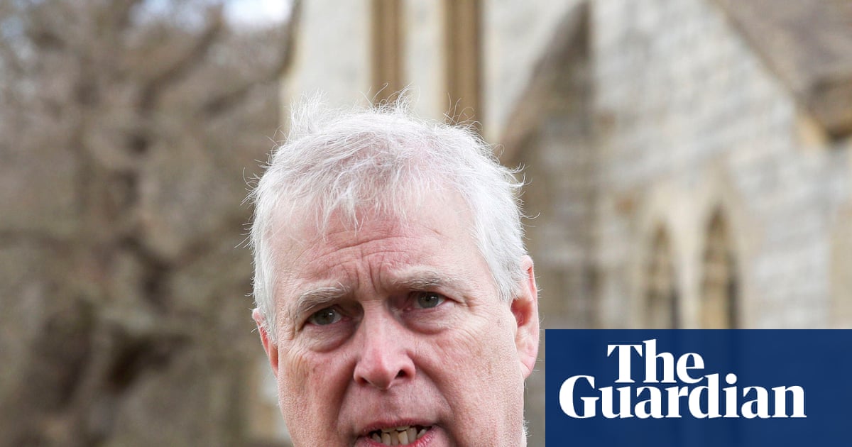
Andrew invited Epstein to Buckingham Palace after child sex offender’s release, files suggest | Andrew Mountbatten-Windsor
January 31, 2026
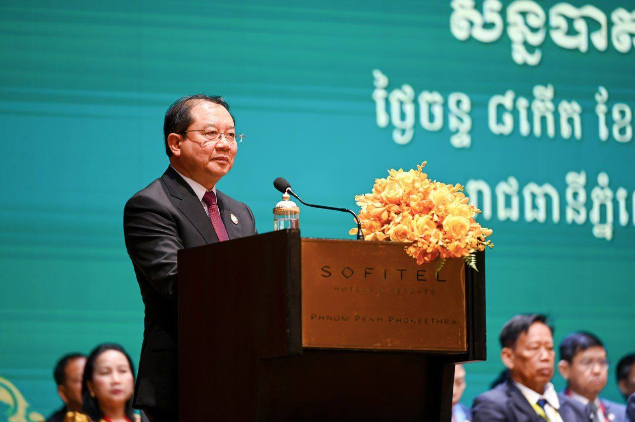
Cambodia Heightens Medical Preparedness as Nipah Virus Raises Concerns in the Region
January 31, 2026
Get your tickets for Sunday’s Champions Cup final | News
January 31, 2026
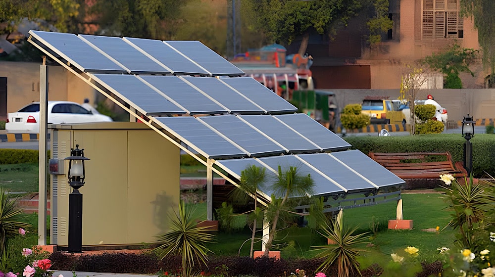
Solar Net-Metering Consumers to Witness Reduction in Benefits
January 31, 2026
Thousands flee northwest Pakistan after mosques warn of possible military action – Reuters
January 31, 2026
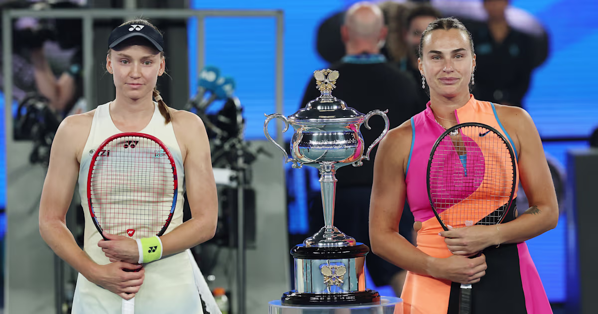
Australian Open 2026 women’s singles final: World No. 1 Aryna Sabalenka meets Elena Rybakina in 2023 rematch
January 31, 2026
Hexcel (HXL) Valuation Check As Recent Share Price Momentum Meets Conflicting Fair Value Signals
January 31, 2026
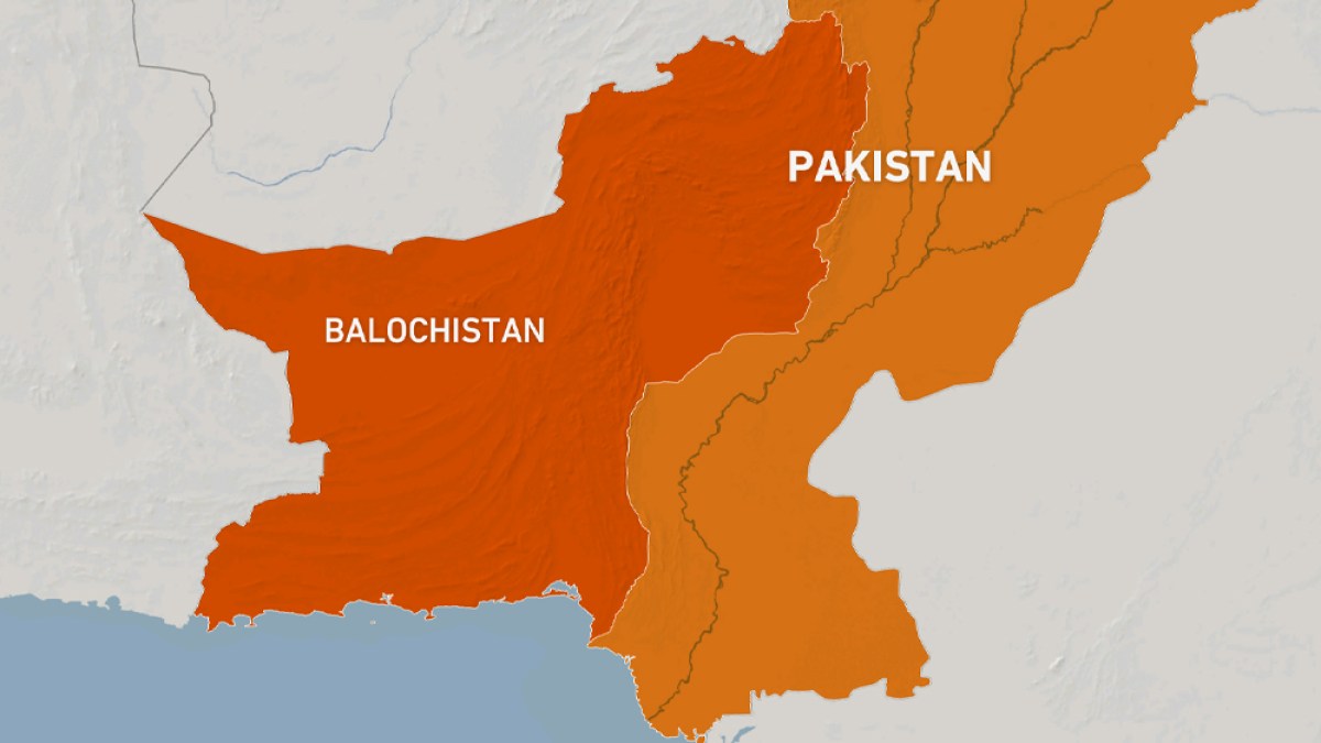
Suspected separatists kill 8 Pakistani policemen in ‘coordinated’ attacks | Conflict News
January 31, 2026
Epstein files latest: photos appear to show former prince Andrew crouching over female | Jeffrey Epstein
January 31, 2026