Qaasid News
Download Our App
Latest News from Pakistan
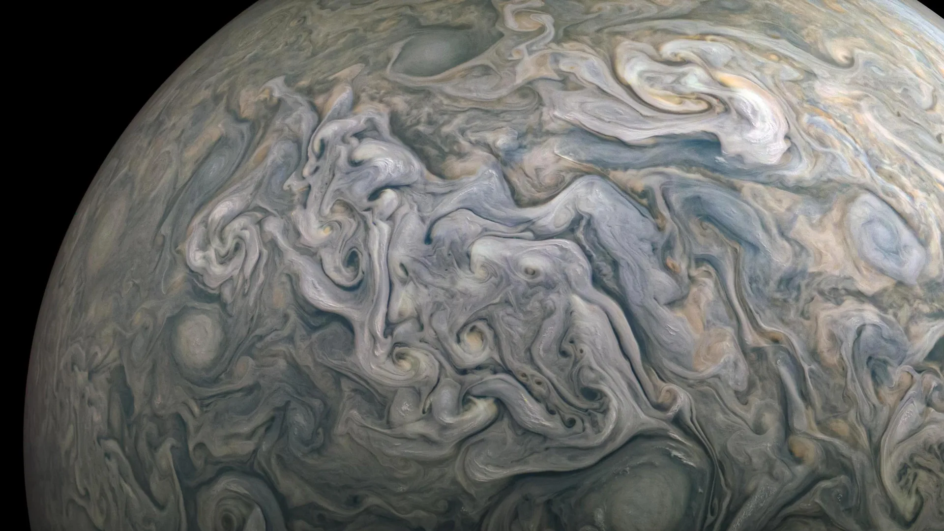
Jupiter’s clouds are hiding something big
January 31, 2026
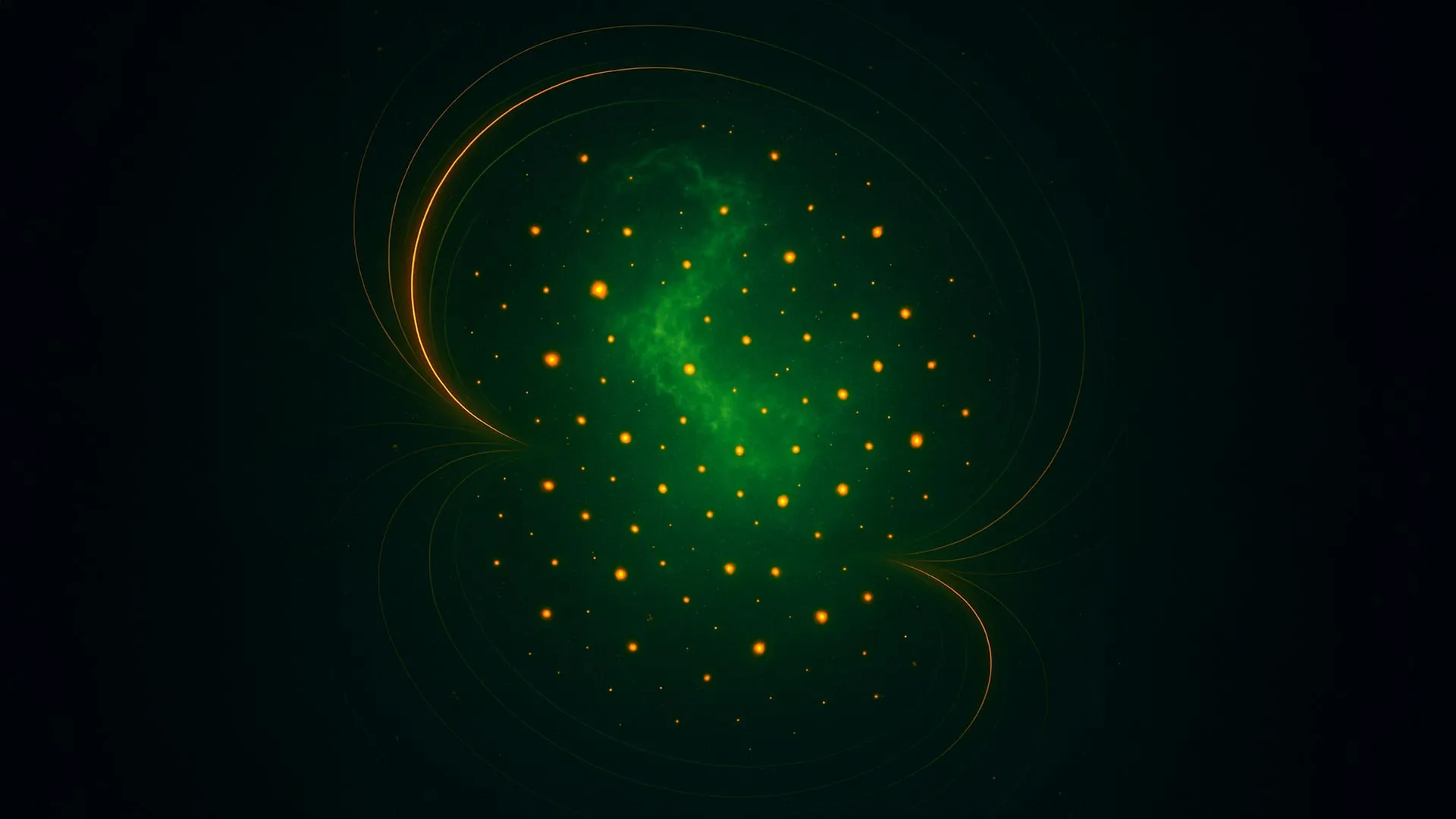
Weak magnetism causes big changes in a strange state of matter
January 31, 2026
What AI Predicts About the Future of the Stock Market — and Your Wallet
January 31, 2026
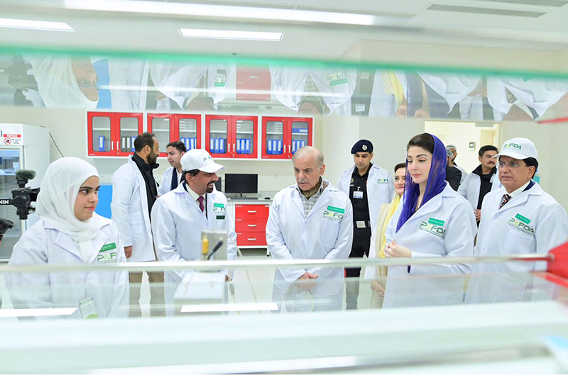
Prime Minister Muhammad Shehbaz Sharif and Chief Minister Punjab Maryam Nawaz Sharif visiting PAFDA Laboratory and receiving a briefing about the operations
January 31, 2026
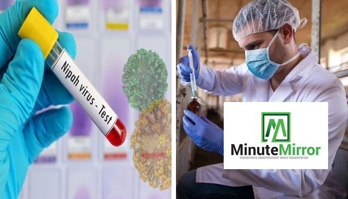
Nipah virus outbreak in Malaysia traced from bats to pigs to humans
January 31, 2026
You've Got Just One More Day to Update Your PC to Windows 11 Pro for $10 – PCMag
January 31, 2026
War against terrorism to continue till elimination of last terrorist: Naqvi – RADIO PAKISTAN
January 31, 2026
Iran: Blast rips through building in port city of Bandar Abbas, state media says
January 31, 2026
Pakistan PM inaugurates Punjab food, agriculture and drug authority
January 31, 2026
Is Campbell’s (CPB) Share Price Slide Creating A Long Term Opportunity For Investors
January 31, 2026