Qaasid News
Download Our App
Latest News from Pakistan
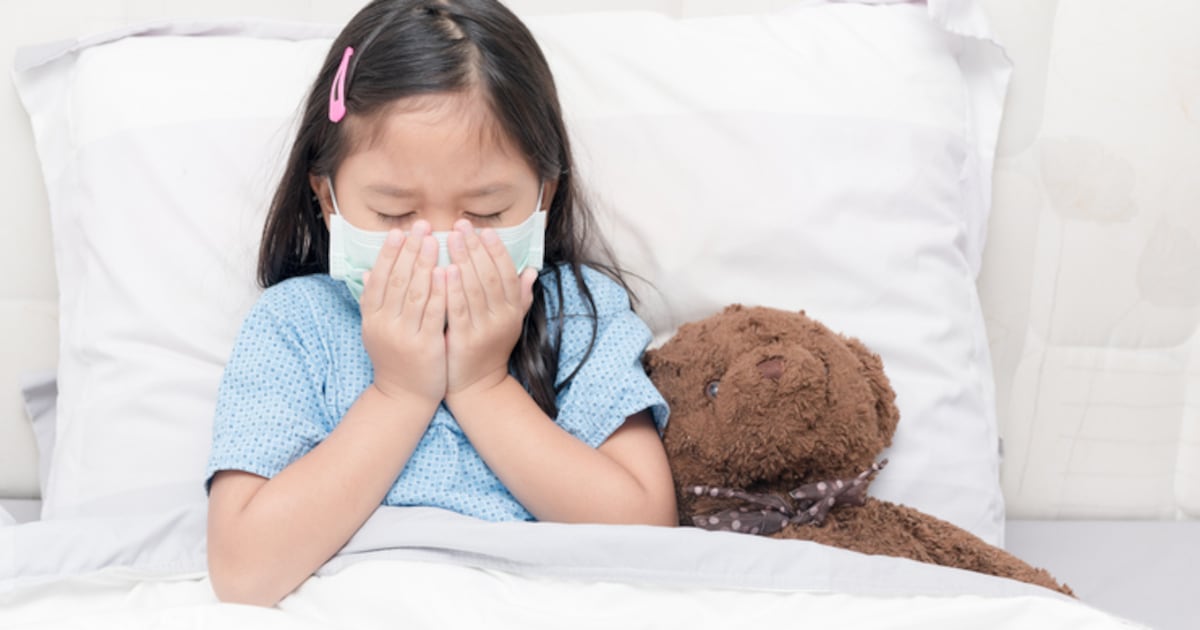
Probiotics alleviate allergic rhinitis in kids
December 22, 2025
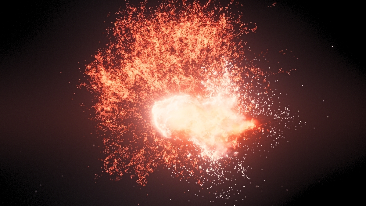
Asteroids Seen Colliding Near a Neighboring Star 25 Light-Years Away : ScienceAlert
December 22, 2025
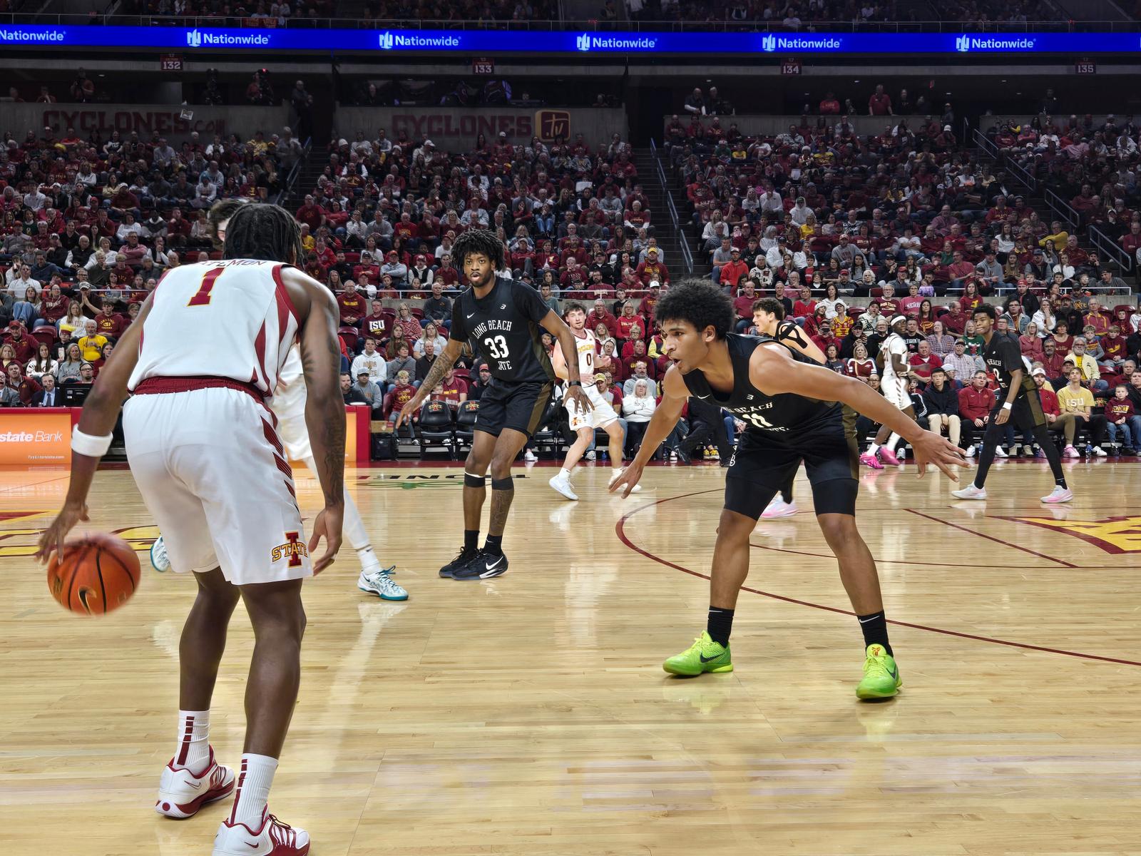
Long Beach State Falls At No. 4 Iowa State
December 22, 2025
Karachi Climate March 2025 demands end to fossil projects
December 22, 2025
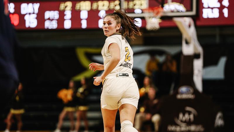
Women’s basketball closes non-conference play at No. 21 Ohio State
December 22, 2025
Actor James Ransone, known for his roles in ‘The Wire,’ ‘It: Chapter Two,’ dead at 46
December 22, 2025
Washington 90-50 Pacific (Dec 21, 2025) Game Recap – ESPN
December 22, 2025
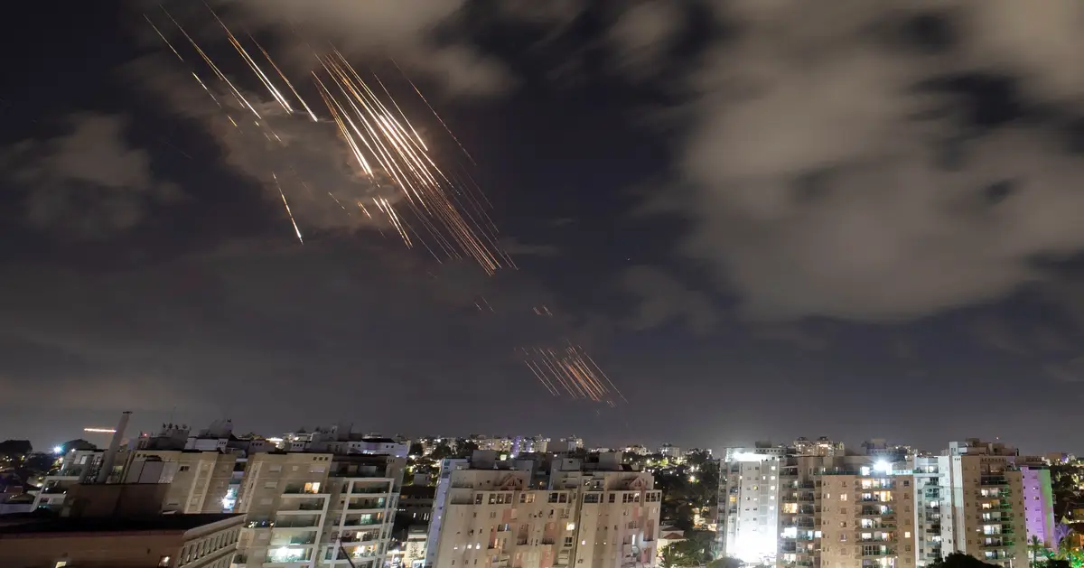
Israel warns US Iran missile drills may be cover for surprise attack – Axios
December 22, 2025
ISU MBB vs. LBSU Box Score – Iowa State Athletics
December 22, 2025
Men’s Basketball Closes Out 2025 At Saint Joseph’s
December 22, 2025