Qaasid News
Download Our App
Latest News from Pakistan
KP CM Afridi reaches SC to request CJP to facilitate meeting between Imran Khan and physicians – Dawn
January 30, 2026
Study links weekly nightmares to accelerated ageing and early death
January 30, 2026
PAG Completes Sale of Anyang Nitrogen Fertilizer to Indorama Corporation
January 30, 2026
President, PM vow to eradicate menace of terrorism – RADIO PAKISTAN
January 30, 2026
Self-adhesive halogen-free conductive textile gaskets
January 30, 2026
Award-winning Palestinian journalist Bisan Owda’s TikTok account restored after reported ban – Images Dawn
January 30, 2026
Security forces kill 41 terrorists in Balochistan – RADIO PAKISTAN
January 30, 2026
Julie Campiche: Unspoken review – a harpist’s tender, quietly radical hymn to women who endure | Jazz
January 30, 2026
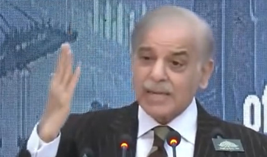
PM announces Rs4.4 cut in industrial electricity prices
January 30, 2026
SpaceX launches Starlink satellites from two coasts in two days
January 30, 2026